
Analysis of Major Histocompatibility Complex Class I
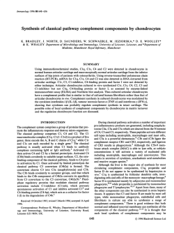
From www.bloodjournal.org by guest on February 6, 2015. For personal use only. Analysis of Major Histocompatibility Complex Class I Expression on Reed-Sternberg Cells in Relation to the Cytotoxic T - c e l l Response in Epstein-Barr Virus -Positive and -Negative Hodgkin’s Disease By J.J. Oudejans, N.M. Jiwa, J.A. Kummer, A. Horstman, W. Vos, J.P.A. Baak, Ph.M. Kluin, P. van der Valk, J.M.M. Walboomers, and C.J.L.M. Meijer To get insight into the failure of the immune system to eradicate Epstein-Barr virus (EBV) harboring Hodgkin and ReedSternberg cells (H-RS cells), expressing the latent membrane protein 1 (LMPl), we analyzed major histocompatibility complex (MHC) class I expression on H-RS cells in relation to the presence of activated cytotoxic cells, ie, granzymeB-expressing lymphocytes. H-RS cells in EBV+ cases of Hodgkin‘s disease (HD) were found to express significantly higher levels of MHC class I heavy- and light-chain molecules compared with EBV- HD cases. When low levels of MHC class I expression were found (mainly in EBV- cases), these were not associated with low levels of the transporter protein associated with antigen presentation 1 (TAP-1). The rel- atively high levels of MHC class I expression in H-RS cells in EBV+ HD cases were accompanied by significantly higher numbers of activated cytotoxic T lymphocytes (CTLs) as shown by the presence of increased numbers of CD8 and granzyme B+ lymphocytes. However, these cells were only sporadically detected in the close vicinity of the H-RS cells. These data suggest that mechanisms other than downregulation of MHC class I or TAP-1 expression on H-RS cells are involved in the failure of the immune system to eradicate EBV harboring H-RS cells. Probably, the function of activated CTLs is locally inhibited by the H-RS cells or by reactive cells in the vicinity of the H-RS cells. 0 1996 by The American Society of Hematology. T and natural killer (NK) cells. Recently, monoclonal antibodies (MoAbs) became available that react specifically with g r a n ~ y m e s .These ’~ serine proteases are found in the cytoplasmic granules of CTLs and NK cells. Exocytosis of these granules in the cytoplasm of the target cell will lead to induction of DNA fragmentation and apoptosis of the target ce~~.lc.16 Because expression of these proteins is restricted to NK cells and activated CTLs.” antibodies directed against granzymes are an excellent tool for in vivo detection of these cells. CTLs recognize endogenously processed antigens, such as viral proteins and oncogene products, only when presented at the cell surface as peptides bound to the MHC-I molecules.” A reduction or lack of MHC-I surface expression can render aberrant cells resistant to CTLs, and may provide a selective growth advantage for these cells. Such an immune escape mechanism has been demonstrated for cervical carcinomas that harbor the human papilloma virus.” In these carcinomas MHC-I downregulation was associated with the loss of function of one of the transporter proteins associated with antigen presentation (TAP- 1 ).2(’ Lack of peptide transport by TAP will inhibit peptide loading of empty class I molecules, thereby severely reducing the complex stability. Consequently, steady-state levels of MHC-I will be reduced. A similar mechanism might be involved in the immune escape of EBV-harboring neoplastic cells in EBV’ cases of HD. The activation of CTLs depends on the presence of certain cytokines, among others, IL-2 and IFN-y,”.” which are secreted by activated T-helper type 1 cells (Thl cells). Activation of these T-helper cells depends on antigen presentation in the context of MHC class I1 (MHC-11) molecules by antigen presenting cells.23Occasionally, other nonlymphoid cells like neoplastic epithelial cells have been described to express MHC-11, which might assist in the onset of the cellular immune response.” The aim of the study was to get insight in the cellular immune response against H-RS cells by analyzing the cellular composition around H-RS cells in relation to the expression of MHC-I, MHC-11, and TAP- 1 on H-RS cells in EBV’ and EBV- cases of HD. HE DIAGNOSIS OF Hodgkin’s disease (HD) is based on the presence of typical Reed-Stemberg cells (RS cells) and their mononuclear variants, Hodgkin cells (H cells), in an appropriate cellular environment of “reactive” cells.’ The presence of Epstein-Barr virus (EBV) in the neoplastic cells has been shown in approximately 40% of all cases of HD, depending on histologic type.* In these EBVharboring cells expression of a restricted set of viral latent genes was detected, ie, EBV nuclear antigen 1 (EBNAI) and three latent membrane proteins (LMPI , LMP2A, and LMP2B).3-6Despite the fact that LMPl and LMP2 contain epitopes recognized by cytotoxic T lymphocytes (CTLs) when presented in the context of appropriate major histocompatibility complex class I (MHC-I) molecule^,^^' HD patients apparently cannot raise an effective immune response against these EBV-harboring neoplastic cells. There is some evidence that patients with HD exhibit a defect in cell-mediated immunity.’,’”These defects include an impaired response to T-cell mitogens” and a decreased capacity of T cells to respond in an autologous mixed lymphocyte response.‘*Moreover, in vitro studies showed a decreased synthesis of interleukin-2 (IL-2) and interferon-? (IFN-y) (both cytokines with a T-cell-stimulating effect) in untreated patients with HD.” However, the contribution of this immune defect to the pathogenesis of HD is still obscure. The major effector cells in cellular cytotoxicity are CTLs From the Department of Pathology, Free University Hospital Amsterdam, and the Department of Pathology, University Hospital Leiden, Leiden, The Netherlands. Submitted August 24, 1995; accepted December 11, 1995. Supported by u grant from the Dutch Cancer Society (VU 94749). Address reprint requests to J.J. Oudejuns, MD, Department oj Pathology, Free University Hospital, PO Box 7057, 1007 MB Amsterdam, The Netherlands. The publication costs of this article were dejrayed in part by page charge payment. This article must therefore be hereby marked “advertisement” in accordance with 18 U.S.C. section 1734 solely to indicate this fact. 0 1996 by The American Society of Hematology. 0006-4971/96/8709-0044$3.00/0 3844 Blood, Vol 87,No 9 (May 1). 1996:pp 3844-3851 From www.bloodjournal.org by guest on February 6, 2015. For personal use only. 3845 MHC CLASS I EXPRESSION ON RS CELLS The results show that, compared with EBV- cases of HD, H-RS cells in EBV+ cases express high levels of MHC-I. Although these high MHC-I expression levels were accompanied by significantly higher numbers of activated CTLs (ie, CD8 and granzyme B+ cells), these cells were only sporadically detected in the close vicinity of the H-RS cells. These data suggest that the failure of the immune system to eradicate EBV-harboring H-RS cells is due to local inhibition of the function of activated CTLs by H-RS cells or by reactive cells in the vicinity of H-RS cells. MATERIALS AND METHODS Table 2. MoAbs Used in This Study Antibody Antigen Titer P/Fx HCA2 HCIO B2M W6/32 LN3 HLA-A HLA-B, C µglobulin MHC-I complex TAP-I TAP-I CD3 CD3 1:400 1:50 P/F P/F OPD4 CD45R05 1:25 P/F HLA-DR 1:500 1:1,000 1:40/1:100 1:25 1:25§ P/F P/F P/F F P/F Pretreatment1 10 min 95°C TUFS - 10 min 100°C lead thiocyanate 10 min 95°C TUF 10 min 100°C citrate 10 min 100°C water, trypsin 10 min 100°C citrate 10 min 100°C citrate CD8 CD8 1:511 P/F Tissues. Patients with HD were selected from the files of the Department of Pathology of the Free University Hospital Amsterdam GrB7 Granzyme B 1:50 P/F (Amsterdam, The Netherlands; N = 34) and the Department of Pathology, University Hospital Leiden (Leiden, The Netherlands; N 1:lOO LMP-I P/F = 29). Cases were classified according to the Rye clas~ification.~~ SI2 1:lOO CSI-4 LMP-1 P/F Staging of the patients was done according to the Ann Arbor classificationZ6Patient and tumor characteristics are summarized in Table * P, paraffin sections; F, frozen sections. 1. Representative tissue specimens were fixed in buffered formaldet Only for formalin-fixed, paraffin-embedded tissue sections. hyde or in a sublimate formaldehyde mixture. Paraffin-embedded *Target Unmasking Fluid, Kreatech, Amsterdam, The Netherlands. tissues were cut to 5-pm thick sections and mounted on 3-amino5 Incubations were performed overnight. propyl-triethoxy-silane (APES; Sigma, St Louis, M0)-coated slides 11 Strains mainly CD4’ cells, is not reactive with monocytes. for RNA in situ hybridization (RISH) and on poly-L-lysine-coated slides for immunohistochemical staining. When available, frozen tissue sections, fixed in pure acetone for 15 minutes, also were endogenous peroxidase was blocked, and sections were pretreated investigated. In total, frozen tissue sections of 9 EBV’ and 9 EBVaccording to the appropriate protocol for the different antibodies. cases could be examined. Histologic features and EBV status have Frozen sections were not pretreated, but transferred after acetone partly been described elsewhere.” fixation to phosphate-buffered saline (PBS). Both paraffin and frozen Detection of the presence of EBV in H-RScells. The presence sections were subsequently washed, preincubated with normal seof EBV in H-RS cells was determined by RISH using the abundantly rum, and incubated with the primary antibody. To visualize the transcribed noncoding EBV small RNAs (EBERI and 2) as has been antibodies, a routine ABC immunoperoxidase method was used. The described previouslyz7and by the detection of LMPl using two sets following antibodies were used: MoAb HCA2, reactive with HLAof MoAbs (CSI-4: DAKO, Glostrup, Denmark and S12: Organon A locus productsz9; MoAb HC 10, preferentially recognizing HLATeknika, Oss, The Netherlands).” B/C locus products2’; polyclonal anti-µglobulin B2M, recImmunohistochemistry. Immunohistochemical analysis for MHC ognizing free and complexed @,-microglobulin (DAKO); MoAb expression, phenotypic lymphocyte markers, and granzyme B exLN3 (Biotest Seralc, Brussels, Belgium), specific for HLA-DR; W6/ pression was performed as described p r e v i o ~ s l y ?Briefly, ~ formalin32 (Seralab, Sussex, UK), recognizing HLA-A, -B, and -C locus fixed, paraffin-embedded sections were deparaffinized with xylene, products, complexed to µglobulin; polyclonal antihuman Table 1. Patient and Tumor Characteristics EBV Status Negative (n = 381 Age (range) Sex M/F Histology NS MC LP LD Unclassifiable Stage of presentation Stage I Stage II Stage 111 Staae IV 36, 7 ( 14-73) 19/19 30 5 2 1 0 Positive In = 25) 38, 9 (10-78) 17/8 16 6 1 0 2 6 27 15 1 4 2 7 1 Abbreviations: NS, nodular sclerosing; MC, mixed cellularity; LP, lymphocyte predominant; LD, lymphocyte depleted. TAP- 1 Iy; polyclonal anti-CD3 (DAKO); because no CD4-specific MoAbs are available for paraffin-embedded material, the OPD4 MoAb (DAKO) was used. This MoAb preferentially detects CD4+ T helper cells, but was officially assigned to the CD45RO cluster‘o.”; MoAb anti-CD8 (a generous gift from Dr D.Y. Mason, Oxford, UK); and MoAb GrB7, raised against recombinant granzyme B protein and specific for human granzyme B.14 MoAb GrB7 reacts with isolated granzyme B from activated cytotoxic lymphocytes on immunob10t.I~In addition, GrB7 detects granzyme-B-expressing cells in routinely formalin-fixed paraffin-embedded tissue sections.3z Pretreatment and incubation conditions are summarized in Table 2. Phenotypical analysis of grunzyme-B-expressing cells. In five cases, double stainings were performed for either granzyme B and CD3, granzyme B and OPD4, or granzyme B and CD8. After boiling in a citrate buffer for 10 minutes, sections were incubated with GrB7, followed by incubation with biotin-labeled goat-antimouse IgG2a (Southem Biotechnology Associates Inc, Birmingham, AL). Subsequently, sections were incubated with peroxidase-conjugated streptavidin. The peroxidase was visualized by incubation for 10 minutes in 0.2 mg/mL DAB, 0.002% H202,0.07% NiCll in 50 mmol/L TrisHCI, pH 7.6. After blocking the remaining peroxidase activity with 0.3% H202-methanol for 15 minutes, sections were incubated with either CD3-, OPD4-, or CD8-specific antibodies. Subsequently, the sections were incubated with either biotin-labeled swine-antirabbit From www.bloodjournal.org by guest on February 6, 2015. For personal use only. 3846 OUDEJANS ET AL (DAKO) for CD3 or biotin-labeled goat-antimouse-IgG1 (DAKO) for OPD4 or CD8, followed by incubation with the streptavidinbiotin-horseradish peroxidase complex. Visualization was done with diaminobenzidene (DAB)/H202, resulting in a clear brown staining signal. Subsequently, silver enhancement of the DAB-nickel precipitate was performed as described previo~sly,’~ resulting in a black staining signal for granzyme-B expression. Negative controls included simultaneously processed sections with omission of the GrB7 and CD3-, OPD4- or CD8-specific antibodies, respectively. Interpretation of immunohistochemical stainings. Evaluation of the staining levels was performed as described el~ewhere’~:only those tissue sections in which the surrounding reactive cells showed homogeneous staining were analyzed. The staining intensities observed in surrounding lymphocytes and histiocytes with the different MHC markers was considered to be 2+ (moderate staining intensity). If the staining intensity was weaker, the staining was scored as 1+; if stronger it was scored as 3 + . TAP- 1 staining was scored as negative or positive. Using this protocol, the stainings were analyzed independently by three observers. In case of disagreement the staining results were reanalyzed by the observers until consensus was reached. Quantification of the relative numbers of OPD4, CD8, and GrB7+ lymphocytes was performed using a commercially available interactive video-overlay based measuring system (Q-PRODIT; Leica, Cambridge, UK). The microscopal image is recorded by the video camera and displayed on the computer screen. Selected corresponding areas in the sections stained with the respective antibodies were demarcated. In these areas 10 to 20 fields of vision were systematically randomly selected using an automatic scanning stage controlled by Q-PRODIT. In these fields 200 to 400 lymphocytes were scored positive or negative for OPD4, CD8, and GrB7 using a point sampling method with a Weibel test grid.35A 40X objective was used. This gives a final magnification of 1,200X on the computer screen, and appeared the most suitable for this purpose. The number of OPD4, CD8, and granzyme B+ cells was expressed as percentage of all lymphocytes as judged by morphology. To see whether the percentage of granzyme-B-expressing cells was merely a reflection of the number of neoplastic cells present, the number of CD30’ H-RS cells was estimated on several representative areas of the tumor. Cases were divided into four categories; less than 1 neoplastic cell per medium power field (mpf), 1 to 3 per mpf, 3 to 10 per mpf, and more than 10 per mpf. Statistical analysis. Categorical data were analyzed using the Pearson x2 test or the Fisher’s exact test when appropriate. The Mann-Whitney U-test was used to compare group means and the Spearman test was used to test correlations between the different variables, considering P < .05 as significant. All analyses were performed using the SPSS statistical software (SPSS Inc, Chicago, ILl RESULTS MHC-I, MHC-II, and TAP-I expression in H-RS cells. Frozen as well as formalin-fixed, paraffin-embedded tissue sections were available from 18 cases of HD (9 EBV+ and 9 EBV- cases). In these cases, no major discrepancies in immunohistochemical staining intensities between frozen and paraffin-embedded tissue sections were observed. Because the availability of frozen material was limited and interpretation of the immunohistochemical staining on frozen tissue sections was relatively difficult, we choose to expand the study on paraffin-embedded tissue sections only. The results of MHC-I, MHC-11, and TAP-1 expression in H-RS cells in relation to EBV status of these HD cases are Table 3. MHC-I Expression on H-RS Cells in EBV- and EBV’ Cases of HD EBV Status HLA-A 0 1 2 3 ni HLA-BIC 0 1 2 3 ni &Microglobulin 0 Negative Positive n n = 25 0 1 6 16 2 n = 25 0 2 12 11 0 n = 25 0 1 14 10 0 n = 9 0 0 9 0 n = 25 1 9 10 5 0 n = 19 17 0 2 n n 1 2 3 ni W6l32 0 1 2 3 HLA-DR 0 1 2 3 ni TAP-1 Positive Negative ni n n n 38 12 13 6 1 6 = 38 21 12 2 3 0 = 38 6 16 15 1 0 = 9 1 1 7 0 = 38 6 11 17 3 1 = 29 28 1 0 = P Value‘ All cases P < ,001 NS P < ,001 MC P = .03 All cases P < ,001 NS P < .001 M C P = .01 All cases P < ,001 NS P < .001 MC P = .04 ns ns ns Abbreviations: ni, not interpretable; ns, not significant. * As determined by Pearson x 2 test, by Fisher‘s exact test. summarized in Table 3 . In all tissue sections the reactive nonneoplastic cells served as a positive control for staining with the MHC-I-specific antibodies. Expression of MHC-I heavy chains was predominantly observed on the cell membrane. This is in agreement with a previous report showing intense membranous reactivity of both the HCA2 and HClO antibodies by immunoelectron micro~copy.~~ In most EBV+ cases, even at low magnification, the atypical cells were easily recognized because the intensity of the immunohistochemical staining was stronger than in the reactive cells (see Fig 1A and B). This in contrast to the EBV- group where the staining intensity of H-RS cells was frequently weaker than in the surrounding reactive cells (Fig 1E and F). Cases in which reactive cells were not clearly positive using the different anti-MHC and TAP-1 antibodies were considered as not interpretable. In the 25 EBV’ cases strong staining of the H-RS cells (staining intensity 2 or 3 ) was observed in 22, 23, and 24 cases for HLA-A, HLA-B/C, and ¯oglobulin, respectively, whereas in the 35 EBV- cases strong staining was From www.bloodjournal.org by guest on February 6, 2015. For personal use only. MHC CLASS I EXPRESSION ON RS CELLS 3847 . Fig 1. Three cases of HD showing differences in MHC-Iexpression. Hematoxilin counterstaining. Original magnificetion ~600. EBV+ HD expressing high levels of HLA-A (A) and HLA-B/C IB, staining intensity scored as 3+1. EBV+HD expressing normal levels of HLA-A IC) and HLA-B/C ID, staining intensity scored as 2+1. EBV-HD expressing no detectable levels of HLA-A (El and HLA-B/C IF, staining intensity scored as 01. I Fig 2. TAP-1 expression in HD. Clear cytoplasmic staining is observed in the H-RS cells end in most surrounding lymphocytes. Fig 3. Phenotype of granzyme-B-expressing cells. Black staining indicates granzyme-B expression, brown staining indicates either OPD4 or CD8 expression. Hematoxilin counterstaining. Original magnification x 900. [AI EBV+ HD case, showing many granzyme B+/CD8+cells. (B) EBV- HD case, showing many OPD4+ cells, granzyme B+ cells are OPD4-. From www.bloodjournal.org by guest on February 6, 2015. For personal use only. 3848 only observed in 7,5, and 16 cases, respectively ( P < ,001). These differences were still significant when nodular sclerosing (NS) and mixed cellularity (MC) HD were analyzed separately (see Table 3), excluding the possibility that differences in MHC-I expression are primarily related to histologic features. In contrast, no differences between EBV+ and EBV- cases were observed for MHC-I1 expression using the LN3 antibody recognizing HLA-DR. In both EBV+ and negative HD cases, strong expression of HLA-DR in H-RS cells was detected in approximately 50% of cases. TAP-1 expression was detected in the cytoplasm of H-RS cells in nearly all cases of HD tested (Fig 2). In only 1 EBV- case no TAP- 1 positive H-RS cells could be detected, whereas the surrounding reactive cells showed clear positive signals. This TAP- 1 case was also negative for HLA-A and HLA-B/C, whereas ¯oglobulin was clearly detectable. Detection of MHC-I in H-RS cells using the W6/32 antibody. In cases where frozen material was available (9 EBV' and 9 EBV- cases), expression of MHC-I molecules was also studied using the W6/32 antibody and compared to the staining intensities obtained with HLA-A-, HLA-B/ C-, or P2-macroglobulin-specific antibodies. In all 9 EBV' HD cases comparable strong staining intensities on H-RS cells were observed with both the W6/32 antibody and the other MHC-I-specific antibodies. In the EBV- cases H-RS cells were sometimes stained more intensely using W6/32 compared with the other MHC-I-specific antibodies. In 2 of 9 EB- cases low expression of all MHC-I-specific markers was observed, including W6/32. In the remaining 7 cases with clear W6/32+ signals, 3 cases showed clear staining signals for HLA-A using the HCA2 MoAb, 4 cases for HLAB/C using the HClO MoAb, and 5 cases for ,&-microglobulin. In situ detection of (activated) cytotoxic cells. In all 45 tested cases both OPD4 and CD8+ lymphocytes were observed. Identical areas on consecutive tissue sections were selected for quantification. In the EBV' cases of HD the OPD4/CD8 ratio was significantly lower ( 1 3) than in the EBV- cases (2.9, P = ,004, see Table 4). The relatively low OPD4/CD8 ratio in EBV+ cases was mainly caused by a significantly higher mean percentage of CD8' cells (34% L' 21%, P = .002). Identical levels of significance were obtained when statistical analysis was restricted to NS and MC HD cases. In addition, a significant correlation was observed between the number of CD8' cells and expression of HLAA ( r = .31, P = .04), HLA-B/C ( r = .45, P = .002), and µglobulin ( r = .35, P = .02) on H-RS cells, irrespective of EBV status. In 57 of 61 cases tested, granzyme B' small reactive lymphocytes were found. As expected, a granular staining pattern was observed in these cells using the GrB7 antibody (Fig 3). The percentage of lymphocytes expressing granzyme B ranged from less than 10% in the majority of cases to more than 20% in 5 cases. However, granzyme B+ cells were only sporadically detected in the close vicinity of the H-RS cells. The mean percentage of granzyme B+ cells was significantly higher in the EBV' cases (1 1.4%) compared with the EBV- cases (6.5%, P = .02, see Table 4). When statistical analysis was restricted to NS and MC and HD OUDEJANS ET AL Table 4. Quantification of Reactive Lymphocytes in EVB- and EBV' Cases of HD EBV Status Percentage of OPD4' lymphocytes Mean (SD) Percentage of CD8lymphocytes Mean (SD) OPD4lCD8 ratio Mean (SD) Percentage of granzyme B ' lymphocytes Mean (SD) Negative Positive n = 27 48% (18) n = 18 38% Ail cases P = ,057 (11) NS and MC P = .060 n = 27 21% (14) n = 27 2.9 (2.2) n = 18 34% (15) n = 18 1.3 (0.7) n=33 6.5% (5.8%) n=24 11.4% All cases P = .015 (8.7%) NS and MC P = .04 PValue' All cases P = ,002 NS and MC P = ,002 All cases P = ,004 NS and MC P = .005 * A s determined by Mann-Whitney U-test. cases, the mean percentages were 11.1 and 6.3 for EBV' and EBV- cases, respectively ( P = .04). Also irrespective of EBV status, the number of GrB7' lymphocytes correlated well with expression levels of HLA-A ( r = .45, P = ,003) and HLA-B/C ( r = .41, P = .006), whereas no significant correlation was found with P2 microglobulin ( r = .14, P = .I@. The estimated number of H-RS cells per medium power field was not found to correlate with the number of activated CTLs ( r = .13, P = .26), suggesting that relatively high numbers of activated CTLs cannot simply be explained by the presence of high numbers of neoplastic cells only. Phenotype qf grunzyme-B-expressing cells. GranzymeB expression has been detected in activated CTLs and in NK cells." Double-staining experiments were performed to determine whether granzyme-B-expressing cells were either T-helper cells (CD3+/OPD4+),CTLs (CD3'/CD8+), or NK cells (CD3 -). The results showed that the large majority of granzyme-B-expressing cells were CD3'/CD8+/OPD4(Fig 3A and B), with sporadic GrB7+/OPD4' and GrB7'/ CD3 cells. This indicates that the majority of granzymeB-expressing cells were activated CTLs. This was further substantiated by the strong correlation between the number of GrB7+ cells and the number of CD8+ cells ( r = .48, P ,001). Clinical features. No major differences were found in clinical presentation between EBV+ and E B V cases (Table 1). In both groups, patients usually presented with stage I or stage I1 disease at time of diagnosis with tumor localizations in one or several lymph nodes in the neck region. = DISCUSSION We have shown that H-RS cells in EBV' HD cases express significantly higher levels of MHC-I compared with EBV- cases. High MHC-I expression levels were accompanied by higher numbers of activated CTLs. No such differences were observed in MHC-I1 expression. In a significant number of cases downregulation of MHC- From www.bloodjournal.org by guest on February 6, 2015. For personal use only. MHC CLASS I EXPRESSION ON RS CELLS 3849 cells were rarely present in the close vicinity of the H-RS I expression on the surface of H-RS cells was found. MHC-I downregulation was also observed by Poppema and V i s ~ e r . ~ ~cells suggests that activated CTLs are either inhibited in their cytotoxic function by H-RS cells or by reactive cells in However, in our study, these decreased levels of MHC-I the vicinity of H-RS cells or fail to recognize the antigens expression were mainly observed in the EBV- cases of HD, presented by MHC-I molecules on the surface of the H-RS whereas Poppema and Visser reported a lack of MHC-I excells. pression levels in nearly all cases. These discrepancies might Interestingly, similar conclusions were drawn in a recent partly be explained by the fact that, except for W6/32, differarticle by Frisan et al.42 In their study, tumor-infiltrating ent antibodies were used. That, in contrast to Poppema and lymphocytes isolated from patients with EBV' HD lacked Visser, our study was performed not on frozen material but EBV-specific cytotoxicity, whereas EBV-specific cytotoxicprimarily on paraffin-embedded material cannot explain the ity was detected in CTL precursors derived from the blood discrepancies, because in a small group (n = 18) we found of one of these patients. In line with our data, this lack of comparable results on both frozen and paraffin-embedded EBV-specific cytotoxicity was not associated with decreased tissue sections. numbers of CTLs present in the biopsy specimens of these The presence of HD cases harboring H-RS cells with low EBV' HD patients. levels of MHC-I expression can well be explained by a Several mechanisms might explain this ineffective CTL positive selection for these cells in immunocompetent indiresponse. First, alterations in EBV-encoded proteins may viduals, as has been suggested before.3hHowever, the mechaffect antigen presentation or modify immunogenic epitopes anism leading to this partial or complete loss of MHC-I that are relevant for T-cell recognition. It has been shown expression has yet to be unravelled. Unlike cervical carcinothat in a murine model system, tumor cells expressing a mas, where downregulation of MHC-I expression was mutated form of LMPl, isolated from a nasopharyngeal carstrongly associated with absence of the TAP-1 transporter," cinoma biopsy sample, were not recognized by cytotoxic T no such downregulation of TAP-1 was observed in HD cases ~ e l l s . 4In~ a proportion of cases of HD, the LMPl gene was with low expression levels of MHC-I. In a recent report, a dissociation of MHC-I and TAP expression was also found found to harbor similar mutations.44 in certain cell lines.37Although expressing the appropriate Another mechanism could be that EBV-encoded proteins MHC-I molecule, endogenous processing of specific epiare recognized by activated CTLs, but that the function of topes was impaired in these cell lines, whereas no apparent these CTLs is inhibited by certain cytokines secreted by the abnormalities were observed in the function of TAP-I and H-RS cells or by reactive cells in the vicinity of H-RS cells. Among cytokines that inhibit cell-mediated cytotoxicity are TAP-2.'7 Our data indicate that the level of MHC-I expression on IL- 1045-48 and transforming growth factor-@(TGF-p),49.50 of H-RS cells and the number of activated cytotoxic cells preswhich TGF-@expression has been detected in an HD-derived ent at the tumor site is related to the presence of EBV in Hcell line.51Interestingly, LMPl has been shown to upregulate RS cells. If the CTLs are directed against the H-RS cells, IL-10s2 and one of many EBV genes (BCRFl) encodes a the differences in numbers of activated CTLs might be exprotein that is highly homologous to IL-10.s3 Recently, explained by the presence of "non-self," ie, viral antigens in pression of BCRFl as well as IL-10 has been reported in EBV' HD and the absence of such viral antigens in EBVEBV' cases of HD.54 Thus, expression of either TGF-P or cases. Putative targets for a CTL response in EBV' HD viral or human IL- 10 in H-RS cells could explain the failure are LMPl and LMP2?.9.38..39 However, the possibility that of the activated CTLs to eradicate EBV-harboring H-RS another, yet unknown virus is present in H-RS cells of EBVcells. In conclusion, we have shown that H-RS cells in EBV' HD cannot be excluded. HD cases express relatively high levels of MHC-I, which Apart from the ability of LMPl to function as target for are accompanied by increased numbers of CD8 and granCTLs, the results of several reports have shown that LMPl zyme B+ cells. Because activated CTLs were only sporadiis able to increase the overall immunogenicity of EBV-harcally detected in the close vicinity of the H-RS cells, we boring cells through upregulation of molecules in the antigen suggest that local factors (ie, cytokines) expressed by H-RS presentation pathway.@"' LMPl was shown to be able to cells or reactive cells in the vicinity of H-RS cells inhibit upregulate expression of the TAP proteins and of MHC-I the function of activated CTLs, resulting in an inefficient molecules, which was accompanied by restoration of CTL immune response against H-RS cells. recognition of endogenously processed viral antigen^.^' Still, different from the cases with low levels of MHC-I expression in H-RS cells, the failure of the immune system ACKNOWLEDGMENT to eradicate the neoplastic cells in cases where H-RS cells The authors thank Gemt Meijer for his excellent assistance in display normal or high levels of MHC-I expression and harstatistical analysis of the data, Prof H.L. Ploegh for critically reading bor relatively high numbers of activated CTLs remains difthe manuscript, and Dr J.M. Middeldorp for providing the SI2 antificult to explain. Activation of cytotoxic cells depends on body. specific MHC-I-restricted antigen recognition and on the presence of costimulatory cytokines secreted by Th- 1 cells. REFERENCES Because activated CTLs were present, inhibition of the func1. Kadin ME: Hodgkin's disease: Immunobiology and pathogenetion of cytotoxic cells probably occurs after these cells have sis, in Knowles DM (ed): Neoplastic Hematopathology. Baltimore, been activated. However, the observation that granzyme B' MD, Williams and Wilkins, 1992, p 535 From www.bloodjournal.org by guest on February 6, 2015. For personal use only. 3850 2. Herbst H, Niedobitek G: Epstein-Barr virus in Hodgkin’s disease. Epstein-Barr Virus Report 1:31, 1994 3. Deacon EM, Pallesen G , Nieboditek G, Crocker J, Brooks L, Rickinson AB, Young LS: Epstein-Barr virus and Hodgkin’s disease: Transcriptional analysis of virus latency in the malignant cells. J Exp Med 177:339, 1993 4. Joske DJL, Emery-Goodman A, Bachmann E, Bachmann F, Odermatt B, Knecht H: Epstein-Barr virus burden in Hodgkin’s disease is related to membrane protein gene expression but nor to active viral replication. Blood 80:2610, 1992 5. Herbst H, Dallenbach F, Hummel M, Niedobitek G, Pileri S, Mueller-Lantzsch N, Stein H: Epstein-Barr virus latent membrane protein expression in Hodgkin and Reed-Stemberg cells. Proc Natl Acad Sci USA 88:4766, 1991 6. Grasser FA, Murray PG, Kremmer E, Klein K, Remberger K, Feiden W, Reynolds G, Niedobitek G, Young LS, Mueller-Lantzsch N: Monoclonal antibodies directed against the Epstein-Barr virusencoded nuclear antigen 1 (EBNA 1): Immunohistologic detection of EBNAI in the malignant cells of Hodgkin’s disease. Blood 84:3792, 1994 7. Masucci M, Emberg I: Epstein-Barr virus: Adaptation to a life within the immune system. Trends Microbiol 2:125, 1994 8. Khanna R, Burrows SR, Kurilla MG, Jacob CA, Misko IS, Sculley TB, Kieff E, Moss DJ: Localization of Epstein-Barr virus cytotoxic T cell epitopes using recombinant vaccinia: Implications for vaccine development. J Exp Med 176:169, 1992 9. Murray R, Kurilla M, Brooks J, Thomas W, Rowe M, Kieff E, Rickinson A: Identification of target antigens for the human cytotoxic T-cell response to Epstein-Barr virus (EBV): Implications for the immune control of EBV-positive malignancies. J Exp Med 176157, 1992 10. Slivnick DJ, Ellis TM, Nawrocki JF, Fisher RI: The impact of Hodgkin’s disease on the immune system. Semin Oncol 17:673, 1990 11. Levy RA, Kaplan HS: Impaired lymphocyte function in untreated Hodgkm’s disease. N Engl J Med 290: 181, 1974 12. Engleman EG, Benike CJ, Hoppe RT, Kaplan HS, Berberich FR: Autologous mixed lymphocyte reaction in patients with Hodgkin’s disease. Evidence for a T cell defect. J Clin Invest 66:149, 1980 13. Ford RJ, Tsao J, Kouttab NM, Sahasrabuddhe CG, Metha SR: Association of an interleukin abnormality with the T-cell defect in Hodgkin’s disease. Blood 64:386, 1984 14. Kummer JA, Kamp AM, van Katwijk M, Brakenhof JP, Radosevic K, van Leeuwen AM, Borst J, Verwey CL, Hack C E Production and characterization of monoclonal antibodies raised against recombinant human granzymes A and B and showing cross reactions with the natural proteins. J Immunol Methods 163:77, 1993 15. Shiver JW, Lishan SU, Henkart PA: Cytotoxicity with target DNA breakdown by rat basophilic leukaemia cells expressing both cytolysin and granzyme A. Cell 71:315, 1992 Wesselschmidt RL, Shresta S, Russell JH, Ley TJ: 16. Heusel JW, Cytotoxic lymphocytes require granzyme B for the rapid induction of DNA fragmentation and apoptosis in allogenic target cells. Cell 76977, 1994 17. Garcia-Sanz JA, Macdonald HR, Jenne DE, Tschopp J, Nabholz M: Cell specificity of granzyme gene expression. J Immunol 145:3111, 1990 18. Monaco JJ: A molecular model of MHC class-I-restricted antigen presentation. Immunol Today 13:173, 1992 19. Cromme FV,Airey J, Heemels MT, Ploegh HL, Keating PJ, Stem PL, Meijer CJLM, Walboomers JMM: Loss of transporter protein, encoded by the TAP-1 gene, is highly correlated with loss of HLA expression in cervical carcinomas. J Exp Med 179:335, I994 OUDEJANS ET AL 20. Trowsdale J, Hanson I, Mockridge I, Beck S, Townsend A, Kelly A: Sequences encoded in the class I1 region of the MHC related to the ABC superfamily of transporters. Nature 348:741, 1990 21. Paul WE, Seder RA: Lymphocyte responses and cytokines. Cell 76:24 1, 1994 22. Biron CA: Cytokmes in the generation of immune responses to, and resolution of, virus infection. Curr Opin Immunol 6:530, 1994 23. Unanue, ER: Cellular studies on antigen presentation by class I1 MHC molecules. Cum Opin Immunol 4:63, 1992 24. Glew SS, Duggan-Keen M, Cabrera T, Stem PL: HLA class I1 antigen expression in human papillomavirus-associated cervical cancer. Cancer Res 52:4009, 1992 25. Lukes RJ, Craver LF, Hall TC, Rappaport H, Ruben P: Report of the nomenclature committee. Cancer Res 26:1311, 1966 26. Carbonne PP, Kaplan HS, Musschof K, Smithers DW: Report of tbe committee on Hodgkin’s disease staging classification. Cancer Res 31:1860, 1971 27. Jiwa NM, Kanavaros P, van der Valk P, Walboomers JMM, Horstman A, Vos W, Mullink H, Meijer CJLM: Expression of c-myc and bcl-2 oncogene products in Reed Stemberg cells independent of presence for EBV. J Clin Pathol 4621 I , 1993 28. Jiwa NM, Oudejans JJ, Dukers DF, Vos W, Horstman A, van der Valk P, Middeldorp JM, Kluin PhM, Walboomers JMM, Meijer CJLM: Immunohistochemical demonstration of different latent membrane protein-1 epitopes of Epstein-Barr virus in lymphoproliferative diseases. J Clin Pathol 48:438, 1995 29. Stam NJ, Vroom TM, Peters PJ, Pastoors EB, Ploegh HL: HLA-A and HLA-B-specific monoclonal antibodies reactive with free heavy chains in Westem blots, in formalin fixed, paraffin embedded tissue sections and in cryo-immuno-electron microscopy. lnt Immunol 2:113, 1990 30. Yoshino T, Mukuzono H, Aoki H, Takahashi K, Takeuchi T, Kubonishi I, Ohtsuki Y, Motoi M, Akagi T: A novel monoclonal antibody (OPD4) recognizing a helperhnducer T cell subset. Am J Pathol 134:1339, 1989 31. Poppema S, Lai R, Visser L: Monoclonal antibody OPD4 is reactive with CD45R0, but differs from UCHLl by the absence of monocyte reactivity. Am J Pathol 139:725, 1991 32. Kummer JA, Kamp AM, Tadema TM, Vos W, Meijer CJLM, Hack E Localization and identification of granzymes A and B expressing cells in normal human lymphoid tissue and peripheral blood. Clin Exp Immunol 100:164, 1995 33. Merchentaler I, Stankovics J, Gallyas F: A highly sensitive one-step method for silver intensification of the nickel-diaminobenzidine end product of peroxidase reaction. J Histochem Cytochem 37:1563, 1989 34. Jiwa NM, Oudejans JJ, Bai MC, van den Brule AJC, Horstman A, Vos W, van der Valk P, Kluin PhM, Walboomers JMM, Meijer CJLM: Expression of bcl-2 protein and transcription of the Epstein-Barr virus bcl-2 homologue BHRFl in Hodgkin’s disease: Implications for different pathogenic mechanisms. Histopathology 26547, 1995 35. Fleege JC, van Diest PJ, Baak JPA: Computer assisted efficiency testing of different sampling methods for selective nuclear graphic tablet morphometry. Lab Invest 63:270, 1990 36. Poppema S, Visser L: Absence of HLA class I expression by Reed-Stemberg cells. Am J Pathol 145:37, 1994 37. Rowlan-Jones S, Powis SH, Sutton J, Mockridge 1, Gotch FM,Murray N, Hill AB, Rosenberg WM,Trowsdale J, McMichael AJ: An antigen processing polymorphism revealed by HLA-B8restricted cytotoxic T lymphocytes which does not correlate with TAP gene polymorphism. Eur J Immunol 23:1999, 1993 38. Lee SP, Thomas WA, Murray RJ, Khanim F, Kaur S, Young From www.bloodjournal.org by guest on February 6, 2015. For personal use only. MHC CLASS I EXPRESSION ON RS CELLS LS, Rowe M, Kurilla M, Rickinson AB: HLA A2.1 restricted cytotoxic T cells recognizing a range of Epstein-Barr virus isolates through a defined epitope in latent membrane protein LMP2. J Virol 67:7428, 1993 39. Thorley-Lawson DA, Israelson ES: Generation of specific cytotoxic T cells with a fragment of the Epstein-Barr virus encoded p63Aatent membrane protein. Proc Natl Acad Sci USA 84:5384, 1987 40. De Campos-Lima PO, Torsteinsdottir S, Cuoma L, Klein G, Sulitzeana D, Masucci G: Antigen processing and presentation by EBV carrying cell lines: Cell phenotypes dependence and influence of the EBV encoded LMPl. Int J Cancer 53:856, 1993 41. Rowe M, Khanna R, Jacob CA, Argaet V, Kelly A, Powis S, Belich M, Croom-Carter D, Lee S, Burrows SR, Trowsdale J, Moss DJ, Rickinson AB: Restoration of endogenous antigen processing in Burkitt’s lymphoma cells by Epstein-Barr virus latent membrane protein- 1 : Coordinate up-regulation of peptide transporters and HLA-class I antigen expression. Eur J Immunol 25:1374, 1995 42. Frisan T, Sjoberg J, Dolcetti R, Boiocchi M, De Re V, Carbone A, Brautbar C, Battat s, Biberfeld P, Eckman M, Ost A, Christensson B, Sundstrom C, Bjokholm M, Pisa P, Masucci MG: Local suppression of Epstein-Barr virus (EBV)-specific cytotoxicity in biopsies of EBV-positive Hodgkin’s disease. Blood 86:1493, 1995 43. Trivedi P, Hu L, Chen F, Christensson B, Masucci M, Klein G, Winberg G: The EBV encoded membrane protein LMPl from a nasopharyngeal carcinoma is nonimmunogenic in a murine model system, in contrast to a B-cell derived homologue. Eur J Cancer 30A:84, 1994 44. Knecht H, Bachmann E, Brousset P, Sandvej K, Nadal D, Bachmann F, Odermatt BF, Delsol G, Pallesen G: Deletions within the LMPl oncogene of Epstein-Barr virus are clustered in Hodgkin’s disease and identical to those observed in nasopharyngeal carcinoma. Blood 82:2937, 1993 45. Modlin RL, Nutman TB: Type 2 cytokines and negative immune regulation in human infections. Cum Opinion Immunol 5:511, 1993 385 1 46. Taga K, Tosato G: IL-10 inhibits human T-cell proliferation and IL-2 production. J Immunol 148:1 143, 1992 47. Ding L, Shevach E M IL-10 inhibits mitogen induced Tcell proliferation by selectively inhibiting macrophage costimulatory function. J Immunol 148:3133, 1992 48. Matsuda M, Salazar F, Petersson M, Masucci G, Hansson J, Pisa P, Zhang QJ, Masucci MG, Kiessling R: Interleukin 10 pretreatment protects target cells from tumor- and allo-specific cytotoxic T cells and downregulates HLA class I expression. J Exp Med 180:2371, 1994 49. Kerhl JH, Wakefield LM, Roberts AB, Jakowlew S, AlvarezMon M, Derynck R, Spom MB, Fauci AS: Production of transforming growth factor-beta by human T-lymphocytes and its potential role in the regulation of T-cell growth. J Exp Med 163:1037, 1986 50. Rook AH, Kerhl JH, Wakefield LM, Roberts AB, Spom MB, Burlington DB, Lane HC, Fauci AS: Effects of transforming growth factor B on the functions of natural killer cells: Depressed cytolytic activity and blunting of interferon responsiveness. J Immunol 136:3916, 1986 51. Newcom SR, Kadin ME, Ansari AA, Diehl V: L-428 nodular sclerosing Hodgkin’s cell secretes a unique transforming growth factor-beta active at physiologic pH. J Clin Invest 83:1915, 1988 52. Nakagomi H, Dolcetti R, Bejarano MT, Pisa P, Kiessling R, Masucci MG: The Epstein-Barr virus latent membrane protein-I (LMPI) induces interleukin- 10 production in Burkitt lymphoma lines. Int J Cancer 57:240, 1994 53. Moore KW, Vieira P, Fiorentina DF, Trounstine ML, Khna TA, Mosmann TR: Homology of cytokine synthesis inhibitory factor (IL-10) to the Epstein-Barr virus gene BCRFl. Science 248:1230, 1990 54. Ohshima K, Suzumiya J, Akamatu M, Takeshita M, Kikuchi M: Human and viral interleukin-10 in Hodgkin’s disease, and its influence on CD4+ and CD8+ T lymphocytes. Int J Cancer 62:5, 1995 From www.bloodjournal.org by guest on February 6, 2015. For personal use only. 1996 87: 3844-3851 Analysis of major histocompatibility complex class I expression on Reed- Sternberg cells in relation to the cytotoxic T-cell response in Epstein- Barr virus-positive and -negative Hodgkin's disease JJ Oudejans, NM Jiwa, JA Kummer, A Horstman, W Vos, JP Baak, PM Kluin, P van der Valk, JM Walboomers and CJ Meijer Updated information and services can be found at: http://www.bloodjournal.org/content/87/9/3844.full.html Articles on similar topics can be found in the following Blood collections Information about reproducing this article in parts or in its entirety may be found online at: http://www.bloodjournal.org/site/misc/rights.xhtml#repub_requests Information about ordering reprints may be found online at: http://www.bloodjournal.org/site/misc/rights.xhtml#reprints Information about subscriptions and ASH membership may be found online at: http://www.bloodjournal.org/site/subscriptions/index.xhtml Blood (print ISSN 0006-4971, online ISSN 1528-0020), is published weekly by the American Society of Hematology, 2021 L St, NW, Suite 900, Washington DC 20036. Copyright 2011 by The American Society of Hematology; all rights reserved.


© Copyright 2026