
Investigating developmental delay/impairment
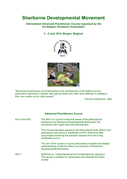
SYMPOSIUM: SPECIAL NEEDS
Investigating developmental
delay/impairment
child’s difficulties are temporary and will “catch up”. A recent
international consensus conference has proposed early developmental impairment (EDI) as a more appropriate diagnostic term.
EDI is defined by the group as persistent significant limitations in
two or more developmental domains (motor, communication,
cognitive skills, social skills, emotional regulation/behavioural
skills, self care skills) with onset before the age of five and not
better explained by another established developmental disorder.
The group recommends that children are reassessed by school
age or before the age of nine to review the diagnosis.
We will use the term early developmental impairment for the
purposes of this review.
Lesley A B McDonald
Alison C Rennie
Abstract
Pre-school children presenting to developmental paediatric services
because of concerns that they are not peer-equivalent is a well recognised
clinical scenario, and yet the approach to investigation varies widely. Evaluation depends on thorough history taking, careful clinical examination
and astute observation of social and play skills. An investigative pathway
needs to be evidence-based but also pragmatic; tailored to the child
whilst acknowledging the benefit of validated screening tests. Although
the overall positive yield is small, it must not be forgotten that negative
test results also have value. Both clinician and parents will be reassured
by the exclusion of genetic, metabolic and structural aetiologies in their
search for answers. It is increasingly acknowledged that there are more
subtle presentations of recognised disorders and the new generation of
genetic tests and neurological imaging is allowing earlier and more accurate diagnosis. This may afford opportunities for amelioration of the
condition in the affected child, together with more accurate genetic counselling for the family and indeed the child themselves. The search for an
answer should never stop.
Diagnosis
Concerns about a child’s developmental progress might be raised
by parents, health visitors or general practitioners. Education
staff in the pre-school setting may be the first to highlight
developmental concerns, particularly as families have less
contact with health visitors than in the past.
Referral to secondary level Community Child Health services
is recommended for detailed evaluation of all children with
developmental concerns, as an underlying aetiology may be
identified irrespective of the severity of impairment. It is reported
that a diagnostic cause can be identified in up to 40e60% of
cases, and early diagnosis may improve outcome.
A detailed history and careful physical examination are the
most important components of the assessment of the child with
EDI. One third of aetiological diagnoses made are on the basis of
history and examination findings alone.
Keywords aetiology; array-based comparative genomic hybridisation
(array CGH); early developmental impairment; global developmental
delay; guidelines; investigation; multiplex ligation-dependent probe
amplification (MLPA); neuroimaging; MRI diffusion tensor imaging;
telomeres
History
A thorough history is essential in the evaluation of children with
EDI. Detailed information regarding the pregnancy, delivery and
postnatal period should be sought. The mother should be asked
about bleeding, gestational diabetes, infection and medical
conditions during her pregnancy. Antenatal smoking, alcohol
consumption and prescribed and illicit drug use should be
documented. Birth details should include onset of labour, mode
of delivery, any complications, birth weight, APGAR scores and
need for admission to special care. Clear evidence of a neonatal
encephalopathy and a significant motor disorder is required
before attributing problems to the perinatal period.
The child’s medical history should be sought, including
medications and immunisations.
A full developmental history is required. The age and reason
for initial concern should be recorded, along with documentation
of milestone attainment. It is important to establish if there has
been any developmental regression. The child’s current performance in each domain should be assessed. Parents should also
be asked about social interaction and play skills, feeding
patterns, and the presence or absence of seizures. A sleep and
behaviour history is critical.
The family history is important and should include a threegeneration pedigree with note of parental consanguinity,
history of miscarriages or neonatal deaths, and family ethnic
origin.
Features in the history which are predictive of aetiological
determination are female gender, abnormal pre or perinatal
Definition
Global developmental delay (GDD) is commonly encountered in
paediatric practice, affecting 1e3% of children under the age of
five. It has traditionally been defined as significant delay in two
or more developmental domains (gross and fine motor; speech
and language; cognition; personal and social; activities of daily
living) with “significant” indicating performance two or more
standard deviations below the mean on developmental screening
or assessment tests.
There is agreement however that GDD is an unhelpful term,
not least because the word delay implies to parents that their
Lesley A B McDonald MBChB MRCP(UK) MRCPCH is an Associate Specialist in
Community Paediatrics at the West Centre, Children’s Community
Health and Care, 60 Kinfauns Drive, Glasgow, UK. Conflict of interest:
none.
Alison C Rennie MBChB MRCP(UK) FRCPCH is a Consultant Paediatrician in
Neurodisability at Southbank Child Centre, 207 Old Rutherglen Road,
Glasgow, UK. Conflict of interest: none.
PAEDIATRICS AND CHILD HEALTH 21:10
443
Ó 2011 Elsevier Ltd. All rights reserved.
SYMPOSIUM: SPECIAL NEEDS
history, antenatal toxin exposure, absence of autistic features,
positive family history, parental consanguinity and developmental regression.
(MLPA) adds to this detection rate by identifying tiny chromosome deletions and duplications at selected sites such as chromosome telomeres and the sites of known microdeletion and
microduplication syndromes such as Angelman/PradereWilli
syndromes, Di George syndrome, SmitheMagenis syndrome and
the MECP2 microduplication syndrome.
It is now commonly agreed that the best test to detect chromosome imbalances in children with developmental impairments is microarray-based comparative genomic hybridisation
(array CGH). This high resolution whole genome survey examines the patient’s chromosomes for tiny deletions or duplications
of genetic material (also known as copy number variants or
CNVs) as small as 100 kb. When array CGH is applied to children
with EDI and apparently normal G-banded karyotype, 10e15%
will have a pathogenic genomic imbalance of 150e15 000 kb.
This greatly improved detection rate, together with the rapidly
falling cost of the array CGH analysis, means that most genetic
laboratories will soon replace karyotyping with array CGH as the
first line investigation of EDI. However, the present wording of
laboratory reports is often quite technical and genetics-trained
clinicians are required to explain terminology and implications
of genomic imbalances to families. A particular interpretive
problem concerns small, non-pathogenic CNVs that commonly
need to be distinguished from pathogenic CNVs. Family studies
are required and even then the results may not be straightforward. Communicating to families such complex results, often of
uncertain significance, can be very challenging especially if one
or both parents has a learning disability. It should be noted that
array CGH cannot detect balanced chromosome rearrangements
such as inversions or translocations, nor will it detect small
intragenic mutations.
The DECIPHER website (http://decipher.sanger.ac.uk) is
becoming a very useful clinical resource that is gathering clinical
data on the effects of novel genomic imbalances. The Unique Rare
Chromosome Disorder Support Group (http://www.rarechromo.
org/html/home.asp) also produces excellent leaflets on
commoner microdeletion and microduplication syndromes, as
well as a leaflet that describes the array CGH technique.
Physical examination
All children referred with concerns about their development
should have a careful physical examination performed. Height,
weight and head circumference should be measured and plotted
on appropriate growth charts. Parental head circumference
should also be measured and plotted and compared to the child’s
centile to ensure that relative micro or macrocephaly is not
overlooked.
The child should be undressed and inspected carefully for
neurocutaneous stigmata such as pale patches and cafe au lait
spots. Dysmorphic features of the face, ears, hands and feet
should be looked for. The spine should be examined for abnormalities and the abdomen palpated to exclude organomegaly.
A full neurological examination should be performed,
including assessment of cranial nerve function. Tone, power and
reflexes should be examined with any asymmetry or focal findings noted. The quality of the child’s movements and gait should
be observed and recorded.
An eye examination is recommended and may require referral
to an ophthalmologist. Assessment of hearing and vision is also
necessary. Several studies have shown that children with EDI are
at risk of sensory impairment, with disorders of vision in
13e50% and audiological impairments in up to 18%.
A formal developmental assessment should be conducted, for
example Schedule of Growing Skills or Griffiths Mental Development Scales, in order to accurately determine the child’s
current developmental profile.
Features on physical examination which are predictive of
aetiological determination include microcephaly, macrocephaly,
focal neurological findings and the presence of dysmorphic
features.
Investigations
Subsequent to history and examination, if a specific condition is
suspected then investigations should be targeted accordingly.
One third of diagnoses are made in this way, using investigations
to confirm clinical suspicion. Laboratory investigations alone
account for a further one third of diagnoses made. Evidence
suggests that if diagnosis is not apparent after history and
examination, routine karyotyping, fragile X analysis and neuroimaging can result in a diagnosis in one sixth of all cases.
Neuroimaging
Neuroimaging may detect abnormalities in up to 50% of children
with neurodevelopmental disability, with a variable detection
rate dependent on factors such as selection criteria and imaging
method used. The yield is increased by the presence of abnormal
findings on physical examination, for example microcephaly or
focal neurological signs. One paper reported a yield of 13.9%
from imaging performed on a screening basis, compared to
41.2% if carried out due to a clinical indication.
MRI is preferable to CT for the evaluation of children with
EDI. MRI findings may include cerebral injury, cerebral malformation, or cerebral dysgenesis, however not all abnormalities
found are aetiological in nature. A recent review reported that
although abnormal MRI findings were found in approximately
30% of patients with developmental delay or learning disability,
these led to an aetiology or syndrome diagnosis in fewer than 4%
of cases.
MRI is therefore not recommended as a first line investigation,
but is useful when used selectively in the presence of abnormal
head size, seizures or focal neurological findings.
Genetics
Genetic abnormalities are among the commonest identifiable
causes of EDI. Congenital malformation, microcephaly, short
stature and dysmorphism are suggestive features but not specific
or obligatory. Associated sensory impairments, unusual behaviour patterns and a family history of a particular condition may
also indicate a syndrome diagnosis and prompt referral to the
genetics clinic.
As far as genetic investigations are concerned, there is
consensus that chromosome analysis and Fragile X testing can be
employed as screening tests with a detection rate of about 3%.
Targeted Multiplex Ligation-Dependent Probe Amplification
PAEDIATRICS AND CHILD HEALTH 21:10
444
Ó 2011 Elsevier Ltd. All rights reserved.
SYMPOSIUM: SPECIAL NEEDS
Further advances in neuroimaging may detect more subtle
anomalies. Diffusion tensor imaging is a new MRI technique
which uses the pattern of diffusion of water molecules to provide
information on tissue structure and white matter tracts. In one
study of 20 children with global delay of unknown aetiology and
normal MRI, the arcuate fasciculus was absent bilaterally in nine
and on the left in another two. In contrast, this tract was present
bilaterally in all typically developing children studied. It appears
that this tract has a central role in both language and cognitive
development and further research is required.
Further metabolic testing should be considered if there is
a family history of a disorder or of developmental impairment.
Other features in the history which warrant consideration include
parental consanguinity, developmental regression, congenital
ataxia or dysequilibrium, epilepsy or episodic decompensation.
Examination findings which may raise clinical suspicion include
coarse facial features, growth failure, hepatosplenomegaly, and
ophthalmological or retinal abnormalities.
Metabolic investigations should also be considered if neonatal
screening has not been done, for example in migrant families
from developing nations.
If metabolic testing is being undertaken, blood should be sent
for lactate, amino acids, ammonia, very long chain fatty acids,
carnitine, homocysteine and disialotransferrins. A urine sample
should also be obtained for organic acids, orotate, glycosaminoglycans and oligosaccharides.
Biochemical and haematological investigations
There are case reports in the literature of boys with Duchenne
muscular dystrophy presenting with early developmental
impairment. It is therefore important to measure creatine kinase
at an early stage to prevent late or missed diagnosis.
Children with developmental problems may have significantly
higher blood lead concentration than the general childhood population. We recommend that lead toxicity should be screened for
routinely as this neurotoxin may contribute further to impairment
and is treatable. Other groups suggest targeting to those with risk
factors such as pica, old housing and poor socioeconomic status.
Biotinidase deficiency is a treatable disorder which may
present as EDI with no other signs or symptoms. Many countries
screen for this disorder routinely in the neonatal period, as it is
known that early diagnosis and treatment improves outcome.
This disorder has a higher incidence in Celtic populations and
travelling communities.
Abnormalities of plasma calcium may be helpful in the identification of conditions such as Di George syndrome, William’s
syndrome and pseudohypoparathyroidism.
There is no evidence to support the use of thyroid function
testing on a screening basis and some centres state testing is not
necessary if newborn screening has been done, unless there are
systemic features of hypothyroidism. This condition is however
easily treatable with significant implications if the diagnosis is
missed. In addition, many chromosome abnormalities are associated with an increased risk of hypothyroidism, for example
trisomy 21 (Down’s syndrome), 45X (Turner syndrome) and
22q11 deletion (Di George syndrome). For these reasons we have
included thyroid function testing as a first line investigation.
Recent reports in the literature suggest that creatine deficiency
disorders may be associated with early developmental impairment. It may be necessary to consider routine testing for these
disorders in the future.
Iron deficiency anaemia may be associated with developmental impairment and is easily measured and treated, so should
be screened for routinely.
Neurophysiology
EEG should not be performed routinely for all children with EDI.
If the history is suggestive of seizures or a neurodegenerative
disorder then EEG should be considered. It is also important in
the evaluation of children with a history of regression in
language skills, in order to exclude or diagnose LandaueKleffner
syndrome.
Guidelines
An evidence-based guideline for investigating children with GDD
was published in 2006. Publication predated the proposed
change in terminology from GDD to EDI. Updated guidance is
shown in Figure 1.
Management
Management of children with EDI may be influenced by the
results of any investigations or underlying diagnoses made. For
many there will be no cause found for their difficulties; however
the benefits of negative testing should not be underestimated as
this reassures the family and clinician that serious underlying
disorders have been excluded. Management will then focus on
maximising the child’s developmental potential. This will vary
between children depending on their individual needs, but may
include multidisciplinary working with colleagues in physiotherapy, occupational therapy and speech and language therapy.
Involvement of education services at an early stage is also useful,
in particular for assessment by educational psychology and
possible input from a peripatetic pre-school teaching service.
Referral to social work should also be considered for family
support and assistance with income maximisation, including
disability living allowance and Family Fund application as
appropriate. Regular review and documentation of developmental progress by the paediatrician is recommended.
Metabolic investigations
The evidence available does not support the use of metabolic
testing on a screening basis. The yield is 1% or less, with a high
frequency of non-specific, non-diagnostic abnormalities detected.
Investigations should be selective and targeted, with a low
threshold for testing if a metabolic disorder is suspected clinically. We have however included urate measurement in our first
line investigations as this is an easy way to diagnose purine
disorders, which may present as isolated EDI, and is more stable
than both ammonia and lactate.
PAEDIATRICS AND CHILD HEALTH 21:10
Prognosis
Prognosis may also be influenced by the outcome of assessment
and investigations, which may in addition give information
about recurrence risks for the family.
For the child with early developmental impairment with no
cause found, giving an accurate prognosis may prove difficult.
445
Ó 2011 Elsevier Ltd. All rights reserved.
SYMPOSIUM: SPECIAL NEEDS
Specialist Children's Services
Department of Community Child Health
Early Developmental Impairment in Pre School Children
Guidelines for Investigation
Early developmental impairment is defined as persistent significant limitations in two or more
developmental domains. Investigations should be considered only after a thorough history and
examination have been performed. These guidelines are not intended for isolated speech & language
or motor problems, or for children with autism.
If diagnosis not apparent after history and examination, proceed as follows:
First line
Chromosomes
Fragile X
Telomeres
Learning disability MLPA
U&E
Creatine kinase
Lead
Thyroid function tests
Urate
Full blood count
Ferritin
Biotinidase
Second line
Neuroimaging
Abnormal head size
Seizures
Focal neurology
Metabolic
Family history
Consanguinity
Regression
Organomegaly
Coarse features
Blood
Lactate
Amino acids
Ammonia
VLCFA
Carnitine
Homocysteine
Urine
Organic acids
Orotate
Gags
Oligosaccharides
MRI
CT (bones,
calcification)
EEG
Speech regression
Seizures
Neurodegenerative disorder
Genetics
Dysmorphism
Abnormal growth
Sensory impairment
Odd behaviour
Family history
Consider
24hr EEG
Figure 1
Such children require to be followed up at least until school
entry, as the phenotype may change over time and a diagnosis
may become apparent. In any event, the term EDI should not stay
with the child indefinitely. Reassessment is required at early
primary school age to determine whether impairments can be
better explained by another established diagnosis or descriptor,
such as learning difficulties of unknown aetiology.
PAEDIATRICS AND CHILD HEALTH 21:10
Recent and ongoing developments in genetic and neuroimaging techniques are resulting in improved diagnostic yield
and better understanding of the underlying disorders associated with early developmental impairment. More subtle
presentations of recognised disorders are increasingly being
identified by newer screening techniques, allowing earlier
diagnosis.
446
Ó 2011 Elsevier Ltd. All rights reserved.
SYMPOSIUM: SPECIAL NEEDS
Srour M, Mazer B, Shevell M. Analysis of clinical features predicting
etiologic yield in the assessment of global developmental delay.
Pediatrics 2006; 118: 139e45.
Sundaram S, Sivaswamy L, Makki M, Behen M, Chugani H. Absence of
arcuate fasciculus in children with global developmental delay of
unknown etiology: a diffusion tensor imaging study. J Pediatr 2008;
152: 250e5.
van Karnebeek C, Jansweijer M, Leenders A, Offringa M, Hennekam R.
Diagnostic investigations in individuals with mental retardation:
a systematic literature review of their usefulness. Eur J Med Genet
2005; 13: 6e25.
van Karnebeek C, Scheper F, Abeling N, et al. Etiology of mental retardation in children referred to a tertiary care center: a prospective
study. Am J Ment Retard 2005; 110: 253e67.
Williams J. Global developmental delay e globally helpful? Dev Med Child
Neurol 2010; 52: 227.
Children and families deserve a systematic and thorough
assessment and investigative process. Clinicians should aspire to
a uniform approach to ensure best practice standards are
followed.
A
FURTHER READING
Essex C, Roper H. Late diagnosis of Duchenne’s muscular dystrophy
presenting as global developmental delay. BMJ 2001; 323: 37e8.
Francoeur E, Ghosh S, Reynolds K, Robins R. An international journey in
search of diagnostic clarity: early developmental impairment. J Dev
Behav Pediatr 2010; 31: 338e40.
King M, Stephenson J. Delayed Development. In: King M, Stephenson J,
eds. A Handbook of Neurological Investigations in Children. London:
Mac Keith Press, 2009: 223e7.
McDonald L, Rennie A, Tolmie J, Galloway P, McWilliam R. Investigation of
global developmental delay. Arch Dis Child 2006; 91: 701e5.
Moeschler J. Genetic evaluation of intellectual disabilities. Semin Pediatr
Neurol 2009; 15: 2e9.
Ozmen M, Tatli B, Aydinli N, Caliskan M, Demirkol M, Kayserili H. Etiologic
evaluation in 247 children with global developmental delay at
Istanbul, Turkey. J Trop Pediatr 2005; 51: 310e3.
Paciorkowski A, Fang M. Chromosomal microarray interpretation: what is
a child neurologist to do? Pediatr Neurol 2009; 41: 391e8.
Shevell M, Ashwal S, Donley D, et al. Practice parameter: evaluation of
the child with global developmental delay. Neurology 2003; 60:
367e80.
Shevell M. Global developmental delay and mental retardation or intellectual disability: conceptualization, evaluation and etiology. Pediatr
Clin N Am 2008; 55: 1071e84.
Shevell M, Majnemer A, Rosenbaum P, Abrahamowicz M. Etiologic yield of
subspecialists’ evaluation of young children with global developmental delay. J Pediatr 2000; 136: 593e8.
PAEDIATRICS AND CHILD HEALTH 21:10
Practice points
C
C
C
C
C
447
The term global developmental delay is unhelpful and should
be replaced by early developmental impairment.
Paediatricians should review departmental practice to ensure
best evidence guidelines are being followed.
Be aware of new investigative techniques and their relevance
in the evaluation of children with EDI.
It is important to review diagnoses (and non-diagnoses) in
older children who have not had the benefit of newer tests.
The search for an underlying aetiology should be an ongoing
process.
Ó 2011 Elsevier Ltd. All rights reserved.


© Copyright 2026