
Silicon nanostructures for bioapplications
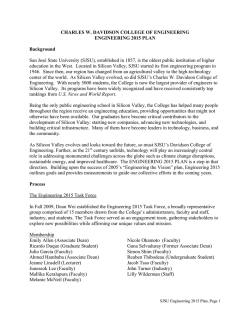
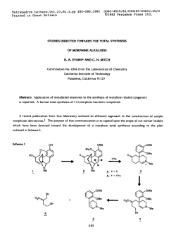
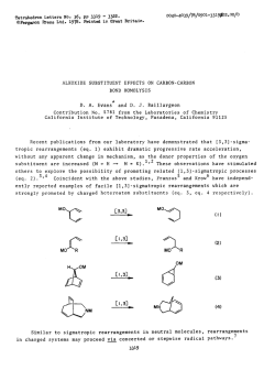
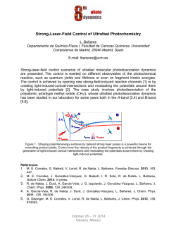
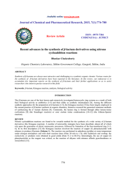
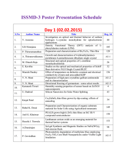
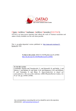
Nano Today (2010) 5, 282—295 available at www.sciencedirect.com journal homepage: www.elsevier.com/locate/nanotoday REVIEW Silicon nanostructures for bioapplications Yao He a, Chunhai Fan c, Shuit-Tong Lee b,∗ a Institute of Functional Nano & Soft Materials (FUNSOM) and Jiangsu Key Laboratory for Carbon-based Functional Materials & Devices, Soochow University, Suzhou, Jiangsu 215123, China b Center of Super-Diamond & Advanced Films (COSDAF) and Department of Physics & Materials Science, City University of Hong Kong, 83 Tat Chee Avenue, Kowloon Tong, Hong Kong SAR, Hong Kong, China c Laboratory of Physical Biology, Shanghai Institute of Applied Physics, Chinese Academy of Sciences, Shanghai 201800, China Received 6 May 2010; received in revised form 10 June 2010; accepted 28 June 2010 Available online 27 July 2010 KEYWORDS Silicon; Quantum dots; Nanowires; Bioimaging; SERS; Biosensor Summary There has been rapidly increasing interest in design and synthesis of siliconbased nanostructured materials for bioapplications. In this review, we focus on recent research progress in design, synthesis and bioapplications of two silicon-based nanostructures, zero-dimensional silicon quantum dots and one-dimensional silicon nanowires. These two low-dimensional silicon nanomaterials have found important applications in ultrasensitive biomolecular detection and fluorescent cellular imaging. We further highlight major challenges and promises in this area. © 2010 Elsevier Ltd. All rights reserved. Introduction Nano-biotechnology is a promising and interdisciplinary research involving chemistry, physics, biology and medicine [1,2]. Particularly, the utilization of nanostructured materials in biology and biomedicine is recognized as one of the fast moving and exciting interfaces in nanobiotechnology, and has been extensively explored [3—7]. It is critically important to develop and fabricate novel functional nanomaterials with well-defined structures for nano-biotechnology [5—7]. During the past decade, a great number of nanomaterials and nanofabrication techniques have been elegantly developed, imparting momentum to ∗ Corresponding author. Tel.: +852 2784 4209; fax: +852 2784 4696. E-mail addresses: [email protected] (Y. He), [email protected] (C. Fan), [email protected] (S.-T. Lee). the development of nano-biotechnology and opening up new avenues in biological and biomedical studies [1—4]. To date, many interesting nanomaterials, such as fluorescent quantum dots [5], magnetic nanoparticles [6] and carbon nanotubes [7], have been studied and utilized in widespread nano-bioapplications (e.g., biological imaging, biosensor, disease diagnosis and therapy). Their attractive optical, magnetic and mechanical properties provide new platforms for studying complicated biological processes that are difficult to access with conventional techniques [3—7]. Silicon nanomaterials are a type of important nanomaterials with attractive properties including excellent electronic/mechanical properties, favorable biocompatibility, huge surface-to-volume ratios, surface tailorability, improved multifunctionality, as well as their compatibility with conventional silicon technology [8—11]. Consequently, there has been great interest in developing functional silicon nanomaterials for various applications ranging from electronics to biology. To meet increasing demands of 1748-0132/$ — see front matter © 2010 Elsevier Ltd. All rights reserved. doi:10.1016/j.nantod.2010.06.008 Silicon nanostructures for bioapplications silicon-based applications, silicon materials of various nanostructures (e.g., nanodot [12,13], nanowire [14,15], nanorod [16,17] and nanoribbon [18,19]) have been developed, among which silicon quantum dots (SiQDs) and silicon nanowire (SiNWs) are of our primary interest [12—15,20—24]. Quantum confinement phenomenon in SiQDs can increase the probability of irradiative recombination via direct band gap transition [15,26], leading to improvement of fluorescent intensity and the prospect of optical applications. Due to their excellent biocompatibility and noncytotoxic property, SiQDs are also considered as promising fluorescent biological probes for in vivo and in vitro imaging [27—31]. On the other hand, SiNWs could act as a general platform for greatly enhanced surface-enhanced Raman scattering (SERS) studies. Significantly, SiNWs decorated with metal nanoparticles, e.g. silver nanoparticles, possess high enhancement factor (EF) of up to 107 —109 . Such SiNWs-based highly efficient SERS-active substrates could be utilized as biosensors for ultrasensitive detection of biomolecules (e.g., DNA and protein) [32—36]. In this review article, by paying particular attention to the SiQDs and SiNWs, we summarize recent research progress in synthesis and bioapplications of these lowdimensional silicon nanomaterials, based particularly on recent progress of our laboratory. In brief, in the following sections, we first introduce methods for preparing SiQDs and SiNWs, and categorize representative synthesis strategies. Then we summarize typical examples of SiQDs/SiNWs-based bioapplications that were reported recently, and describe in detail about the biological applications of SiQDs or SiNWs as fluorescent bioprobes or biosensors, respectively. In the final section, we discuss challenges and perspectives for the silicon-relative biological applications in the future. This review intends to take SiQDs and SiNWs as models and discuss topics ranging from their synthesis to their biological applications, with the hope to outline these exciting achievements as the starting points in the realm of silicon-based nano-biotechnology. Due to page limitation, this review will not discuss the applications of silicon nanostructures in other important areas, such as energy, catalysis and optoelectronics. Synthesis of SiQDs and SiNWs Synthesis of SiQDs Bulk silicon is considered as one of the most important materials in semiconductor microelectronics industry; nevertheless, its optical properties are poor because of its indirect band gap, leading to the slow electron-hole radiative recombination [37]. Notably, intense room temperature photoluminescence could be observed from nanocrystalline silicon, which is generally believed to result from a combination of quantum confinement effects. For examples, recombination rates of electrons and holes are distinctively enhanced due to the increased overlap of the electron and hole wave functions confined in nanodots with diameters smaller than 5 nm [25,26]. Such observations have intrigued intense studies on this fluorescent SiQDs-relative field, since the first reports of room temperature light emission from porous silicon in the early 1990s [38,39]. 283 Over the past decade, a variety of fabrication techniques have been developed for preparation of silicon quantum dots, including solution-phase reductive [40,41], plasma-assisted aerosol precipitation [42,43], microemulsion [44], mechanochemical [45], laser ablation [46] and sonochemical synthesis [47]. Particularly, the solution-phase reduction synthesis strategy was developed by Kauzlarich’s group in 1990s [40], involving reduction of silicon halides (e.g., SiCl4 ) in organic solution. This strategy has been well-established for producing silicon quantum dots, which could be readily prepared under mild reaction conditions (e.g., room temperature and low pressures) [40,41]. Moreover, this method leads to silicon nanoparticles terminated with alkyl groups, hydrogen or other chemical species. The ability for facile surface chemical modification provides a new way to study the relationship between surface effects and optical properties of the SiQDs, and more importantly, offers opportunity to functionalize the SiQDs according to different requirements [40]. Alternatively, plasma-assisted aerosol precipitation provides a way to production of silicon nanoparticles with high efficiency and yield [42,43]. This method is also very rapid as compared to the relatively timeconsuming liquid-phase approaches (e.g., solution-phase reductive, microemulsion and sonochemical synthesis). Typically, luminescent silicon nanocrystals of 2—8 nm can be synthesized via a single-step continuous flow nonthermal plasma process within milliseconds [42]. While luminescent intensity of silicon is enhanced at the nanoscale, SiQDs usually possess inferior optical properties (quantum yield <10%) to semiconductor II—VI and III—V QDs (e.g. CdSe, CdSe/ZnS QDs) with direct band gaps (quantum yield 80—90%) [48,49]. Recent theoretical studies reveal that optical properties of SiQDs are significantly influenced by surface oxidation. Particularly, oxygen bonded to the surface of silicon quantum dots often reduces the band gap and limits luminescent emission, resulting in low quantum yield [20—22,50,51]. Consequently, it is essential to avoid surface oxidation to improve the quantum yields of SiQDs [50—52]. Indeed, Kortshagen and co-workers recently reported successful preparation of SiQDs with remarkably high ensemble quantum yields exceeding 60% by using plasma-assisted synthesis with strict removal of oxygen and elaborate surface passivation [43], which provides an excellent example. It demonstrates that SiQDs could possess quantum yields as high as II—VI QDs under optimum conditions. Our group recently developed a polyoxometalateassisted electrochemical etching method for synthesizing SiQDs with controllable luminescent colors (e.g., blue, orange and red) [53]. This design adopts an electrochemical cell with graphite as anode and silicon wafer as cathode (Fig. 1a). H3 PMo12 O40 (POM) is introduced into our electrochemical etching system as catalyst due to its unique properties. Significantly, highly monodispersed SiQDs with a narrow size distribution are directly synthesized without further separation via this electrochemical method. Moreover, SiQDs with controllable sizes are readily achieved by adjusting the current density (Fig. 1b). Higher current density yields small-size particles, while lower density yields larger particles. Also of note, the resultant SiQDs display serial PL maximum emission wavelengths from 450—700 nm. Fig. 1b and c inset displays SiQDs with sizes of ∼1, ∼2, ∼3 and ∼4 nm emitting a blue peak at 450 nm, a green peak 284 Y. He et al. Figure 1 (a) Schematics for the POMs-assisted electrochemical etching process. (b) TEM picture of serial sizes of SiQDs and their corresponding luminescence colors under UV irradiation (inset). (c) Typical PL spectra of SiQDs with sizes from ∼1 to ∼4 nm. (Reprinted with permission from [53], 2007 American Chemical Society). at 520 nm, a red band at 640 nm and an infrared band at 740 nm, respectively. Synthesis of SiNWs SiNWs are well-known one-dimensional nanomaterials with many attractive electronic properties [14,15]. Several techniques have been developed to fabricate SiNWs, such as metal-catalyzed vapor—liquid—solid (VLS) [54,55], oxideassisted growth (OAG) [14,56], laser ablation [57,58] and thermal evaporation with catalyst [59,60]. Particularly, the OAG and VLS approaches are widely recognized as two classic methods for synthesizing SiNWs. In 1998, the groups of Lieber [61] and Lee [62] independently reported the method of laser ablation-assisted VLS growth for the synthesis of single-crystal SiNWs with diameters of 6—20 nm and lengths ranging from 1 to 30 m [61,62], realizing the first large-quantity fabrication of SiNWs [62]. Thereafter, the VLS method has been extensively optimized to be a well-established approach for preparing SiNWs [54—56,61,62]. In a typical VLS growth, nanometerdiameter metals or metal compounds (e.g., Fe, Au) act as catalysts that effectively define the diameter of wires. Once metal catalysts induce SiNW growth, SiNWs would continue to grow as long as the Si vapor source continues. In the VLS process, different reaction temperatures are required to produce SiNWs when varying the catalytic metals because of their distinct melting temperatures. For example, 1150 or 370 ◦ C is favorable for preparation of SiNWs when using Fe or Au as the catalyst, respectively [63]. On the other hand, the OAG method is considered as another established strategy for preparation of the SiNWs, which is well complementary to and coexistent with the metal catalyst VLS approach. Instead of metal catalysts used in the VLS method, oxides play an important role in inducing the nucleation and growth of nanowires in the OAG process. Consequently, SiNWs free of metal impurities are readily achieved via the OAG technique. Besides, with comparison to the metal-catalytic VLS growth, the OAG method has three additional advantages [56]. First, large-scale synthesis of SiNWs can be realized by simple vaporization and condensation of readily available silicon monoxide or a mixture of Si and SiO2 powders. Second, myriad structural morphologies such as rods, chains and ribbons could be produced. Third, ultrasmall-diameter SiNWs can be readily prepared. For instance, significantly, SiNWs with a diameter of ∼1 nm have been successfully synthesized via the OAG approach by our group (Fig. 2) [14]. In contrast, the smallest SiNW diameter with the metal-catalyzed VLS approach is larger than 4 nm [56]. Nevertheless, the VLS method still possess the unique characteristics that the diameter and growth alignment of the nanowires can be more readily controlled by using uniformly distributed metal seeds with well-designed sizes on the substrate [56,59—62]. Peng et al. recently developed a HF-etching-assisted nanoelectrochemical method to produce SiNWs [64—70]. The whole process does not require high temperature, vacuum, templates, complex equipment, or hazardous silicon precursors. In this method, noble metal atoms deposited from HF solution on the silicon wafer surface could form nuclei that behave as a cathode, and the area surrounding these nuclei behaves as an anode, and can subsequently be etched and dissolved into the solution by the galvanic cell reaction [64,65,70]. As a result, silicon nanowire arrays could be readily grown on silicon wafers in noble metal HF solution via selective etching, based on the principle of micro-electrochemical redox reaction. For instance, large-area growth of ordered silicon nanowire arrays on silicon wafers can be prepared in an aqueous HF solution containing silver nitrate at room temperature. Moreover, the SiNWs of controllable lengths are finely Silicon nanostructures for bioapplications 285 Figure 2 Constant-current scanning tunneling microscopy (STM) image of a SiNW prepared via OAG method. The wire axis is along the [1 1 2] (left) and [1 1 0] (right) direction. Scale bar: 1 nm. (Reprinted with permission from [14], 2003 Science). achieved through adjusting the reaction conditions (e.g., etching time, precursor concentration) (Fig. 3) [64—70]. biology, particularly fluorescence bioimaging and ultrasensitive biosensing. We will summarize their interesting bioapplications in this section (Fig. 4). Bioapplications of SiQDs and SiNWs SiQDs-based fluorescent biological probes SiQDs and SiNWs are attractive low-dimensional silicon nanostructures that have found important applications in Fluorescent biological probes are powerful tools for biological and biomedical studies [71]. A high-performance Figure 3 SEM images of silicon nanowires arrays prepared via metal-assisted chemical etching method. SiNWs with controllable lengths of 2 m (a), 4 m (b), 6 m (c) and 10 m (d), could be readily achieved through adjustment of etching time. 286 Y. He et al. Figure 4 Future prospects of silicon nanomaterials. Left: four issues (large-scale synthesis, surface modification method, biosafety-relative investigation and manipulation of intrinsic properties) are essential to biological applications of silicon nanomaterials. Right: a new era of silicon-based bioapplications, especially on fluorescence bioimaging and ultrasensitive biosensor, is expected in the near future. Figure 5 (a) Schematic mode of synthesis of silicon-based nanospheres. The whole process is performed under N2 -protection environment. To simplify the model and make it easier to comprehend, only five SiQDs are shown in one nanosphere. (b) TEM images of three nanospheres with a size of ∼60 nm (top left), ∼120 nm (top right) and ∼200 nm (bottom left), as well as HRTEM image (bottom right) of a single SiQD inside the as-prepared nanospheres. (c) Photoluminescent spectra of the free-standing SiQDs and as-prepared nanospheres, and the corresponding photograph of the luminescence from the samples of the free-standing SiQDs and nanosphere solution under irradiation by 365 nm light from UV lamp (inset). To ensure the objective comparison, the samples of SiQDs and nanosphere solution were directly extracted respectively from the original solution in the initial or the final state of the whole process shown in Fig. 1c. Neither samples received any post-treatment. (d) Temporal evolution of the absorption and corresponding PL spectra of the as-prepared nanospheres over one-half year in ambient air condition without any special protection. (Reprinted with permission from [30], 2009 Wiley). Silicon nanostructures for bioapplications fluorescent cellular probe should be water-dispersible, highly fluorescent, anti-photobleaching and biocompatible [4,5]. Organic dyes and fluorescent proteins have been widely used in biological studies. Notwithstanding, these 287 dyes and proteins usually suffer from severe photobleaching that hampers long-term imaging in vitro or in vivo [5]. Over the past decade, semiconductor II—VI QDS have been developed as high-performance fluorescent biological Figure 6 (a) Schematic illustration of silicon nanospheres conjugating with goat-anti-mouse IgG. The carboxylic acid groups of nanospheres readily reacted with the amino groups of IgG by using EDC and NHS as zero-length cross-linkers (Figure is not to scale). (b) Optical micrographs of the prepared silicon bioconjugates under UV (365 nm) irradiation. The prepared bioconjugates preserve stable and bright fluorescent intensity for over 1 month (right). (Reprinted with permission from [31], 2009 American Chemical Society). Figure 7 (a) Dual-color cellular imaging photos. The HEK293 cells are distinctively labeled by the bioconjugates (left: green) and Hoechst (middle: blue); right: bright field image. (b) The bioconjugates fluorescently label the cells incubated with monoclonal mouse anti-actin antibody. Left: 488 nm excitation; middle: superposition of fluorescence and transillumination images; and right: bright field image. (c) Comparison of fluorescent signals of HEK293T cells imaging with the nanospheres (top) and FITC (bottom) excited by different wavelengths. (d) Temporal evolution of fluorescence of the HEK293T cells labeled with the as-prepared nanospheres (top) and FITC (bottom). The nanospheres and FITC were both excited at 488 nm by argon laser with 8 s dwell time and ∼15 mW power. (Parts (a) and (b) reprinted with permission from [31], 2009 American Chemical Society; parts (c) and (d) reprinted with permission from [30], 2009 Wiley). 288 probes because of advantages such as size-tunable emission color, strong fluorescence and high resistance to photobleaching [72]. However, the potential toxicity problem of the II—VI QDs associated with release of heavy metal ions (e.g. Cd ions) has not yet been fully addressed, which limits their widespread biological and medical applications [73]. Consequently, novel fluorescent probes with robust photostability, strong fluorescence and favorable biocompatibility are still urgently required to satisfy various requirements of biological studies. Recent exciting progress on SiQDs synthesis provides promising new solutions for a range of applications such as SiQDs-based light-emitting diodes [74] and SiQDs lasers [75]. Due to their biocompatibility and low toxicity, fluorescent SiQDs are potentially ideal fluorescent probes for biological and biomedical studies. Nevertheless, most as-prepared SiQDs are not well water-dispersible since their surfaces are covered by hydrophobic moieties (e.g., styrene, alkyl and octene) [40—43]. Extensive efforts have been undertaken to realize aqueous dispersibility of SiQDs. In 2004, Ruckenstein and Li developed a UV-induced graft polymerization for surface modification of SiQDs [27]. These SiQDs became well water-dispersible with the grafting of a water-soluble poly(acrylic acid) (PAAc) layer. In addition, the grafted PAAc also improved the photoluminescence stability of the SiQDs. Moreover, high density of carboxylic acid moieties of PAAc could be used to immobilize biomolecules (e.g., protein). Photo-stability comparison of SiQDs and four types Y. He et al. of organic dyes (e.g., Alexa 488, Cy5, fluorescein isothiocyanate (FITC) and laser dye styryl (LDS751)) demonstrated superior resistance to photobleaching than the conventional organic dyes. Such modified SiQDs with quantum yield of 24% were employed as biological probes for cell imaging, suggesting potential bioimaging applications of modified SiQDs. Tilley and co-workers later reported a room temperature synthesis for preparing water-dispersed SiQDs that exhibited strong blue photoluminescence [28]. In their method, a platinum chemical was utilized as catalyst for initiating reaction between Si—H surface bonds of the SiQDs and C C bonds of allylamine. The resultant blue-emitting SiQDs became hydrophilic because their surfaces were modified with allylamine. In addition to the good aqueous dispersibility as well as relatively high quantum yield (∼10%), the allylaminecapped SiQDs possessed robust storage- and photo-stability. They kept stable optical properties for several-month and long-time (more than 1 h) UV irradiation. As a comparison, the photoluminescence from rhodamine 6G dropped by 60% under the same illumination conditions. Sato and Swihart utilized photoinitiated hydrosilylation to successfully attach propionic acid (PA) to the surface of SiQDs, thereby producing water-dispersible, PA-terminated SiQDs with average diameter of less than 2.4 nm [76]. Compared to the former two reports showing water-dispersed SiQDs of a single emission color (red or blue), this work is significant because the size and corresponding PL emission color of SiQDs could be controlled by varying conditions. PA- Figure 8 (a) SEM images of as-prepared SiNWs with lengths larger than 2 mm. (b) TEM and high-resolution TEM (inset) images of a single silicon nanowire with a diameter of 35 nm (c) TEM image of a silicon nanowire coated with Au nanoparticles. (d) Cyclic voltammograms of BSA with a concentration of 0 M (a) and 0.38 M (b). pH = 7, scan rate: 100 mV s−1 . (e) Peak current vs. concentration of BSA showing a linear range beyond 1 M. (Reprinted with permission from [85], 2005 American Institute of Physics). Silicon nanostructures for bioapplications terminated SiQDs with continuous luminescent color from yellow to green were readily synthesized in their work. Recently, Erogbogbo et al., Swihart et al. revealed that most of the modified SiQDs often showed obvious PL degradation especially in biological media with different pH, despite their high storage- and photo-stability in water [29]. Low pH stability would severely hinder their broad applications in biology. For example, conjugation of SiQDs with antibodies would be technically difficult if they were instable at neutral and alkaline pH environment. They further developed a new kind of SiQDs encapsulated by phospholipid micelles. Significantly, such micelle-encapsulated SiQDs kept stable optical properties under various biologically relevant conditions of pH values (4—10) and temperatures (20—70 ◦ C). However, the quantum yield of such micelle-encapsulated SiQDs dramatically decreased from 17 to 2% after the encapsulation process [29]. Very recently, Tilley and co-workers systematically investigated the chemical reactions on molecules attached to the surface of SiQDs, and further developed a multi-stepped chemical method for surface modification of SiQDs [77]. This stepwise approach offers new opportunities to prepare the SiQDs with diverse and desirable functionalities. This study sheds new insight into biological applications of silicon quantum dots. 289 Lee and co-workers recently presented an EtOH/H2 O2 assisted oxidation method to synthesize water-dispersed Si/SiOx Hy core/shell quantum dots with a Si core of different controlled diameters [78]. Significantly, this method allows for fine tuning emission wavelengths of QDs, producing seven luminescent colors from blue to red. On the basis of such studies [53,78] and theoretical prediction [20—22], we developed a new class of fluorescent silicon nanospheres (SiNSs) that each containing several hundreds of SiQDs (Fig. 5). The as-prepared nanospheres possesses excellent aqueous dispersibility, strong fluorescence (quantum yield: ∼15%), robust photo-stability and favorable biocompatibility [30]. Further, we developed a novel kind of water-dispersed oxidized SiNSs (O-SiNSs) prepared via thermal oxidation of the precursor SiNSs [31]. The quantum yield of the O-SiNSs was dramatically increased to as high as 25%. More significantly, O-SiNSs are stable under highpower UV irradiation and in acidic-to-basic environments covering pH = 2—12. This extremely high pH stability leads to facile conjugation of nanospheres with antibodies, resulting in brightly luminescent silicon bioconjugates for immunofluorescent bioimaging (Fig. 6). Our cellular experiments well demonstrated the great promise for real-time and long-term bioimaging with our silicon nanospheres (Fig. 7) [30,31]. Figure 9 (a) Schematic mode of the SiNW optical sensor. (b) Relative fluorescence intensity of the QlOEt-modified SiNWs (68 g/ml: 9 × 10⁻⁶ M) in the presence of various metal ions alone (20 M, red) and interfering ions with Cu (II) (20 M, green). EtOH—water solution (30%) of 0.05 M HEPES buffer (pH 7.0). ex = 324 nm, em = 490 nm. Fluorescence spectra (c) and titration curve (d) of QlOEtmodified SiNWs (68 g/ml: 9 × 10⁻⁶ M QlOEt) with Cu (II). EtOH—water solution (30%) of 0.05 M HEPES buffer (pH 7.0). ex = 324 nm, em = 490 nm. (Reprinted with permission from [90], 2008 American Chemical Society). 290 SiNWs-based biosensor SiNWs biosensor One-dimensional nanomaterials (e.g., carbon nanotubes (CNTs) and SiNWs) have been employed as an effective substrate for various sensing applications because of their favorable biocompatibility, convenient surface modifica- Y. He et al. tion, huge surface-to-volume ratios, fast response and good reproducibility [7,14,15,79]. These unique properties make CNTs and SiNWs attractive in the development of field-effect transistor (FET), chemical and biological sensors [80—90]. For example, Dai and co-workers developed CNT-based FET for sensing of proteins or detecting protein-protein interactions in the range of 100 pM to 100 nM [81]. Ryon and Choi modified CNT biosensor to enable detection sensitivity to Figure 10 (a) TEM image of AgNPs-modified SiNWs, (b) HRTEM image of AgNPs-modified SiNW, the Moiré fringes marked with arrow indicates that Ag nanoparticles are embedded in the surface. Raman spectra obtained from Ag-modified SiNWs coated with 25 l of (c) 1 × 10⁻¹⁶ M R6G solution, (d) 1 × 10⁻¹⁶ M crystal violet solution, (e) 1 × 10⁻¹⁴ M nicotine solution and (f) 1 × 10⁻⁸ mg/ml CT DNA solution in water. Curves in (1) and (2) are, respectively, the Raman spectrum collected from R6G powder and solid CT DNA. (Reprinted with permission from [97], 2008 American Institute of Physics). Silicon nanostructures for bioapplications 1 pM protein [82]. Lieber and co-workers reported SiNWsbased FET for spatially resolved, high-sensitivity detection [83]. Li et al. fabricated SiNWs sensor for high-sensitivity and sequence-specific DNA detection with a detection limit of 25 pM [84]. Lee and co-workers have investigated SiNWs for detection of various biological molecules and metal ions [85—90]. Notably, an individual strand of OAG-growth SiNWs was fabricated into a sensor with proper surface modification (e.g., decorated with gold nanoparticles) taking advantage of the large dimensionality of SiNWs of >2 mm in lengths and diameters of 35 nm. The SiNW sensor could 291 sensitively detect a typical kind of protein (bovine serum albumin (BSA)) via cyclic voltammetric (CV) detection (Fig. 8) [85]. The SiNWs sensor could also be used for highly sensitive and reproducible detection of glucose, hydrogen peroxide, and pesticide via electrochemical methods, due to the high electrical conductivity of the modified SiNWs [86—88]. More recently, SiNWs FET were developed for detection of toxic heavy metal cations at low concentrations (e.g., 10−7 M Hg2+ and 10−4 M Cd2+ ) via current change of the FET [89]. In addition, Lee and coworkers designed a new SiNWs optical sensor for high sensitive and specific detection of Cu2+ , an important ele- Figure 11 Characterization of SERS-active substrate for immunoassay: (a) and (b) are respectively top and cross-section SEM images of as-prepared SiNWs array, (c) and (d) are respectively TEM and HRTEM images of AgNPs coated SiNWs and (e) Raman spectra of mIgG, gamIgG and their corresponding controls. (Reprinted with permission from [98], 2008 American Institute of Physics). 292 ment for hemopoiesis, metabolism, growth and immune system. This optical sensor was fabricated via covalent modification of SiNWs with fluorescein, i.e. N-(quinoline8-yl)-2-(3-triethoxysilyl- propylamino)-acetamide (QlOEt). Fluorescence intensity of the QlOEt-modified SiNWs gradually decreased with increasing Cu (II) concentration, since the 8-aminoquinoline derivative could effectively coordinate with Cu2+ , leading to fluorescence quenching of QlOEt. As a result, Cu2+ at low concentration of 10−8 M was selectively detected despite in the presence of interference metal ions (e.g., Zn2+ , K+ , Ca2+ , Fe2+ and Na+ ) (Fig. 9) [90]. These systematical studies suggest that SiNWs are promising, effective electrical and optical signal transducer which could be exploited in widespread sensing applications. SiNWs-based SERS biosensor Surface-enhanced Raman scattering (SERS) is an attractive phenomenon raising scattering cross-sections of molecules residing at or near the surface of roughened or nanostructured materials to ∼1014 compared to normal Raman signals [91—94]. This huge enhancement factor is widely ascribed to the strong light-induced electric field at locations in metallic nanostructured spaces, dubbed as ‘‘hot spots’’. SERS features narrow Raman bands that minimize background signals and favor multiplexing assays. Moreover, Raman scatter- Y. He et al. ing is highly resistant to interferences of exoteric factors (e.g., humidity, oxygen and foreign species), indicating SERS technique is particularly suitable for various applications in diverse environments. As a consequence, SERS offers promising opportunities for ultrasensitive chemical or biochemical analysis on single-molecule level [93,94]. A series of ultrasensitive SERS-based biosensors has been developed over the past few years [93—96]. Particularly, metal (e.g. gold and silver) nanoparticles (AgNPs or AuNPs) are wellestablished as SERS substrate for biomolecular detection. Notwithstanding, the enhancement factor (EF) of these metal nanoparticles often only reaches 104 —105 , which is far lower than the maximum EF value [95,96]. Recently, Lee and co-workers reported that SiNWs decorated with meal (e.g., silver and gold) nanoparticles can serve as highly effective SERS-active substrate with a high EF of up to 109 for detection of various biological molecules, including DNA, proteins. They showed that AgNPs coated OAG-grown SiNWs (SiNWs@AgNPs) as substrates could enable sensitive SERS detection of various chemical and biological moieties at extremely low concentrations (e.g., 25 l 1 × 10⁻¹⁶ M Rhodamine 6G, 1 × 10⁻¹⁶ M crystal violet, 1 × 10⁻¹⁴ M nicotine and 1 × 10⁻⁸ mg/ml DNA) (Fig. 10) [97]. Interestingly, AgNPs-coated SiNWs array (etched on a Si wafer) for SERS-active substrate could provide ultrasensitive protein detection and immunoassay. Under optimized condition, trace amounts (4 ng) of mouse immunoglobulin Figure 12 Raman spectra of different amounts of Sudan dyes on the SiNWs@AgNPs SERS substrate. The red curves show the Raman spectra from 10−13 mol Sudan dye molecules, and the green curves show the Raman spectra from 10−16 or 10−17 mol Sudan dye molecules. (Reprinted with permission from [99], 2010 American Chemical Society). Silicon nanostructures for bioapplications G (mIgG) and goat-anti-mouse immunoglobulin G (gamIgG) were readily detected (Fig. 11) [98]. Sudan dyes down to several tens of molecules (e.g., Sudan I—IV and G) were detected by using the resultant SiNWs array-based SERS substrate (Fig. 12) [99]. Recently, Lee and co-workers designed a SiNWs-based sandwich structural DNA sensor based on the SiNWs@AgNPs nanostructure immobilized with capture probe DNA. A remarkably low DNA concentration down to 0.1 fM could be detected via this new SiNWs-based biosensor [100]. The exceptional ability of metal nanoparticles coated SiNWs as SERS-active substrate has also been reported for detecting other biological molecules. Zhang et al. reported silver-coated silicon nanowire arrays, prepared by a chemical etching method, as a stable and efficient SERS-active substrate for sensitive molecular detection [34]. Calcium dipicolinate, a biomarker for anthrax, was readily detected at low concentration of ∼10−6 mol. Galopin et al. explored the VLS-grown SiNWs substrates capped with Ag NPs as ultrasensitive SERS interfaces with a high EF value of ∼108 , allowing a low detection limit of 10−9 M for R6G [35]. Fang et al. developed a simple method to metalize SiNWs via deposition of metal ions on the top of the SiNWs [36]. Particularly, an extremely strong Raman signal was observed and a detection limit of ∼600 molecules was reached when the resultant metalized SiNWs was employed as a SERS substrate. While the exceptional SERS activity of SiNWs@AgNPs appears to be widely observed, however the enhancement mechanisms of SiNWs for SERS are not well understood. We proposed that the quasi two-dimensional array arrangement as well as the unique pseudo three-dimensional arrangement of AgNPs on the SiNWs surface as two contributing factors for the high SERS enhancement efficiency [97—100]. In addition, a large amount of SERS hot spots are prone to be generated on such nanowire-based substrates, which may contribute to the observed ultrahigh enhancement factor of the SiNWs decorated with metal NPs [101]. We envision the waveguide and microcavity effects of SiNWs substrate may additionally play a role in the SERS effect. Large SERS effect has also been observed from AgNPs-coated AgVO3 nanoribbons [102]. This raises the question if the substrate material and shape is also important. These open questions demand investigations and understanding, which are essential for further development of the SiNWs-based SERS technique. Conclusion and perspectives In the past few years, there have been considerable advances in the synthesis and bioapplications of silicon quantum dots and silicon nanowires. Several efficient synthetic strategies including solution-phase reduction, plasma-assisted aerosol precipitation, microemulsion, mechanochemical and chemical etching synthesis have been developed to synthesize highly fluorescent SiQDs so far. Importantly, SiQDs become well water-dispersed after appropriate surface modification with either hydrophilic molecules or polymers. The favorable biocompatibility as well as the robust photo-stability renders SiQDs promising candidates for fluorescent biological probes. While there have been several exciting reports on the utility of SiQDs for biological imaging, further efforts are necessary to mod- 293 ify and tailor SiQDs architectures to fit the demands of biological and biomedical applications. One big challenge remaining is the development of economic and facile strategies for the large-scale synthesis of highly luminescent SiQDs with controllable colors, which is the fundamental basis for their widespread bioapplications. Furthermore, effective methods of surface modification are required to further improve aqueous dispersibility and optical properties of SiQDs. Moreover, a number of biological parameters have to be satisfactorily addressed before eventual in vivo and in vitro applications. While silicon is expected to be biocompatible, it is critically important to carry out systematic studies to assess their biosafety, including biodistribution and interactions between SiQDs and biomolecules, cells and animals for in vivo bioimaging. Three strategies have been well-established for synthesizing well-defined SiNWs, metal-catalyzed vapor—liquid—solid (VLS), oxide-assisted growth (OAG) and metal-assisted chemical etching. Each approach has its own advantages. In VLS method, the diameter and growth alignment of the nanowires can be readily controlled by using uniformly distributed metal seeds with well-designed sizes on the substrate. The OAG method is favorable for large-scale synthesis of SiNWs without metal contamination, and capable of producing ultrasmall-diameter (∼1 nm) SiNWs exhibiting quantum confinement effects. The third method provides a facile wafer-scale production of robust SiNWs without the need of high temperature, vacuum, templates, complex equipment and hazardous Si precursors. Recent investigations demonstrated that SiNWs could act as efficient SERS-active substrate with a large EF factor of 107 —109 . Such SiNWs-based SERS biosensor can provide highly sensitive and specific detection of various biomolecules (e.g., DNA and protein). Despite the progress, there remain a number of open questions on how to design sophisticated SiNWs-based SERS-active architectures, as well as how to achieve reproducible and sensitive signals in SERS detection. Additionally, satisfactory SERS mechanisms are absent and urgently require systematic investigations, which are highly essential for developing ultrasensitive and robust SiNWs-based SERS biosensors. Also of note, we have preliminarily and systematically investigated the cytotoxicity of SiNWs on different cellular lines, revealing that SiNWs prepared through metal-assisted chemical etching method [103] are indeed compatible with cells. We expect that it will be a rewarding direction to develop a SiNWs-based platform for cellular studies, which is potentially a new way to study cellular morphology, behavior and physiological functions at the nanoscale [104—106]. Acknowledgements This work was supported by the Research Grants Council of HKSAR (CityU5/CRF/08 and N CityU108/08), Ministry of Health (2009ZX10004-301), National Basic Research Program of China (2007CB936000) and NSFC (30900338, 20725516, 90913014) 973 (2010CB934502). The authors would like to acknowledge significant contributions from Dr. K.Q. Peng, Dr. M.W. Shao, Dr. Z.H. Kang, Dr. J. Antonio Zapien, Dr. Y.Y. Su, Dr. M.L. Zhang, Dr. X. Fan, Dr. S. Su, Dr. T.T. Xu and Mr. C.W.C. Michael. 294 References [1] K. Riehemann, S.W. Schneider, T.A. Luger, B. Godin, M. Ferrari, H. Fuchs, et al., Angew. Chem. Int. Ed. 48 (2009) 872. [2] H. Hong, Y. Zhang, J.T. Sun, W.B. Cai, Nano Today 4 (2009) 399. [3] D.A. Rothenfluh, H. Bermudez, C.P. O’Neil, J.A. Hubbell, Nat. Mater. 7 (2008) 248. [4] M. De, P.S. Ghosh, V.M. Rotello, Adv. Mater. 20 (2008) 4225. [5] X. Michalet, F.F. Pinaud, L.A. Bentolila, J.M. Tsay, S. Doose, J.J. Li, et al., Science 307 (2005) 538. [6] J.J. Won, M. Kim, Y.W. Yi, Y.H. Kim, W. Jung, T.K. Kim, Science 309 (2005) 121. [7] K. Kostarelos, A. Bianco, M. Prato, Nat. Nanotechnol. 4 (2009) 627. [8] L. Pavesi, L.D. Negro, C. Mazzoleni, G. Franzo, F. Priolo, Nature 408 (2000) 440. [9] Z.F. Ding, B.M. Quinn, S.K.P. Haram, E. Lindsay, B.A. Korgel, A.J. Bard, Science 296 (2002) 1293. [10] J.E. Allen, E.R. Hemesath, D.E. Perea, J.L. Lensch-Falk, Z.Y. Li, F. Yin, et al., Nat. Nanotechnol. 3 (2008) 168. [11] G.F. Grom, D.J. Lockwood, J.P. McCaffrey, H.J. Labbé, P.M. Fauchet, B. White Jr., et al., Nature 407 (2000) 358. [12] J.D. Holmes, K.J. Ziegler, R.C. Doty, L.E. Pell, K.P. Johnston, B.A. Korgel, J. Am. Chem. Soc. 123 (2001) 3743. [13] M. Cavarroc, M. Mikikian, G. Perrier, L. Boufendi, Appl. Phys. Lett. 89 (2006) 013107. [14] D.D.D. Ma, C.S. Lee, F.C.K. Au, S.Y. Tong, S.T. Lee, Science 299 (2003) 1874. [15] V. Schmidt, J.V. Wittemann, S. Senz, U. Gosele, Adv. Mater. 29 (2009) 2681. [16] J.G. Fan, X.J. Tang, Y.P. Zhao, Nanotechnology 15 (2004) 501. [17] D. Zschech, D.H. Kim, A.P. Milenin, R. Scholz, R. Hillebrand, C.J. Hawker, et al., Nano Lett. 7 (2007) 1516. [18] A.J. Baca, M.A. Meitl, H.C. Ko, S. Mack, H.S. Kim, J.Y. Dong, P.M. Ferreira, J.A. Rogers, Adv. Funct. Mater. 17 (2007) 3051. [19] H.C. Ko, A.J. Baca, J.A. Rogers, Nano Lett. 6 (2006) 2318. [20] Q.S. Li, R.Q. Zhang, T.A. Niehaus, Th. Frauenheim, S.T. Lee, J. Chem. Theory Comput. 3 (2007) 1518. [21] X. Wang, R.Q. Zhang, T.A. Niehaus, Th. Frauenheim, J. Phys. Chem. C 111 (2007) 2394. [22] Q.S. Li, R.Q. Zhang, S.T. Lee, T.A. Niehaus, Th. Frauenheim, Appl. Phys. Lett. 92 (2008) 053107. [23] J.S. Jie, W.J. Zhang, K.Q. Peng, G.D. Yuan, C.S. Lee, S.T. Lee, Adv. Funct. Mater. 18 (2008) 3251. [24] C.S. Guo, L.B. Luo, G.D. Yuan, X.B. Yang, R.Q. Zhang, J. Zhang, S.T. Lee, Angew. Chem. Int. Ed. 121 (2009) 10080. [25] W.L. Wilson, P.F. Szajowski, L.E. Brus, Science 262 (1993) 1242. [26] N.M. Park, C.J. Choi, T.Y. Seong, S.J. Park, Phys. Rev. Lett. 86 (2001) 1355. [27] Z.F. Li, E. Ruckenstein, Nano Lett. 4 (2004) 1463. [28] J.H. Warner, A. Hoshino, K.J. Yamanoto, R.D. Tilley, Angew. Chem. Int. Ed. 44 (2005) 4550. [29] F. Erogbogbo, K.T. Yong, I. Roy, C.X. Xu, P.N. Prasad, M.T. Swihart, ACS Nano 2 (2008) 873. [30] Y. He, Z.H. Kang, Q.S. Li, C.H.A. Tsang, C.H. Fan, S.T. Lee, Angew. Chem. Int. Ed. 48 (2009) 128. [31] Y. He, Y.Y. Su, X.B. Yang, Z.H. Kang, T.T. Xu, R.Q. Zhang, et al., J. Am. Chem. Soc. 131 (2009) 4434. [32] W.N. Leng, A.A. Yasseri, S. Sharma, Z.Y. Li, H.Y. Woo, D.J. Vak, et al., Anal. Chem. 78 (2006) 6279. [33] M. Becker, V. Sivakov, G. Andra, R. Gerger, J. Schreiber, S. Hoffmann, et al., Nano Lett. 7 (2007) 75. [34] B.H. Zhang, H.S. Wang, L.H. Lu, K.L. Ai, G. Zhang, X.L. Cheng, Adv. Funct. Mater. 18 (2008) 2348. Y. He et al. [35] E. Galopin, J. Barbillat, Y. Coffinier, S. Szunerits, G. Patriarche, R. Boukherroub, ACS Appl. Mater. Int. 7 (2009) 1396. [36] C. Fang, A. Agarwal, E. Widjaja, M.V. Garland, S.M. Wong, L. Linn, et al., Chem. Mater. 21 (2009) 3542. [37] L.E. Brus, P.J. Szajowski, W.L. Wilson, T.D. Harris, S. Schuppler, P.H. Citrin, J. Am. Chem. Soc. 117 (1995) 2915. [38] L.T. Canham, Appl. Phys. Lett. 57 (1990) 1046. [39] A.G. Cullis, L.T. Canham, Nature 335 (1991) 335. [40] C.S. Yang, R.A. Bley, S.M. Kauzlarich, H.W.H. Lee, G.R. Delgado, J. Am. Chem. Soc. 121 (1999) 5191. [41] R.K. Baldwin, K.A. Pettigrew, E. Ratai, M.P. Augustine, S.M. Kauzlarich, Chem. Commun. (2002) 1822. [42] L. Mangolini, E. Thimsen, U. Kortshagen, Nano Lett. 5 (2005) 655. [43] D. Jurbergs, E. Rogojina, L. Mangolini, U. Kortshagen, Appl. Phys. Lett. 88 (2006) 233116. [44] R.D. Tilley, K. Yamanoto, Adv. Mater. 18 (2006) 2053. [45] A.S. Heintz, M.J. Fink, B.S. Mitchell, Adv. Mater. 19 (2007) 3984. [46] D. Riabinina, C. Durand, M. Chaker, F. Rosei, Appl. Phys. Lett. 88 (2006) 073105. [47] N. Arul Dhas, C. Paul Raj, A. Gedanken, Chem. Mater. 10 (1998) 3278. [48] Y. He, L.M. Sai, H.T. Lu, M. Hu, W.Y. Lai, Q.L. Fan, et al., Chem. Mater. 19 (2007) 359. [49] Y. He, H.T. Lu, L.M. Sai, Y.Y. Su, M. Hu, C.H. Fan, et al., Adv. Mater. 20 (2008) 3416. [50] Z. Zhou, L. Brus, R. Friesner, Nano Lett. 3 (2003) 163. [51] A. Puzder, A.J. Williamson, J.C. Grossman, G. Galli, J. Am. Chem. Soc. 125 (2003) 2786. [52] M.V. Wolkin, J. Jorne, P.M. Fauchet, G. Allan, C. Delerue, Phys. Rev. Lett. 82 (1999) 197. [53] Z.H. Kang, C.H.A. Tsang, Z.D. Zhang, M.L. Zhang, M.B. Wong, J.A. Zapien, et al., J. Am. Chem. Soc. 129 (2007) 5326. [54] Y.Y. Wu, P.D. Yang, J. Am. Chem. Soc. 123 (2001) 3165. [55] M.C. Putnam, M.A. Filler, B.M. Kayes, M.D. Kelzenberg, Y.B. Guan, N.S. Lewis, et al., Nano Lett. 8 (2008) 3109. [56] R.Q. Zhang, Y. Lifshitz, S.T. Lee, Adv. Mater. 15 (2003) 635. [57] Y.F. Zhang, Y.H. Tang, H.Y. Peng, N. Wang, C.S. Lee, I. Bello, S.T. Lee, Appl. Phys. Lett. 75 (1999) 1842. [58] Y.H. Tang, Y.F. Zhang, N. Wang, C.S. Lee, X.D. Han, I. Bello, S.T. Lee, J. Appl. Phys. 85 (1999) 7981. [59] W.S. Shi, H.Y. Peng, Y.F. Zheng, N. Wang, N.G. Shang, Z.W. Pan, C.S. Lee, S.T. Lee, Adv. Mater. 12 (2000) 1343. [60] H. Pan, S. Lim, C. Poh, H. Sun, X. Wu, Y. Feng, J. Lin, Nanotechnology 16 (2005) 417. [61] A.M. Morales, C.M. Lieber, Science 279 (1998) 208. [62] Y.F. Zhang, Y.H. Tang, N. Wang, D.P. Yu, C.S. Lee, I. Bello, S.T. Lee, Appl. Phys. Lett. 72 (1998) 1835. [63] J.T. Hu, T.W. Odom, C.M. Lieber, Acc. Chem. Res. 32 (1999) 435. [64] K.Q. Peng, Y.J. Yan, S.P. Gao, J. Zhu, Adv. Mater. 14 (2002) 1164. [65] K.Q. Peng, Y. Wu, H. Fang, X.Y. Zhong, Y. Xu, J. Zhu, Angew. Chem. Int. Ed. 44 (2005) 2737. [66] K.Q. Peng, Y. Xu, Y. Wu, Y.J. Yan, S.T. Lee, J. Zhu, Small 1 (2005) 1062. [67] K.Q. Peng, J.J. Hu, Y.J. Yan, Y. Wu, H. Fang, Y. Xu, S.T. Lee, J. Zhu, Adv. Funct. Mater. 16 (2006) 387. [68] K.Q. Peng, M.L. Zhang, A.J. Lu, N.B. Wong, R.Q. Zhang, S.T. Lee, Appl. Phys. Lett. 90 (2007) 162123. [69] K.Q. Peng, A.J. Lu, N.B. Wong, R.Q. Zhang, S.T. Lee, Adv. Funct. Mater. 18 (2008) 3026. [70] K.Q. Peng, X. Wang, S.T. Lee, Appl. Phys. Lett. 92 (2008) 163103. [71] N.U. Nienhaus, Angew. Chem. Int. Ed. 47 (2008) 8992. [72] X.Y. Wu, H.J. Liu, J.Q. Liu, K.N. Haley, J.A. Treadway, J.P. Larson, et al., Nat. Biotechnol. 21 (2003) 41. Silicon nanostructures for bioapplications [73] Y.Y. Su, Y. He, H.T. Lu, L.M. Sai, Q.N. Li, W.X. Li, et al., Biomaterials 30 (2009) 19. [74] B.H. Kim, C.H. Cho, S.J. Park, N.M. Park, G.Y. Sung, Appl. Phys. Lett. 89 (2006) 063509. [75] R.A. Soref, Thin Solid Film 294 (1997) 325. [76] S. Sato, M.T. Swihart, Chem. Mater. 18 (2006) 4083. [77] A. Shiohara, S. Hanada, S. Prabakar, K. Fujioka, T.H. Lim, K.J. Yamanoto, et al., J. Am. Chem. Soc. 132 (2010) 248. [78] Z.H. Kang, Y. Liu, C.H.A. Tsang, D.D.D. Ma, X. Fan, N.B. Wong, et al., Adv. Mater. 21 (2009) 661. [79] R.H. Baughman, A. Zakhidov, W.A. de Heer, Science 297 (2002) 787. [80] F.S. Lu, L.R. Gu, M.J. Meziani, X. Wang, P.G. Luo, C.M. Veca, et al., Adv. Mater. 21 (2009) 139. [81] R.J. Chen, H.C. Choi, S. Bangsaruntip, E. Yenilmez, X.W. Tang, Q. Wang, et al., J. Am. Chem. Soc. 126 (2004) 1563. [82] H.R. Ryon, H.C. Choi, J. Am. Chem. Soc. 128 (2006) 2188. [83] F. Patolsky, B.P. Timko, G.H. Yu, Y. Fang, A.B. Greytak, G.F. Zheng, et al., Science 313 (2006) 1100. [84] Z. Li, Y. Chen, X. Li, T.I. Kamins, K. Nauka, R.S. Williams, Nano Lett. 4 (2004) 245. [85] M.W. Shao, H. Yao, M.L. Zhang, N.B. Wong, Y.Y. Shan, S.T. Lee, Appl. Phys. Lett. 87 (2005) 183106. [86] S. Su, Y. He, M.L. Zhang, K. Yang, S.P. Song, X.H. Zhang, et al., Appl. Phys. Lett. 93 (2008) 023113. [87] W.W. Chen, H. Yao, J.J. Zhu, M.S. Yang, S.T. Lee, Appl. Phys. Lett. 88 (2006) 213104. [88] M.W. Shao, Y.Y. Shan, N.B. Wong, S.T. Lee, Adv. Funct. Mater. 15 (2005) 1478—1482. [89] L.B. Luo, J.S. Jie, W.F. Zhang, Z.B. He, J.X. Wang, G.D. Yuan, et al., Appl. Phys. Lett. 94 (2009) 193101. [90] L.X. Mu, W.S. Shi, J.C.C. Chang, S.T. Lee, Nano Lett. 8 (2008) 104. [91] D.L. Jeanmaire, R.P. Van Duyne, J. Electroanal. Chem. 84 (1977) 1. [92] M.G. Albrecht, J.A. Creighton, J. Am. Chem. Soc. 99 (1977) 5215. [93] S.M. Nie, S.R. Emory, Science 275 (1997) 1102. [94] Y.W.C. Cao, R.C. Jin, C.A. Mirkin, Science 297 (2002) 1536. [95] D.C. Graham, D.G. Thompson, W.E. Smith, K. Faulds, Nat. Nanotechnol. 3 (2008) 548. [96] G. Braun, S.J. Lee, M. Dante, T.Q. Nguyen, M. Moskovits, N. Reich, J. Am. Chem. Soc. 129 (2007) 6378. [97] M.W. Shao, M.L. Zhang, N.B. Wong, D.D.D. Ma, H. Wang, W.W. Chen, et al., Appl. Phys. Lett. 93 (2008) 23318. [98] M.L. Zhang, C.Q. Yi, X. Fan, K.Q. Peng, N.B. Wong, M.S. Yang, S.T. Lee, Appl. Phys. Lett. 92 (2008) 043116. [99] M.L. Zhang, X. Fan, H.W. Zhou, M.W. Shao, J.A. Zapien, N.B. Wong, S.T. Lee, J. Phys. Chem. C 114 (2010) 1969. [100] Y. He, S. Su, T.T. Xu, Y.L. Zhong, C.H. Fan, S.T. Lee, in preparation. [101] M. Becker, V. Sivakov, U. Gosele, T. Stelzner, G. Andra, H.J. Reich, et al., Small 4 (2008) 398. [102] M.W. Shao, L. Lu, H. Wang, S. Wang, M.L. Zhang, D.D.D. Ma, S.T. Lee, Chem. Commun. 20 (2008) 2310. [103] Y. He, Z.Y. Jiang, Y.Y. Su, Y.L. Zhong, C.H. Fan, S.T. Lee, in preparation. [104] S.J. Qi, C.Q. Yi, W.W. Chen, C.C. Fong, S.T. Lee, M.S. Yang, Chem. Bio. Chem. 8 (2007) 1115. [105] C.Q. Yi, C.C. Fong, W.W. Chen, S.J. Qi, S.T. Lee, M.S. Yang, Chem. Bio. Chem. 8 (2007) 1225. [106] W. Kim, J.K. Ng, M.E. Kunitake, B.R. Conklin, P.D. Yang, J. Am. Chem. Soc. 129 (2007) 722. 295 Yao HE received his bachelor degree (2003) in chemistry from Fudan University (China), and continued the graduate study (Master and Doctor) in Fudan University (China) in 2003—2007. He was a visiting scholar for the joint program in Shanghai Institute of Applied Physics, Chinese Academy of Sciences (2005—2007). Afterwards, he worked in the Center of Super-Diamond and Advanced Films (COSDAF) in the City University of Hong Kong, HKSAR, China, as a research fellow. He became an associate professor in Institute of Functional Nano & Soft Materials (FUNSOM) at Soochow University (China, 2009). His research interest focuses on development of functional nanomaterials and exploration of nanomaterials-based bioapplications (e.g., bioimaging and biosensors). Chunhai Fan received his BS (1996) and PhD (2000) degrees in biochemistry and molecular biology from Nanjing University (China). After postdoctoral research at the University of California, Santa Barbara (USA), he became a professor in Shanghai Institute of Applied Physics (SINAP), Chinese Academy of Sciences in 2004. He is now the director of the Division of Physical Biology of SINAP. He was the recipient of the Chinese Chemical Society Prize for Young Scientists (2006) and NSFC Outstanding Young Investigators (2007). His research interests focus on electrochemical and optical biosensors and the design of nanoprobes for biological systems. Shuit-Tong Lee received his BS (1969) from The Chinese University of Hong Kong, HKSAR, China, MS (1971) from University of Rochester (USA), PhD (1974) from the University of British Columbia (Canada), and postdoctoral training (1974—1976) in University of California, Berkeley, all in Chemistry. He worked in the Research Laboratories of Eastman Kodak Co. (USA) in 1976—1994 as Senior Scientist and Project Manager. He joined City University of Hong Kong in 1994 as a Senior Lecturer, and became Chair Professor of Materials Science in 1996 and Founding Director of the Center of Super-Diamond and Advanced Films (COSDAF) in 1998. He is Founding Director of Institute of Functional Nano & Soft Materials (FUNSOM) at Soochow University, Suzhou (China). He was elected a Member of Chinese Academy of Sciences in 2005 and a Fellow of the Academy of Sciences for the Developing World in 2006. His research interests are in nanomaterials and nanotechnology, organic electronics, diamond and superhard coatings, and surface science. He has published over 650 peer-reviewed SCI articles, six book chapters and 20 US patents. He was bestowed the Humboldt Senior Research Award (Germany, 2001), Croucher Senior Research Fellowship (Hong Kong, 2002), two National Natural Science Awards of China (second rank, 2003 and 2005), Scientific and Technological Progress Award of Ho Leung Ho Lee Foundation (Hong Kong, 2008) and Hans Fischer Senior Fellowship of Institute of Advanced Study of Technical University of Munich (Germany, 2009). He is the Associate Editor of Applied Physics Letters and Diamond and Related Materials.
© Copyright 2026