
Plasma Crosslinked Fibrin Polymers: Quantitation
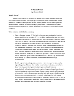
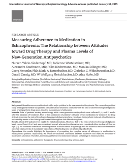
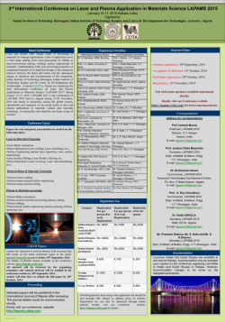
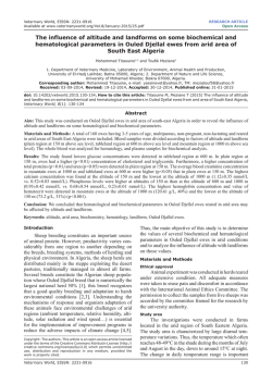
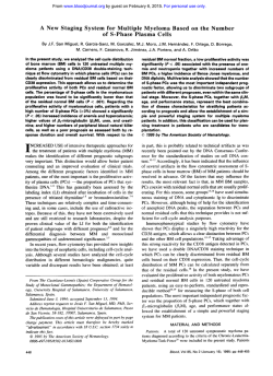
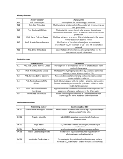
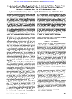
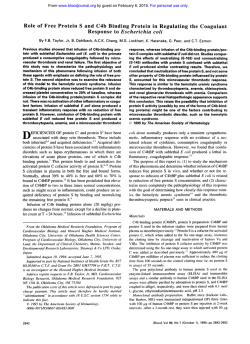
From www.bloodjournal.org by guest on February 6, 2015. For personal use only.
Plasma Crosslinked Fibrin Polymers: Quantitation Based on Tissue Plasminogen
Activator Conversion to D-Dimer and Measurement in Normals and Patients
With Acute Thrombotic Disorders
By Abraham Kornberg, Charles W. Francis, and Victor J. Marder
Plasma crosslinked fibrin polymers (XLFP) are formed as a
result of in vivo hemostatic activation and are elevated in
thrombotic disease. We have investigated the plasmic degradation of plasma XLFP in vitro t o provide information regarding the pattern of crosslinking and the composition of
degradation products. Plasma XLFP were identified by sodium dodecyl sulfate (SDS)-agarose electrophoresis and
Western blotting and quantitated by gel scanning. D-dimer
was measured by enzyme-linked immunosorbent assay and
the results were verified by SDS-polyacrylamide gel electrophoresis and Western blotting of the digests. Complete
degradation of XLFP occurred only after supplementation of
plasma with plasminogen (5 U/mL) and incubation with
recombinant tissue plasminogen activator (rt-PA), indicating
that the normal plasma plasminogen concentration limits
plasmic degradation in vitro. Gel electrophoresis showed
that the principal terminal degradation products of XLDP
were fragments D, DD, and E, indicating that crosslinking
occurred primarily through y chain dimers. After adding a low
concentration of thrombin t o plasma in vitro, XLFP increased
progressively before clotting, and the concentration correlated with the increase in the D-dimer concentration after
degradation (r = .98). Plasma XLFP and D-dimer concentrations in plasmic digests were significantly elevated in patients with stroke (150 f 83 pg/mL and 88 f 32 pg/mL),
myocardial infarction (217 f 110 pg/mL and 84 f 30 pg/
mL), and venous thrombosis (187 f 80 pg/mL and 86 f 19
pg/mL) compared with normals (28 f 12 pg/mL and 25 7
pg/mL). There was a strong correlation between the plasma
concentration of XLFP and the D-dimer immunoreactivity of
plasma after plasmic degradation (r = .87). The results indicate that XLFP in plasma are crosslinked primarily through y
chains and degrade t o fragment DD with plasminogen activation. Also, the immunoreactivity of in vitro plasmic digests of
plasma reflects the concentration of XLFP and may provide a
useful indirect measure of in vivo hemostatic activation in
patients with thrombotic disease.
0 1992by The American Society of Hematology.
H
results indicate that D-dimer is the primary plasmic derivative of XLFP and that the D-dimer concentration in
plasmic digests of plasma provides an indirect measure of
the content of XLFP. Also, the plasma concentration of
XLFP in normals and patients is measured, showing an
increase in patients with thrombosis.
EMOSTATIC activatian is primarily a localized process resulting in fibrin formation at sites of vessel
injury, inflammation, or thrombus formation. However, the
thrombin that is formed results in systemic effects, including release of fibrinopeptide A from fibrinogen and circulation of “soluble fibrin.” The latter is heterogeneous in
composition and reflects a variable extent of polymerization
and crosslinking. Low concentrations of soluble fibrin are
found in normal plasma and increased concentrations are
found in patients with thrombotic disease using several
methods, including gel filtration chromatography,14 affinity
chromatography,5p6high performance liquid chromatography? sodium dodecyl sulfate (SDS)-agarose gel electrophoresis?-1° potentiation of tissue plasminogen activator
(t-PA) activity,” ethanol gelation,’* and protamine sulfate
precipitation.l3
An alternative approach to the evaluation of plasma
soluble fibrin is based on the identification of covalently
crosslinked fibrin polymers (XLFP) resulting from the
action of factor XIII, to crosslink fibrin into dimers and
p01ymers.l~Using electrophoretic techniques, we9 and othe r ~ ~have
~ identified
, ~ ~ - low
~ ~concentrations of XLFP in
normal plasma and elevated concentrations in patients with
thrombotic disease, including acute myocardial infarction
(MI). Crosslinked fibrin polymers contain the yy chain
c r o ~ s l i n k , ~ J ~which
J ~ - ~ is~ resistant to plasmin,zO,21 and
plasmic degradation yields derivatives containing crosslinked
yy chain remnants, including fragment DD.21-z4In plasma
of patients undergoing fibrinolytic therapy, plasma XLFP
are degraded in vivo, contributing to elevated plasma
concentrations of fibrin degradation products.5
In this study, we have investigated the plasmic degradation of XLFP in vitro. Conditions required for complete
degradation of plasma XLFP are identified and relations
between the concentration of XLFP and the D-dimer
immunoreactivity after degradation are characterized. The
Blood, VOI 80,NO3 (August 1). 1992: pp 709-717
*
MATERIALS AND METHODS
Patients and blood samples. Blood was obtained by antecubital
venipuncture from normals and patients, anticoagulated with
sodium citrate (0.4% final concentration), immediately placed on
ice, centrifuged within 1hour of collection at 2,300g for 15 minutes
at 4”C, aliquoted, and stored at -70°C. Samples were thawed
within 7 days and prepared for electrophoresis or plasmic digestion. Acute MI was diagnosed in patiests with persistent chest
pain, an elevated MB fraction of creatine kinase, and electrocardiographic changes of ST segment elevation with subsequent development of significantQ waves (transmural) or ST segment depression
(subendocardial). Stroke was a clinical diagnosis based on the
development of irreversible neurologic signs and symptomsand the
exclusion of other disorders, such as tumors, infections, demyelinating diseases, and trauma, with confirmation by appropriate radio-
From the Hematology Unit, Depaninent of Medicine, University of
Rochester School of Medicine and Dentisty, Rochester, Ny.
Submitted October 25 1991; accepted March 31,1992.
Suppried in pari by Grant No. HL-30616from the National Heart,
Lung and Blood Instihrte, National Institutes of Health, Bethesda,
MD.
Address reprint requests to Charles W. Francis, MD, Hematology
Unit, PO Box 610, University of Rochester Medical Center, 601
Elmwood Ave, Rochester, NY14642.
The publication costs of this article were defrayed in part by page
charge payment. T%is article must therefore be hereby marked
“advertisement” in accordance with 18 U.S.C. section I734 solely to
indicate this fact.
0 1992 by The American Society of Hematology.
0006-4971I92 /8003-OO18$3.OO10
709
From www.bloodjournal.org by guest on February 6, 2015. For personal use only.
710
KORNBERG, FRANCIS, AND MARDER
streptavidin-horseradish peroxidase conjugate (Bethesda Research Laboratories) diluted l:l,OOO in TTBS. The blot waswashed
in TTBS after each step and was developed by submerging it in a
solution of 20 mmol/L. 3.3'-diaminobenzadine tetrahydrochloride
(Sigma) in 'ITBS containing 3.4 mmol/L nickel chloride and
0.009% hydrogen peroxide. The reaction was terminated after
color development ( 5 to 10 minutes) by washingwith water, and the
blot was dried and photographed. For quantitation. gels were
scanned, and protein in bands quantitated by comparison with a
standard curve as described previously.y
graphic or magnetic resonance imaging. Deep vein thrombosis was
diagnosed by venography in patients with compatible clinical
findings.
Fihrinogen and fihrin digests. Fibrinogen (grade L) was purchased from Helena Laboratories (Beaumont, TX) and reconstituted at a concentration of 5 mg/mL in 50 mmol/L Tris, 100
mmollL sodium chloride, pH 7.6. Plasmic digests were prepared by
incubation with plasmin (kindly provided by the Bureau of Biologics Standards, Bethesda, MD) at a concentration of 0.15 Committee on Thrombolytic Agents (CTA) UlmL, at 37°C for 90 minutes.
Digestion was terminated by the addition of aprotinin (Mobay
Chemical Co, New York, NY) to a final concentration of 300
kallikrein inhibitory units (KIU)/mL to inhibit plasmin. Fragment
DD was isolated from a plasmic digest of crosslinked fibrin
prepared as described elsewhere.2hLyophilized finely ground fibrin
was digested by suspending in 50 mmol/L Tris, 100 mmol/L
sodium chloride, 5 mmol/L calcium chloride, pH 7.6, and incubation with 3.4 CTA UlmL plasmin with gentle magnetic stirring at
37°C for 24 hours. Digestion was terminated by the addition of
aprotinin (100 KIUlmL) to inhibit plasmin, and fragment DD was
purified by gel filtration on a column (2.5 x 140 cm) of Sephaclyl
S-300(Pharmacia LKB Biotechnology, Inc, Piscataway, NJ) in 50
mmol/L Tris, 150 mmol/L sodium chloride, 40 mmollL sodium
ethylenediaminetetraceticacid (EDTA), pH 7.6, at a flow rate of
35 mLlh. The protein peak containing fragment DD was identified
by SDS-polyacrylamidegel electrophoresis (SDS-PAGE) of aliquots, p l e d , and stored.
Preparation ofplasma digests. Aliquots of 50 p.L of plasma were
incubated with recombinant t-PA (rt-PA) purchased from Genentech, Inc (South San Francisco, CA) alone or with human gluplasminogen obtained from Sigma Chemical Co (St Louis, MO) at
37°C. and 500 KlUlmL aprotinin was added at intervals to inhibit
plasmin. Normal plasma contains 1 UlmL plasminogen (2.4
FmollL).
Electrophoretic analysis. SDS 2% agarose electrophoresis and
SDS 4% to 10% gradient PAGE were performed as described
e l ~ e w h e r e .Western
~ . ~ ~ blotting was performed using a modification
of the method of Towbin et a12"as described previously.B Immunostainingwas performed by incubating the nitrocellulose paper for 30
minutes at 25°C with the following sequence of antibodies and
reagents: rabbit antihuman fibrinogen antiserum (Cappel Laboratories, Westchester, PA) diluted 1:2,000 in Tween tris-buffered
saline (TTBS); biotinylated goat-antirabbit IgG (Bethesda Research Laboratories, Gaithersburg, MD) diluted 1:1,OOO in TTBS;
1
Trimer
Dimer
--
Monomer-
2
3
4
5
6
D-dimer enzyme-linked immunosorhent assay (ELISA).
Crosslinked fibrin degradation products were measured with an
ELSA (Dimertest; American Diagnostica, Greenwich, CT)using
a monospecific antibody (DDl3B6) reactive with a site associated
with the factor XII1.-mediated yy crosslink and a panspecific tag
antibody (4D2) reactive with fibrin and fibrinogen degradation
Precoated plates were used, and the results were
calculated with a standard curve from 78 to 5,OOO nglmL of purified
fragment DD provided by the manufacturer.
Statistical anulysis. Comparison of means was performed using
the two-tailed t-test. Variance is described as 2SD.
RESULTS
To determine the conditions for in vitro degradation of
XLFP, plasma samples containing varying amounts of
XLFP were incubated in vitro with rt-PA or rt-PA plus
plasminogen and then analyzed by SDS-agarose electrophoresis and Western blotting (Fig 1). Incubation of plasma
with a high concentration of rt-PA (200 pg/mL) resulted in
incomplete degradation of fibrinogen and XLFP (lanes 2,s.
and 8). Bands consistent with fibrinogen degradation products X and Y in addition to fragment D were present after 1
hour of incubation with 200 pg/mL rt-PA in all samples,
and less degraded derivatives larger than fibrinogen were
evident in the digest in lane 5. Supplementation with 1 or 2
U/mL plasminogen resulted in greater, but still incomplete,
degradation with rt-PA (data not shown). However, complete degradation of fibrinogen and XLFP occurred in all
plasma samples after incubation for 1 hour with 25 Fg/mL
rt-PA plus 5 U/mL plasminogen (Fig 1, lanes 3,6, and 9).
After digestion under these conditions, electrophoresis
7
8
9
-w w
cJ @ # 'b
-
-l)
rt-PA (pg/ml)
plasminogen
(U/ml)
so
- -
m -D
0 200 25 0 200 25 0 200 25
0 0 5 0 0 5 0 0 5
'E
+ DD
Fig 1. Plasmic degradation of
plasma XLFP at varying concentrations of rt-PA and plasminogen. Three separate stored, citrated plasma samples were
incubated at 3PC with varying
concentrationsof rt-PA and plasminogen, and the reaction was
terminated after 1 hour by the
additionof aprotinin (500 U/mL).
subjected
An
aliquot from
to SDS
each2%
sample
agarose
was
electrophoresis, followed by
Western blotting with antifibrinogen antiserum. The locations of
the fibrinogen/fibrin monomer
and crosslinked fibrin dimer and
trimer are indicated. Fibrinogen
fragments X, D, and E can be
identified, but fragments DD and
Y overlap with this gel system.
From www.bloodjournal.org by guest on February 6, 2015. For personal use only.
QUANTITATION OF CROSSLINKED FIBRIN POLYMERS
711
II-
)c -Monomer
-0
Pentamer
- Quadramer
- Trimer
- Dimer
.14 .28 .43 .57 .71 .86
1.O
A
Time/ Clotting Time
Fig 2. Formation and degradation of plasma XLFP. (A) Imreaslng
XLFP in plasma after addition of thrombin. Stored, pooled cltrated
plasma was incubated with 0.01 U/mLthrombin (0.9 pmol/L) and 10
mmol/L calcium chloride at 37°C. Aliquots were withdrawn at lntervals and subjected to SDS 2% agarose electrophoresis followed by
Western blotting with antifibrinogen antiserum. The "TimelClotting
Time" value reflects the incubationtime of each sample divided by the
time of first visible fibrin formation at 35 minutes. (B) Plasmic
digestion of plasma containing increasingamounts of XLFP formed by
addition of thrombin. Aliquots of plasma were withdrawn from
plasma samples shown in (A) and incubated with 25 pg/mL rt-PA and
5 U/mL plasminogen for 1 hour at 37°C. Aprotinin (500 U/mL) was
added to each digest, and an aliquot was subjected to SDS 4% to 10%
gradient PAGE, followed by Western blotting with antifibrinogen
antiserum. The .57 time/clotting time value was not analyzed in this
experiment.
-
-DD
-D
showed no bands larger than fragment DD, a heavy band
with the migration of fragment D, and a fainter band
consistent with fragment DD. Because incubation of plasma
with 25 pg/mL rt-PA plus 5 U/mL plasminogen resulted in
apparently complete degradation, all digests in subsequent
experiments were prepared using these conditions.
To determine whether a correlation existed between the
D-dimer immunoreactivityof plasmic digests and the plasma
concentration of XLFP, we prepared plasma containing
varying concentrations of XLFP. A low concentration of
thrombin (0.01 U/mL, 0.9 pmol/L) was added to stored
citrated pooled plasma, resulting in a progressive increase
in XLFP before visible clot formation occurred (Fig 2A).
Whereas the monomer band did not change in intensity
after addition of thrombin, the dimer band increased
slightly at .14 and .28 of the clotting time, and was more
prominent at .43 and .57 of the clotting time. At .71 and .86
of the clotting time, the dimer band was most prominent,
with six to nine polymeric forms. The intensity of all bands
decreased at 1.0 of the clotting time (35 minutes), when
1E
0
.14 .28.43 .71 -86 1.o
TimeXlotting Time
B
fa..it fibrin strands were first visible -.I the plasma and
before solid clot formation occurred.
Incubation of aliquots of the same plasma samples with
rt-PA and plasminogen yielded digests that contained
predominantly fragments D, E, and DD by SDS-PAGE
(Fig 2B). Whereas bands corresponding to fragments D and
E did not change up to .86 of the clotting time, the D-dimer
band increased progressively from .28 to .43 and was most
From www.bloodjournal.org by guest on February 6, 2015. For personal use only.
KORNBERG, FRANCIS, AND MARDER
712
prominent at .71 and .86 of the clotting time, corresponding
to the increase in XLFP (Fig 2A). The intensity of the E, D,
and D-dimer bands decreased at the time of visible fiber
formation (1.0 of the clotting time), in parallel with the
decrease in the monomer and polymeric forms on the
SDS-agarose gel. This correspondence was examined more
closely by comparison of the plasma concentration (pg/
mL) of XLFP as determined by SDS-agarose gel electrophoresis and scanning densitometry and as the ratio of
polymer to monomer bands, with D-dimer concentration in
plasmic digests assayed by ELISA (Fig 3A). Before addition of thrombin to the plasma, XLFP concentration, the
ratio of polymer to monomer, and the digest DD concentration were 180 f 52 pg/mL, .17 f .02, and 44 f 7 pg/mL,
respectively. These values increased 1.3-, 1.2-, and 1.9-fold
at .28 of the clotting time, 2.7-, 2-, and 3.1-fold at .57 of the
clotting time, and to 7.4-, 5.8-, and 7-fold at .86 of the
clotting time. Regression analysis indicated a high correlation between D-dimer immunoreactivity in the plasmic
digests and XLFP expressed as concentration (r = .98) (Fig
3B) or as the ratio of polymer/monomer (r = .80). D-dimer
concentrations in the plasmic digests were also determined
from densitometric scanning of Western blots of SDSpolyacrylamide gels (Fig 2B) and quantitated by comparison with a standard curve of purified fragment DD. Similar
amounts of immunoreactive D-dimer were found in the
digest by ELISA and by densitometric analysis of the gels
(r = .87) (Fig 4).
The D-dimer immunoreactivity in plasmic digests of
1.2
plasma from 12 normals was distributed over a narrow
range, with a mean f SD of 25 2 7 pg/mL (Fig 5). After
collection, maintenance of the blood sample at 4°C for up to
8 hours or at room temperature for up to 4 hours before
centrifugation and preparation of plasmic digests did not
alter the results (data not shown). Similarly, storage of
citrated plasma at -70°C for up to 2 weeks before digestion
did not alter the results obtained by ELISA. The addition of
hirudin (20 U/mL), a specific thrombin inhibitor, or iodoacetamide (10 mmol/L), an inhibitor of factor XI& to
plasma before digestion did not affect results, indicating a
negligible effect of hemostatic activation during plasmic
digestion. The interassay and intraassay coefficients of
variance were both 13% with normal plasma. The concentration of XLFP in plasma from normal individuals was
lower than that in pooled plasma after prolonged storage
(Figs 2 and 3), reflecting storage-induced changes in the
latter.
Increased D-dimer immunoreactivity was found in plasmic digests of plasma from patients with thrombotic disorders (Fig 5). The mean f SD values for 10 patients with
stroke was 88 2 32 pg/mL, for 14 patients with myocardial
infarction 84 2 30 pg/mL, and for six patients with venous
thrombosis 86 f 19 pg/mL. The values were distributed
over a wide range of 32 to 144 pg/mL, but only 4 of the 30
samples from patients were within the range of normals,
and the mean D-dimer concentration of the digested
plasmas in each patient group was significantly higher than
in normal plasma (P < .005).
1200
1.0 1000
.8
800
.6
600
.4
400
,
.2
0
04
0
.2
.4
.6
TimeIClotting Time
.8
1.0
A
0
200
400 600 800 1000
Fibrin Polymer (pglml)
1200 1400
6
Fig 3. Measurementof XLFP in thrombin-treatedplasma samples and D-dimer immunoreactivity of plasmic digests. (A) Thrombin (0.01 U/mL)
and calcium chloride (10 mmol/L) were added to stored, pooled normal plasma, incubated at 37"C, and aliquots withdrawn at the indicated
intervals as in Fig 2A. Samples were subjected to SDS 2% agarose electrophoresis and Western blotting with antifibrinogenantiserum, and the
concentration of plasma fibrin polymer (0-0)
was determined by gel scanning and comparison with a standard curve of purified fibrinogen at
known concentrations. The ratio of polymer/monomer (x-x) was determined directly from the gel scan tracing. Plasmic digests of the same
plasma aliquots were preparedand D-dimer immunoreactivity (0-0) of the digests was measured by ELISA. The values representmean -c SD of
three experiments. (B) Correlation between plasma Concentration of XLFP and D-dimer immunoreactivity in plasmic digests. As determined by
linear regression, the equationfor the line is y = .23X 23 (r = .98).
+
From www.bloodjournal.org by guest on February 6, 2015. For personal use only.
QUANTITATION OF CROSSLINKED FIBRIN POLYMERS
500
713
normals (P < .005) (Table 1).Similarly, the D-dimer immunoreactivity in plasmic digests of plasma from each of the
three patient groups was significantly higher than in normals (P < .005). Just as was found for XLFP prepared in
vitro (Fig 3), linear regression analysis showed a good
correlation (r = .87) between the plasma concentration of
XLFP and the D-dimer immunoreactivity of plasmic digests
(Fig 7), with a best-fit equation of y = .28X + 28. D-dimer
immunoreactivity in the digests measured by ELISA was
closely correlated with D-dimer concentration determined
by band intensity after SDS-PAGE (Fig 6B).
1
400
\
P,
3.
L
DISCUSSION
0
100
200
300
400
DD by ELSA (pg/ml)
Fig 4. Comparison of D-dimer concentration in plasmic digests of
thrombin-treated plasma as measured by ELSA and gel analysis.
Plasma was incubated with 0.01 U/mL thrombin and 10 mmol/L
calcium chloride and aliquots were withdrawn at intervals up to the
time of clot formation as in Fig 1. Digests were prepared and
subjected to SDS 4% to 10% gradient PAGE and Western blotting
with antifibrinogen antiserum. D-dimer concentration in plasmic
digests was measured by ELISA and by gel scanning in comparison
with a standard curve of purifiedfragment DD. The values are derived
from three experiments. The equation of the line as determined by
linear regression is y = .98X 47 (r = .87).
In the present study, we have shown that the D-dimer
immunoreactivity of plasmic digests of plasma reflects the
concentration of plasma XLFP. To obtain this result,
complete degradation to fragment DD was necessary because less complete digests contain heterogeneous mixtures
of larger crosslinked fibrin degradation products with lower
imm~noreactivity.~~
Incubation of plasma with rt-PA at a
high concentration (200 p,g/mL) degraded XLFP incompletely, but the addition of supplemental plasminogen (5
U/mL) to plasma containing rt-PA at a lower concentration
(25 pg/mL) resulted in complete degradation. This is
consistent with a prior report showing incomplete degradation of fibrinogen and XLFP after incubation of plasma in
vitro with plasminogen
and also with the demon-
140
+
The plasma samples from normals and patients that were
used to prepare plasmic digests were also evaluated by
SDS-agarose electrophoresis and Western blotting (Fig
6A). Bands corresponding to XLFP were faint in normals,
but were clearly visible, although to a variable extent, in the
three patient groups. After plasmic degradation, prominent
bands corresponding to fragments D and E were present by
SDS-PAGE and were approximately the same in all samples (Fig 6B). A band corresponding to fragment D-dimer
could not be clearly identified in plasma from normals,
indicating that its concentration was less than 100 p,g/mL,
which is the limit of sensitivity of the electrophoretic
method. The prominence of the fragment DD band by
SDS-PAGE in patient samples corresponded to the intensity of XLFP in the same sample by SDS-agarose electrophoresis. For example, fibrin polymer bands were faint on
the SDS-agarose gel in the patient with stroke in lane 4 of
Fig 6A, and no clearly identifiable D-dimer band was
present in the corresponding SDS-PAGE. In contrast,
XLFP bands were prominent in the samples from a patient
with stroke in lane 7 and with acute MI in lane 10, and the
fragment DD band was easily identified in the corresponding digest samples (Fig 6B).
The concentration of XLFP and the polymer/monomer
ratio in patient samples were significantly higher than in
120
?
100
\
0
a
L
Q
Q
80
60
$
8
20
0
I
I
I
I
Normals
Stroke
I
I
t
Myocardial Deep Vein
Infarction Thrombosis
Fig 5. DD concentration in plasmic digests of plasma from normals
and patients with thrombotic disease. Plasma from normals and
patients was incubated with 25 pg/mL rt-PA and 5 U/mL plasminogen. D-dimer immunoreactivityin the plasmic digests was measured
by ELISA.
From www.bloodjournal.org by guest on February 6, 2015. For personal use only.
714
KORNBERG, FRANCIS, AND MARDER
Normals
Trimer
Myocardial
Infarction
Stroke
1
2
3
4
5
6
7
16
22
28
80 160 150 280
8
9
Deep Vein
Thrombosis
10
11
-
120 170 220 320
2
3
Myocardial
Infarction
Stroke
Normals
4
5
6
7
8 - 9 10 11
220 260 310
A
Fibrin Polymer (pglml)
1
12 13 14
Deep Vein
Thrombosis
12 13 14
I
'"*
- DD
-
I
=- & I ' :
._I
I
I
20 14 31
40 96 102140
DD
88 98140100
97 106106
by ELISA (pg/ml)
stration that plasminogen depletion limits in vitro degradation of plasma clots incubated in plasma with rt-PA.'*
Incomplete degradation of fibrinogen also occurs in vivo,
with a predominance of fragment X during thrombolytic
therapy with streptokinase or t-PA.J3
Table 1. Content of plasma XLFP and of D-Dimer in Plastic Digests of
Plasma From Normals and Patients With Thrombotic Disorders
Normals
In = 12)
Acute MI
In = 14)
Venous
Stroke Thrombosis
In = 101
In = 6 )
Plasma XLFP concentration
(m/mL)
28 f 12 217 f 110 150 f 83 187 f 80
Plasma ratio of polymer/
monomer
.16 f .02 .30 t .1 .28 f .13 .32 f .06
D-dimer concentration in
plasmic digests (rrg/mL) 25 t 7
84 t 30 88 t 32 86 f 19
All values are mean t SD.
B
]E
Flg6. Electrophoretkanalysis of fibrinogenderivin plasma from normals and patients with
thrombotic disease M o m and after degradation. (A)
Untreatedplasma. Plasma from normals and patients
was subjected to SDS 2% agarose electrophoresis
followed bv Western blotting
- with antifibrinwen
antiserum. The plasma concentration of XLFP was
determined by densitometric scanning using a standard curve derived from purified fibrinogen at known
concentrations. (B) Plasma digests. Plasmic digests
of the same plasma samples from normals and patients were prepared and aliquots subjected to SDS
4% to 10% gradient PAGE followed by Western
blotting with antifibrinogen antiserum to determine
D-dimer concentration. The locations of fragments
DD, D, and E are indicated. Bands migrating between
DD and D are not identified but may represent
fragment Y or heterogeneity of DD resulting from
additional proteolytic changes. Ddimer immunoreactivity in the plasmic digests was measured by ELISA.
Using the combination of rt-PA (25 pg/mL) and plasminogen (5 U/mL) to prepare plasmic digests of plasma, we
explored the relationship between plasma XLFP content
and D-dimer immunoreactivity of the digests. Plasma containing varying concentrations of XLFP was prepared by
the addition of a low concentration of thrombin in vitro. A
strong correlation was found between the concentration of
plasma XLFP as determined by SDS-agarose gel electrophoresis and the D-dimer concentration of the plasma
digest as measured by ELISA (r = .98) (Fig 3B). The
reliability of measuring digest D-dimer by ELISA was
confirmedby its significant correlation with D-dimer concentration estimated from Western blots after SDS-PAGE
(r = .87) (Fig 4).
A similar approach was used to evaluate XLFP in plasma
from normals and patients with thrombotic disorders. The
amounts of XLFP and of plasma digest DD were increased
From www.bloodjournal.org by guest on February 6, 2015. For personal use only.
QUANTITATION OF CROSSLINKED FIBRIN POLYMERS
715
I'
"
I
I
0
100
I
I
200
300
I
400
Fibrin Polymer (pg/ml)
Fig 7. Correlation between the plasma concentration of XLFP and
DD immunoreactivityin plasmic digests of plasma from normals and
patients with thrombotic disease. Plasma from normals ( 0 )and
patients with myocardial infarction (0).
stroke (X), and deep vein
thrombosis (A)was subjected to SDS-agarose electrophoresis followed by Western blotting with antifibrinogen antiserum, and the
concentration of XLFP was determined by densitometric scanning of
Western blots using standard curves of purified fibrinogen at known
concentrations. Plasmic digests of the same plasma samples were
preparedand D-dimer immunoreactivitywas measured by ELISA. The
equation for the line is y = .28X 28 (I = .87), as determined by linear
regression.
+
in patients compared with normals (Table l), with a good
correlation between XLFP determined by gel electrophoresis and plasmic digest DD measured by ELISA (Fig 7). The
amount of fragment D D detected by Western blotting of
plasma digests (Fig 6B) corresponded to the amount of
XLFP on SDS-agarose gels (Fig 6A) and to digest DD
measured by ELISA, confirming that the measurement
reflected the D-dimer concentration in the digest derived
from XLFP. Although there was little overlap between the
plasma digest DD values in normals and patients (Fig 5 ) ,
the small number of patients in each group limits conclusions about the diagnostic value of this approach in patients
with thrombotic disease.
The results indicate that D-dimer is the primary
crosslinked plasmic degradation product of XLFP and
thereby provide additional evidence that the yy isopeptide
bond is the principal crosslink in soluble fibrin in plasma.
This view is consistent with prior reports of the pattern of
factor XIIIa crosslinking of fibrin in vitro using purified
p r o t e i n ~ l ~in
, ~ which
~ , ~ ~ y chains are crosslinked more
rapidly than a chains. It is also consistent with several
reports7J5J7Jssuggesting that soluble fibrin contains mainly
yy crosslinks. Shainoff et aP6 have also found crosslinked
fibrin polymers in the plasma of normals and elevated levels
in patients with thrombotic disease similar to those in this
report. However, using direct immunoprobing of gels with
monoclonal antibodies against individual fibrin chains, they
detected both a-Aa and yy crosslinks in the polymers.
Formation of Aa-a and Aa-y crosslinks has been attributed
to tissue transglutaminase activity, whereas yy crosslinks
are a product of factor XIII,.37 Our findings do not exclude
the presence of some fibrin polymers crosslinked through a
or A a chains. However, electrophoretic analysis of digests
(Figs 2B and 6B) and the correlation between the plasma
concentration of XLFP and D-dimer immunoreactivity
(Figs 3 and 7) suggests that D-dimer is the predominant
plasmic degradation product, and indicates that circulating
XLFP are crosslinked primarily through y chains, both in
the in vitro model and in normals and patients with
thrombotic disease.
Consistent with our prior report? we found a mean
plasma concentration of XLFP of 28 p,g/mL in normal
plasma by SDS-agarose electrophoresis. This is associated
with D-dimer immunoreactivity after plasmic digestion of
25 kg/mL (Table 1). Linear regression of the correlation
between plasma XLFP concentration and D-dimer immunoreactivity (Fig 7) derived the equation y = .28X + 28. An
XLFP concentration of 28 pg/mL in normals predicts a
digest D-dimer continuation of 36 kg/mL, close to the
observed mean in normals of 25 pg/mL (Table 1). The
slope of .28 indicates an increase of D-dimer iqmunoreactivity of 280 kg/mL for an increase of 1,000 kg/mL in
plasma XLFP, and is close to the value of .29 predicted on
the relative mass of the D-dimer portion of a fibrin dimer,
which is the predominant polymeric species present before
degradation. This analysis, together with the highly significant correlation between digest DD concentrations measured by ELISA and by SDS-PAGE densitometry, suggest
that increases in D-dimer immunoreactivity in plasma
digests accurately reflect elevated levels of XLFP and prove
that the measurements by the 3B6/4D2 ELISA reflect the
actual concentrations of D-dimer in the plasmic digests of
plasma.
It has been s ~ g g e s t e d ~that
~ 9the
~ D-dimer immunoreactivity using the 3B6/4D2 assay can be falsely elevated in the
presence of high concentrations of fragment D because the
secondary antibody (4D2) reacts with both fibrin and
fibrinogen degradation products. However, prior reports
have found no reaction with the assay at fragment D concentration of up to 200 ~ g / m L , 3which
~ , ~ ~far exceeds the
concentration of fragment D in the sample after dilution.
Further, a concentration of fibrinogen degradation products of 2,200 pg/mL gave a reaction using the assay of only
1,300 ng/mL.39 Therefore, even if there is some level of
crossreactivity or any other effect of fibrinogen degradation
products on the 3B6/4D2 ELISA, the contribution to the
total would be small but could contribute to the positive Y
intercept of the linear regression analysis.
The correlation between plasma XLFP and D-dimer
immunoreactivity both in the in vitro model (Figs 2 and 3)
and in normals and patients (Fig 7) indicates that the
D-dimer level measured by ELISA after plasmic digestion
may be a useful measure of the amount of XLFP in plasma
from normals and patients with thrombotic disorders.
Plasma soluble fibrin has been measured previously by
several techniques, based on its unique physical and chemical properties. Musumeci et a1,2 using gel filtration chromatography, and Edgar et al: using affinity chromatography,
reported levels of 25 p,g/mL and 27 kg/mL, respectively, in
normals, values similar to our findings. Others have reported higher levels in normals of 120 to 184 kg/mL using
gel filtration c h r o m a t ~ g r a p h y ~and
. ~ ,lower
~
levels of 3 to 7
kg/mL using affinity chromatography6 and chromogenic
From www.bloodjournal.org by guest on February 6, 2015. For personal use only.
KORNBERG, FRANCIS, AND MARDER
716
assay." The different concentrations may be attributed to
different molecular species of soluble fibrin measured using
the various methods. Despite these differences, all methods
identify an increase of between 3- and 10-fold in soluble
fibrin in patients with thrombotic disea~e,l-~.~Jl
similar to
our findings (Table 1).However, most nf these techniques
are difficult to perform and impractical for routine clinical
use. Our approach is based on the specificity of D-dimer as
a marker for XLFP20,21,23,29
and the significant correlation
between plasma XLFP and D-dimer immunoreactivity of
plasmic digests. The results indicate that measurement of
D-dimer immunoreactivity in plasmic digests may be useful
in evaluation of hypercoagulable states by providing a
simple and accurate measure of the plasma content of
XLFP.
ACKNOWLEDGMENT
The authors acknowledge the technical assistance of Kristina
Klingbeil and the help of Carol Weed in preparation of the
manuscript.
REFERENCES
1. Fletcher AP, Alkjaersig N, Davies A, Lewis M, Brooks J,
Hardin W, Landau W, Raichle ME: Blood coagulation and plasma
fibrinolytic enzyme system pathophysiology in stroke. Stroke 7:337,
1976
2. Musumeci V, Culasso D, Boni P: A rapid chromatographic
method for quantitation of high molecular weight fibrinogen
derivatives in plasma. Thromb Haemost 39555,1978
3. Neri Serneri GG, Gemini GF, Abbate R, Favilla S: Occurrence of soluble very high molecular weight fibrinogen complexes
in hypercoagulable states. An in v i m and in vivo study on their
features. Thromb Haemost 42:1561,1979
4. Fletcher AP,Alkjaersig NK, Ghani FM, Tulevski V, Owens
0: Blood coagulation system pathophysiology in acute myocardial
infarction: The influence of anticoagulant treatment on laboratory
findings. J Lab Clin Med 93:1054,1979
5. Edgar W, Mckillop C, Howie PW, Prentice CRM: Composition of soluble fibrin complexes in pre-eclampsia. Thromb Res
10567,1977
6. Matthias FR, Reinicke R, Heene DL: Affinity chromatography and quantitation of soluble fibrin from plasma. Thromb Res
10:365,1977
7. Ishikawa A, Iwasaki Y Rapid estimation of soluble fibrin
monomer complexes by high performance liquid chromatography
for the purpose of early detection of DIC. Thromb Res 30:521,
1983
8. Connaghan DG, Francis CW, Lane DA, Marder VJ: Specific
identification of fibrin polymers, fibrinogen degradation products,
and crosslinked fibrin degradation products in plasma and serum
with a new sensitive technique. Blood 65:589,1985
9. Francis CW, Connaghan DG, Scott WL, Marder VJ: Increased plasma concentration of cross-linked fibrin polymers in
acute myocardial infarction. Circulation 75:1170,1987
10. Proietti AB, McGuire M, Bell W: Specific identification of
fibrin(ogen) degradation products in plasma and serum using
blotting and peroxidase labeled antiserum. Am J Hematol34:270,
1990
11. Wiman B, Ranby M: Determination of soluble fibrin in
plasma by a rapid and quantitative spectorphotometric assay.
Thromb Haemost 55:189,1986
12. Kierulf P, Godal HC: Fibrinemia in medical patients screened
by the ethanol test. Acta Med Scand 190185,1971
13. Gurewich V, Hutchinson E Detection of intravascular
coagulation by serial-dilution protamine sulfate test. Ann Int Med
75:895, 1971
14. Chen R, Doolittle R F y-y Crosslinking sites in human
bovine fibrin. Biochemistry 10:4686, 1971
15. Kierulf P: Studies on soluble fibrin in plasma. V. Isolation
and characterization of the clottable proteins obtained from
plasmas upon gelation with ethanol. Thromb Res 4:183,1974
16. Ly B, Jakobsen E: Some aspects of the bonds interlinking
soluble fibrin complexes in human plasma. Thromb Res 6:65, 1975
17. Selmayer E, Muller-Berghaus G: Soluble crosslinked fibrin(ogen) polymers. Thromb Haemost 542304,1985
18. Graeff H, Hafter R, Bachmann L Subunit and macromolecular structure of circulating fibrin from obstetric patients with
intravascular coagulation. Thromb Res 16:313, 1979
19. Ittyerah R, Alkjaersig N, Fletcher A, Chaplin H: Coagulation factor XI11 concentrates in sickle-cell disease. J Lab Clin Med
88546,1976
20. Pizzo SV, Schwartz ML, Hill RL, McKee P A The effect of
plasmin on the subunit structure of human fibrin. J Biol Chem
248:4574,1973
21. Pizzo SV, Taylor LM Jr, Schwartz ML, Hill RL, McKee P A
Subunit structure of fragment D from fibrinogen and cross-linked
fibrin. J Biol Chem 248:4584,1973
22. Francis CW, Marder VJ, Barlow GH: Plasmic degradation
of crosslinked fibrin. Characterization of new macromolecular
soluble complexes and a model of their structure. J Clin Invest
66:1033,1980
23. Gaffney PJ, Brasher M: Subunit structure of the plasmininduced degradation products of crosslinked fibrin. Biochem Biophys Acta 295:308,1973
24. Brenner B, Francis CW, Marder VJ: The role of soluble
crosslinked fibrin in D dimer immunoreactivity of plasmic digests. J
Lab Clin Med 113:682,1989
25. Brenner B, Francis CW, Totterman S, Kessler CM, Rao AK,
Rubin R, Kwaan HC, Gabriel KR, Marder VJ: Quantitation of
venous clot lysis with the D-dimer immunoassay during fibrinolytic
therapy requires correction for soluble fibrin degradation. Circulation 81:1818, 1990
26. Olexa SA, Budzynski Az: Primary soluble plasmic degradation product of human cross-linked fibrin. Isolation and stoichiometry of the (DD)E complex. Biochemistry 18:991,1979
27. Neville DM Jr: Molecular weight determination of proteindodecyl sulfate complexes by gel electrophoresis in a discontinuous
buffer. J Biol Chem 246:6328,1971
28. Towbin H, Staehelin T, Gordon J: Electrophoretic transfer
of proteins from polyacrylamide gels to nitrocellulose sheets:
Procedure and some applications. Proc Natl Acad Sci USA
76:4350,1979
29. Francis CW, Doughney K, Brenner B, Klingbiel K, Marder
VJ: Increased immunoreactivity of plasma after fibrinolytic activation in an anti-DD ELISA system. Role of soluble crosslinked
fibrin polymers. Circulation 79:666, 1989
30. R latt DB, Blake AS, Cottis LE, Massingham DA, Fletcher
WA, Masci PP, Whitaker AN, Elms M, Bunce I, Webber AJ, Wyatt
D, Bundesen PG: An immunoassay for human D dimer using
monoclonal antibodies. Thromb Res 31:767,1983
31. Elms MJ, Bunce IH, Bundesen PG, Rylatt DB, Webber AJ,
Masci PP, Whitaker AN: Measurement of crosslinked fibrin
degradation products-An immunoassay using monoclonal antibodies. Thromb Haemost 50:591,1983
From www.bloodjournal.org by guest on February 6, 2015. For personal use only.
QUANTITATION OF CROSSLINKED FIBRIN POLYMERS
32. Onundarson FT, Francis CW,Marder VJ: Depletion of
plasminogen in vitro or during thrombolytic therapy limits fibrinolytic potential. J Lab Clin Med (in press)
33. Owen J, Friedman KD, Grossman BA, Wilkins C, Berke
AD, Powers ER: Quantitation of fragment X formation during
thrombolytic therapy with streptokinase and tissue plasminogen
activator. J Clin Invest 791642,1987
34. Pissano JJ, Finlayson JS, Peyton MP: Cross-link in fibrin
polymerized by factor XIII: e(y-glutamyl) lysine. Science 1602392,
1968
35. Doolittle RF, Chen R, Lau F Hybrid fibrin: Proof of the
intermolecular nature of y-y crosslinking units. Biochem Biophys
Res Commun 44:94,1971
36. Shainoff JR, Valenzuela R, Urbanic DA, DiBello PM, Lucas
717
FV, Graor R: Fibrinogen Aa and y-chain dimers as potential
differential indicators of atherosclerosis and thrombotic vascular
disease. Blood Coag Fibrinol1:499,1990
37. Murthy SNP, Lorand L Cross-linked Aa-y chain hybrids
serve as unique markers for fibrinogen polymerized by tissue
transglutaminase.Proc Natl Acad Sci USA 87:9679,1990
38. Eisenberg PR, Jaffe AS, Stump DC, Collen D, Bovill EG:
Validity of enzyme-linked immunosorbent assays of cross-linked
fibrin degradation products as a measure of clot lysis. Circulation
82:1159,1990
39. Lawler CM, Bovill EG, Stump DC, Collen DJ, Mann KG,
Tracy RP: Fibrin fragment D-dimer and fibrinogen Bp peptides in
plasma as markers of clot lysis during thrombolytic therapy in acute
myocardial infarction. Blood 76:1341,1990
From www.bloodjournal.org by guest on February 6, 2015. For personal use only.
1992 80: 709-717
Plasma crosslinked fibrin polymers: quantitation based on tissue
plasminogen activator conversion to D-dimer and measurement in
normal and patients with acute thrombotic disorders
A Kornberg, CW Francis and VJ Marder
Updated information and services can be found at:
http://www.bloodjournal.org/content/80/3/709.full.html
Articles on similar topics can be found in the following Blood collections
Information about reproducing this article in parts or in its entirety may be found online at:
http://www.bloodjournal.org/site/misc/rights.xhtml#repub_requests
Information about ordering reprints may be found online at:
http://www.bloodjournal.org/site/misc/rights.xhtml#reprints
Information about subscriptions and ASH membership may be found online at:
http://www.bloodjournal.org/site/subscriptions/index.xhtml
Blood (print ISSN 0006-4971, online ISSN 1528-0020), is published weekly by the American
Society of Hematology, 2021 L St, NW, Suite 900, Washington DC 20036.
Copyright 2011 by The American Society of Hematology; all rights reserved.
© Copyright 2026