
Hepatoprotective activity of Averrhoabilimbi fruit in acetaminophen
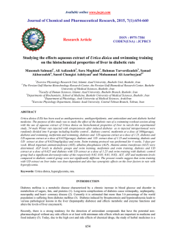
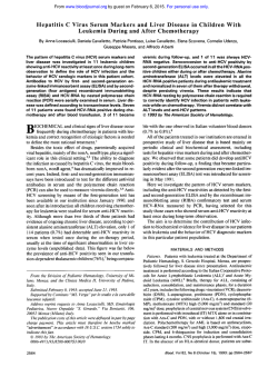
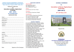
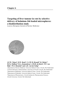
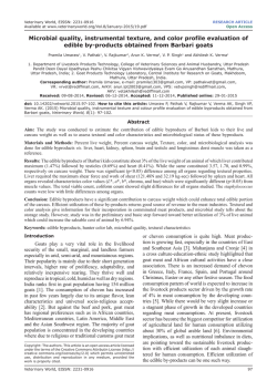
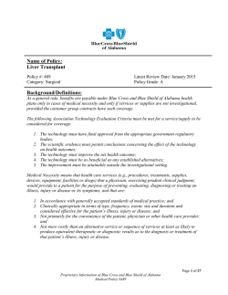
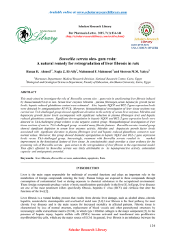
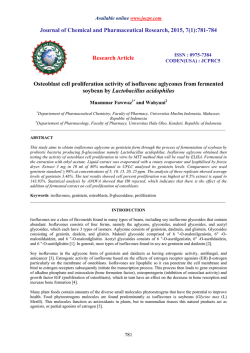
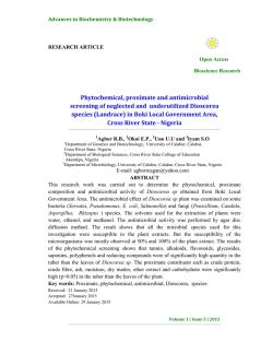
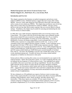
Available online www.jocpr.com
Journal of Chemical and Pharmaceutical Research, 2015, 7(1):535-540
Research Article
ISSN : 0975-7384
CODEN(USA) : JCPRC5
Hepatoprotective activity of Averrhoa bilimbi fruit in acetaminophen induced
hepatotoxicity in wistar albino rats
Thamizh Selvam N., Santhi P. S., Sanjayakumar Y. R., Venugopalan T. N.,
Vasanthakumar K. G. and Swamy G. K.
National Research Institute for Panchakarma, (Central Council for Research in Ayurvedic Sciences, New Delhi
Department of AYUSH, Ministry of Health and Family Welfare, Govt. of India), Thrissur, Kerala, India
_____________________________________________________________________________________________
ABSTRACT
The present study was taken up to evaluate the hepatoprotective activity of Averrhoa bilimbi fruit in wistar albino
rats intoxicated using acetaminophen. The extract was administered at two different concentrations 250 mg/kg.b.wt
and 500 mg/kg.b.wt as lower and higher dose respectively. Silymarin (100 mg/kg.b.wt/) was used as standard
positive control. The liver marker enzymes serum gluatamate oxaloacetate transaminase (SGOT), serum glutamate
pyruvate transaminase (SGPT), Alkaline phosphatise (ALKP) were found to be two fold increased in the disease
control group, and pre-treatment with A. bilimbi inhibited the increase of these enzymes in serum. The
histopathology studies on the tissues of liver, kidney and heart showed safety and efficacy of the test extract. The
present study revealed that A. bilimbi fruits have significant hepatoprotective activity.
Key words: Hepatoprotective activity, Averrhoa bilimbi, acetaminophen intoxication, liver marker enzymes
_____________________________________________________________________________________________
INTRODUCTION
Liver is one of the chief sites for intense metabolism and excretion. It is involved with almost all the biochemical
pathways for growth, fighting against diseases, supply of nutrients and energy provision. But the liver functions are
greatly altered and affected due to varied reasons like environmental toxins, poor drug habits, alcohol and other
ailments [1-3]. So, the liver diseases are one of the leading issues in the world today.
The plant Averrhoa bilimbi commonly known as bilimbi, is essentially tropical tree, less resistant to cold. The
bilimbi tree is long lived, reaches 5 to 10 mt in height. The leaves are alternate, and cluster at branch extremities.
Flowers are found on the trunk and branches. Fruits of bilimbi are too sour to eat raw [4]. The uncooked bilimbi is
prepared relish and served with rice in natives of Kerala, India.
EXPERIMENTAL SECTION
Plant Material
The fruits of A. bilimbi were collected from Western ghat region of Kerala (Thrissur and Palakkad) and the
authentication of the plant was done in the Pharmacy Division, National Research Institute for Panchakarma,
Thrissur.
Preparation of Extract
The fresh fruits were taken (100 gm) and crushed using mixer grinder, the juice was filtered through Whatman filter
paper No.7. The clear filtrate was obtained and was stored in the 4-80 C for experimental purpose.
535
Thamizh Selvam N. et al
J. Chem. Pharm. Res., 2015, 7(1):535-540
______________________________________________________________________________
Chemicals and Reagents:
Chemicals and Reagents of make Transasia, Bayer and Spinreact India were used for the present study.
Phytochemical Studies
The phytochemical analysis of the test extract was carried out as per the standard protocol [5-8].
Animal Experiment:
The animal studies were carried out in the National Research Institute for Panchakarma, Cheruthuruthy as per
CPCSEA guidelines and with the approval of Institutional Animal Ethical Committee.
Six to seven months old Wistar albino rats weighing 150-200 gm were used for the experiment. The animals were
fed with standard laboratory pellet chow (Amrit, Bangalore) and given water ad libitum. All rats were clinically
healthy. The animals were randomly divided into five groups of six animals each and the standard protocol was
used [9-10].
The fruit extract of A. bilimbi, was administered at two different concentrations (250 and 500 mg/kg body weight) as
lower dose (Group IV) and higher dose (Group V) through orally for the period of ten days in test groups. The
disease group (Group II) and test groups received single dose of Acetominophen (paracetamol) at 2.5 gm/kg body
weight. The control group (Group I) received distilled water throughout the experiment and standard group (Group
III) received silymarin 100 mg/kg body weight for the experimental period. The animals were fasted for 24 hrs on
10th day of the experiment and blood sample was collected. The biochemical parameters including liver marker
enzymes were assessed in the blood samples of the test groups, control groups and disease groups [11-14]. At the
end of the experiment, the animals were sacrificed under anaesthesia using diethyl ether and the tissue samples of
liver, kidney and heart were collected for evaluation of histopathological changes in tissue level among all the
experimental groups.
Statistical Analysis
The data were expressed as mean ± SEM and statistically analyzed by one way ANOVA.
RESULTS
The phytochemical analysis of the fruit extract showed the presence of carbohydrates, flavonoids, phenols,
glycosides and amino acids (Table 1).
Table 1 : Phyto-constituents of fruit juice of A. bilimbi
PhytochemicalAnalysis Extract of Syzigium jambos
Carbohydrate
+++
Phenols
+++
Flavonoids
+++
Tannins
+
Steroids
Terpenoids
Alkaloids
+
Glycosides
++
Saponins
Aminoacids
+++
+++ Very strongly present ++ strongly present + Present - Absent
Hepatoprotective Activity
The present study had been attempted to demonstrate the role of hepatoprotective activity of fresh juice of Averrhoa
bilimbi in the experimental animal system. The liver marker enzymes Serum glutamate oxaloacetate transaminase
(SGOT), serum glutamate pyruvate transaminase (SGPT) and alkaline phosphate levels were measured in the serum
samples of all the experimental groups. These liver marker enzymes were found to be increased nearly two to three
fold in the acetominophen intoxicated disease control animals comparing with healthy control group. The results
also showed that there is a significant (p < 0.05 and p < 0.01) decrease in the in SGOT, SGPT and ALKP levels in
the A. bilimbi treated groups, when compared with the disease control. As there is a greater level of efficacy in the
higher dose group (500 mg/kg. b.wt) comparing with lower dose, it is predicted that the efficacy of the extract is
found to be dose dependent. It was also observed that the efficacy of the extract is comparable (Table 2 and
Figure1-2) with standard drug silymarin. The histological study of liver is also supporting these biochemical and
clinical parameters. The histology of liver tissues of disease control group showed extensive areas of haemorrhage
and necrosis in the liver parenchyma and vacuolated cytoplasm in hepatocytes. Kidney tissues of the same group
showed oedema and showed vacuolation in the lining of epithelial cells. The extract treated groups showed
536
Thamizh Selvam N. et al
J. Chem. Pharm. Res., 2015, 7(1):535-540
______________________________________________________________________________
improved and apparently normal architecture of the liver, glomeruli and myocardial tissues. (Figure 3-8). All these
biochemical diagnostic parameters and histological tissue analysis well evidenced the hepatoprotective activity of
Averrhoa bilimbi in experimental animal.
Table 2. Liver marker enzymes level in the serum samples of experimental animals
Liver marker enzymes (IU/L)
SGOT
SGPT
ALP
Healthy Control
133.56 ± 21.61
69.75 ± 8.98
91.82 ± 13.67
Disease Control
358.0 ± 22.14*
293.86 ± 10.19*
266.77 ± 9.75**
*
*
Standard Drug
164.91 ± 23.85
74.58 ± 11.77
117.36 ± 16.47**
A. bilimbi L.D
266.78 ± 14.55* 160.80 ± 16.74*
188.8 ± 35.8*
*
**
A. bilimbi H.D.
233.70 ± 21.14
113.95 ± 14.27
139.29 ± 14.42**
Values are mean ± SEM, n=6 animals in each group. *p < 0.05, **p<0.01 when compared to disease control
Experimental Groups
Figure 1. Status of lipid profile in the A. bilimbi extract administered animals
180
Lipid Profile
160
Units in mg %
140
120
100
80
Cholesterol
60
Triglycerides
40
20
0
Healthy
Control
Deiase
Control
Standard
Group
A. bilimbi L.D A. bilimbi H.D
Experimental Groups
.
Figure 2. Albumin, bilirubin and total proteins levels in the blood samples of A. bilimbi treated experimental animals
9
Biochemical parameters
8
Units in gm %
7
6
5
4
Albumin
3
Total protein
2
Total bilirubin
1
0
Healthy
Control
Deiase
Control
Standard
Group
A. bilimbi L.D A. bilimbi H.D
Experimental Groups
.
Figure. 3. (45X) Acetaminophen treated rat liver showing extensive areas of haemorrhage and necrosis in liver parenchyma.
Hepatocytes showed vacuolated cytoplasm. Collection of inflammatory cells and siderophages observed
537
Thamizh Selvam N. et al
J. Chem. Pharm. Res., 2015, 7(1):535-540
______________________________________________________________________________
Figure 4. (45X) Glomeruli showed oedema. Renal tubules showed vacuolation of the lining epithelia cells. Interstitial tissue showed
focal collections of inflammatory cells and areas of haemorrhage
Figure 5. (45X) Section of heart showing normal endocardium and myocardium
Figure 6. (45X) A. bilimbi treated at 500 mg/kg. Wt showed apparently normal architecture of liver
Figure 7. (45X) Glomeruli of test drug treated group showed normal histology. Interstitial tissue appeared normal
538
Thamizh Selvam N. et al
J. Chem. Pharm. Res., 2015, 7(1):535-540
______________________________________________________________________________
Figure 8. (45X) Heart tissues showed normal endocardium and myocardium
DISCUSSION
The literature review revealed that traditional healers are using this plant for various ailments and natives of Kerala
and some part of Tamil Nadu are using this fruit as food ingredient. The present study was designed and carried out
scientifically to evaluate the hepatoprotective activity of A. bilimbi fruits. Liver damage induced by Acetominophen
(generally known as paracetamol) is one of the commonly used model for the screening of hepatoprotective drugs
[15-17]. Paracetamol is one of the basic medicines used as an antipyretic agent, which is safe in therapeutic doses
but it can produce fatal hepatic necrosis in man and animals [18-20]. The rise in serum SGOT, SGPT, ALKP levels
has been attributed to the damaged structural integrity of liver. It is because of, as these enzymes are belonged to the
cytoplamic location of the liver cells and they are released into circulation after cellular damages [21-22].
Paracetamol induces hepatotoxicity at higher dose by metabolic activation; therefore, it selectively causes toxicity in
liver cells maintaining semi-normal metabolic function [23-25]. The liver protective property of the A. bilimbi was
well proved when the elevated levels of liver markers enzymes SGOT, SGPT, ALKP and bilirubin levels was
brought to down significantly and the efficacy was almost comparable with silymarin. The histology analysis on
the liver, kidney and heart tissues of the experimental animals strongly evidenced that there is safety and efficacy in
the A. bilimbi treated animals. In conclusion, from the overall result of the study it could be inferred that Averrhoa
bilimbi fruit showed the significant hepatoprotective activity. It is assumed that the high contents of phenol group
compounds, flavanoids and amino acids may be attributing for the medicinal property of the A. bilimbi. Further
research on the plants could be extended for the isolation and structure determination of the principle compounds for
this property.
Acknowledgement
Authors are thankful to the Director General, CCRAS, New Delhi and other Staff members of NRIP for their kind
support and encouragement.
REFERENCES
[1] Sharma B, Sharma UK. International Journal of PharmTech Research. 2009;1 (4):1330-1334.
[2] Kshirsagar AD, Mohite R, Aggarwal S, Suralkar UR. Asian Journal of Pharmaceutical and Clinical Research.
2011;4 (3):1-8.
[3] Deepak KD, Veerendra CY, Siva SN, Tirtha G, Rajalinga D, Pinaki S, Bhim CM, Tapan KM. Tropical Journal
of Pharmaceutical Research. 2007; 6(3):755-765.
[4] Warrier PK, Nambiar VPK, Ramankutty C. Orient Longman Publications. 2002; 1:224-226
[5] Maluventhan Viji, Sangu Murugesan. (2010). Journal of Phytology. 2;1:68-77.
[6] Hassan SW, Umar RA, Lawal M, Bilbis LS, Muhammad BY and Dabai YU. (2006). African journal of
Biotechnology. 5;18:1602-1607.
539
Thamizh Selvam N. et al
J. Chem. Pharm. Res., 2015, 7(1):535-540
______________________________________________________________________________
[7] EL- Olemyl MM, AL-Muhtadi FJ, Afifi AA. (1994). Experimental phytochemistry. A Laboratory manual
college of Phramcy, King Saud University. King Saud University Press. 1-34.
[8] Trease GE, Evans WC. (1978) A textbook of Pharmacognosy. 11th Edition. Bailliere Tindall Publication.
London. 530.
[9] Gupta AK, Misra N. (2006) Am J Pharmacol Toxicol 1:1-7.
[10] Thamizh Selvam N, Venkatakrishnan V, Dhamodharan R, Murugesan S, Damodar Kumar S. (2013) . AYU
34:3:305-308.
[11] Girish C, Koner BC, Jayanthi S, Rao KR, Rajesh B, Pradhan SC. Indian Journal of Medical Research. 2009;
129 (5): 569-578.
[12] Gupta AK, Neelam M. American Journal of Pharmacology and Toxicology. 2006; 1 (1):17-20
[13] Thamizhselvam N, Anjusha P, Sanjayakumar YR, Salini KC, Venugopalan TN. International Research
Journal of Pharmacy. 2011; 2(11):116-118.
[14] Roy CK, Kamath JV, Asad M. Indian Journal of Experimental Biology. 2006; 44 (4): 305-311.
[15] Mitchell JR. Jollow DJ, Potter WZ. Gillettee JR, Brodie BN. Acetominophen induced hepatic necrosis. I.
Role of drug metabolism. J Pharmacol. Expl. Therap. 1973; 187: 185-194.
[16] Kuma S and Rex D. Arch. Inter. Med. 1991; 151: 1189-1191.
[17] Erikson L, Broome U, Kahn M, Lindholm M. J Inter Med. 1992; 231: 567-570.
[18] Asha VV, Akhila S, Wills PJ, Subramoniam A. J Ethnopharmacol. 2004; 92: 67-70.
[19] Kumar G, Banu SG, Pappa VP, Sundararajan M, Pandian RM. J Ethnopharmacol. 2004; 92:37-40.
[20] Visen PKS, Shukla GK, Patnaik and Dhawan BN. J Ethnopharmacol. 1993; 40:131-136.
[21] Boyd, EH, Bereczky GM. British Journal of Pharmacology. 1996; 26:606-614.
[22] Dahlin D, Miwa G, Lu A, Nelson S. Proc. Natl. Acad,. Sci. 1984; 81:1327-1331.
[23] Moron MS, Depierre JW, Mannervik B. Biochem. Biophys Acta. 1979; 582: 67-78.
[24] Singh A and Handa SS. J Ethnopharmacol. 1995; 49: 119-126.
[25] Gupta AK, Chitme H, Dass SK, Misra N. J Pharmacol Toxicol. 2006; 1: 82-88.
540

© Copyright 2026