
Effects of the creation of arteriovenous fistula for hemodialysis on
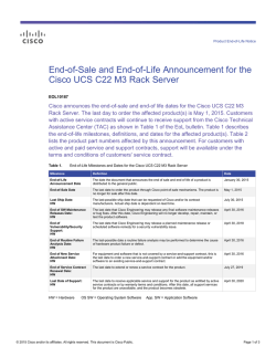
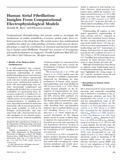
Dialysis Therapies Effects of the Creation of Arteriovenous Fistula for Hemodialysis on Cardiac Function and Natriuretic Peptide Levels in CRF Yoshio Iwashima, MD, Takeshi Horio, MD, Yoichi Takami, MD, Takashi Inenaga, MD, Toshio Nishikimi, MD, Shuichi Takishita, MD, and Yuhei Kawano, MD ● Background: Cardiac failure occasionally is caused by the creation of vascular access for hemodialysis. However, the influence of an arteriovenous (AV) fistula on cardiac function has not been fully elucidated. The present study investigated serial changes in cardiac function and hormonal levels after the AV fistula operation. Methods: Sixteen patients with chronic renal failure underwent echocardiographic studies before and 3, 7, and 14 days after the AV fistula operation. Plasma atrial natriuretic peptide (ANP) and brain natriuretic peptide (BNP) concentrations were measured before and 1, 3, 6, 10, and 14 days after the operation. Results: Creation of an AV fistula produced significant elevations in left ventricular (LV) end-diastolic diameter (؉4%), fractional shortening (؉8%), and cardiac output (CO; ؉15%). In LV inflow velocities measured by Doppler echocardiography, deceleration time of the early diastolic filling wave shortened (؊12%) and the ratio of the peak velocity of early diastolic to atrial filling (E-A ratio) increased (؉18%). The difference in duration of LV inflow and pulmonary venous flow at atrial contraction, a marker of LV end-diastolic pressure, significantly shortened day 14 after the operation (؊37%). That is, creation of an AV fistula induced LV diastolic dysfunction toward a restrictive filling pattern. Both ANP and BNP levels increased after the operation, and maximal percentages of increase were observed after 10 days (ANP, ؉48%; BNP, ؉68%). In the relationship between cardiac function and hormonal response, the increase in CO was associated with elevation of ANP levels (r ؍0.61; P ؍0.01), but not BNP levels. Conversely, the increase in E-A ratio correlated only with BNP level elevation (r ؍0.60; P ؍0.01). Conclusion: Our observations indicate that creation of an AV fistula has significant effects on cardiac systolic and diastolic performance, and ANP release is induced by volume loading, but BNP release is stimulated by LV diastolic dysfunction. Am J Kidney Dis 40:974-982. © 2002 by the National Kidney Foundation, Inc. INDEX WORDS: Arteriovenous (AV) access; cardiac output (CO); diastolic function; natriuretic peptides. C REATION OF AN arteriovenous (AV) fistula for hemodialysis therapy is a technique that provides convenient access to the circulation in patients with end-stage renal disease.1 A number of studies showed that cardiac failure could be induced by creation of an AV fistula for dialysis.2-7 The contribution of an AV fistula to cardiac performance has been studied using different methods.8-17 In many of these studies, patients were already on dialysis therapy, and at some stage, cardiac function was studied From the Department of Medicine, Division of Hypertension and Nephrology, National Cardiovascular Center, Suita; Department of Hypertension and Cardiorenal Medicine, Dokkyo University School of Medicine, Tochigi; and the Third Department of Internal Medicine, University of the Ryukyus School of Medicine, Okinawa, Japan. Received March 26, 2002; accepted in revised form June 17, 2002. Address reprint requests to Takeshi Horio, MD, Division of Hypertension and Nephrology, Department of Medicine, National Cardiovascular Center, 5-7-1, Fujishirodai, Suita, Osaka 565-8565, Japan. E-mail: [email protected] © 2002 by the National Kidney Foundation, Inc. 0272-6386/02/4005-0011$35.00/0 doi:10.1053/ajkd.2002.36329 974 while a short manual compression over the fistula attempted to eliminate the effect of the fistula itself.8-13 Only a few prospective evaluations comparing cardiac performance before and after creation of an AV fistula have been performed in humans.14-17 However, in most of those studies, considerable time (Ն2 weeks) had passed after the creation of the fistula.15-17 Therefore, the short-term effect of AV fistula creation on cardiac function remains to be elucidated. Chronic volume overload by the AV fistula surely is involved in cardiac structural and functional changes, including left ventricular (LV) remodeling in patients with end-stage renal disease on hemodialysis therapy.18,19 However, it also is important to examine serial changes in cardiac function during the early phase in time after fistula creation, apart from its long-term effects, because most of the increase in fistula blood flow occurs within the first 2 weeks of surgery.20 LV systolic function frequently is preserved in patients with congestive heart failure. Previous studies reported that diastolic dysfunction should be considered in patients presenting with heart failure symptoms, but normal systolic func- American Journal of Kidney Diseases, Vol 40, No 5 (November), 2002: pp 974-982 HEMODIALYSIS AV FISTULA AND CARDIAC FUNCTION tion.21-23 Diastolic dysfunction precedes LV systolic impairment and, alone, accounts for approximately 30% to 40% of patients with heart failure.21,22 Thus, it is essential to evaluate LV diastolic dysfunction as a primary cause of cardiac failure in patients with chronic renal failure.18,24 Nevertheless, no study has examined the effect of AV fistula creation on LV diastolic function. Therefore, we conducted the present study to investigate serial changes in cardiac performance, including hormonal responses, before and after creation of an AV fistula in patients with chronic renal failure and determine whether alterations in LV systolic and diastolic function were linked to activation of atrial natriuretic peptide (ANP) and brain natriuretic peptide (BNP) release. METHODS Patients This prospective study involved 20 patients with chronic renal failure admitted to our hospital to create an AV fistula between 1999 and 2000. All had end-stage renal failure and were candidates for chronic hemodialysis treatment. Patients with ischemic heart disease, including myocardial infarction, congestive heart failure, valvular heart disease, or atrial fibrillation, were excluded from this study. All patients had normal cardiac sinus rhythm, and no patient had regional wall-motion abnormalities on echocardiography. All AV fistulas were created by the same surgeon using an end-toside model (radiocephalic fistulae) under local anesthesia. Because our patients had severely decreased renal function before surgery, 4 patients required initiation of dialysis therapy or needed to change medication during the study period, and these patients were excluded from analysis. In the other 16 patients (11 men, 5 women), the present study was completed and analyses were performed. Mean age was 68 Ϯ 11 (SD) years (range, 41 to 80 years). Fifteen patients (94%) were treated with calcium channel blockers; 13 patients (81%), diuretics; 4 patients (25%), -blockers; and 4 patients (25%), other classes of antihypertensive agents. These treatments were not changed during the study period. No patient was administered exogenous erythropoietin before or during the study period. Causes of renal failure were diabetic nephropathy in 5 patients, nephrosclerosis in 5 patients, chronic glomerulonephritis in 4 patients, chronic interstitial nephritis in 1 patient, and unknown in 1 patient. All subjects gave informed consent to participate in the present study. Blood Sampling and Assay for ANP and BNP Blood sampling for measurement of plasma ANP and BNP levels was performed before and 1, 3, 6, 10, and 14 days after the operation. Blood samples were obtained from the brachial vein opposite the side of the operation after a 975 resting period of at least 30 minutes in the supine position. Blood was immediately transferred into chilled glass tubes containing EDTA (1 mg/mL) and aprotinin (500 U/mL).25 After centrifugation for 10 minutes at 4°C, plasma was immediately frozen and stored at Ϫ80°C until assayed. Plasma ANP and BNP concentrations were measured using specific immunoradiometric assay kits (Shiono RIA ANP assay kit and Shiono RIA BNP assay kit; Shionogi Co Ltd, Osaka, Japan), as previously reported.26 Echocardiographic Measurement Echocardiographic studies were performed before and 3, 7, and 14 days after the operation. Echocardiographic measurements, body weight, blood pressure, and heart rate before surgery were measured in the morning after patients had fasted overnight. Comprehensive two-dimensional echocardiography was performed using a cardiac ultrasound unit (Sonos 5500; Hewlett Packard, Andover, MA), as previously described.26 Measurements included left atrial end-systolic dimension (LAD), interventricular septal thickness, posterior wall thickness, LV diameter at end-diastole (LVDd), LV diameter at end-systole (LVDs), and inferior vena cava. Fractional shortening (FS) was calculated as (LVDd Ϫ LVDs)/LVDd. Cardiac output (CO) was calculated using the Teichholz correction of the cube formula.27 To assess LV diastolic function, LV diastolic filling (LV inflow) was examined using Doppler echocardiography. LV diastolic filling pattern was obtained with the sample volume at the tips of the mitral valve in the apical four-chamber view and recorded at end-expiratory phase during quiet breathing.28 Peak velocity of early diastolic filling (E) and peak velocity of atrial filling (A) were recorded, and E-A ratio was calculated. Duration of the A wave also was measured. Deceleration time (DcT) was measured as the time between the top of the E wave and the point at which the descending part of the E wave or its asymptote crossed the zero line. After LV inflow velocities were examined, pulmonary venous flow velocities were obtained from the apical fourchamber view and recorded at end-expiration.28 Left atrial filling from the pulmonary vein is characterized by red signals along the interatrial septum in the upper part of the left atrium in the color Doppler mode. The orifice of the right pulmonary vein is imaged at the bottom of the flame-like red signals, and pulsed Doppler sample volume was set at 0.5 to 1.0 cm into the upper right pulmonary vein. Peak forwardflow velocities during ventricular systole (S) and diastole (D) and peak reverse-flow velocity at atrial contraction (PVa) were measured, and the S-D ratio was calculated. Duration of the PVa wave (PVad) also was measured, and the difference between duration of the mitral A wave and pulmonary reversal wave (Ad-PVad) was calculated. Statistical Analysis Values are expressed as mean Ϯ SE. Unpaired t-test was used for comparison between the two points. Time-dependent changes in variables were evaluated by repeatedmeasure analysis of variance with subsequent Fisher’s multiple comparison test. Relations between variables were assessed using linear regression analysis and Pearson’s cor- 976 IWASHIMA ET AL Table 1. Laboratory Data Before and After the AV Fistula Operation Blood hemoglobin (g/dL) Hematocrit (%) Serum sodium (mEq/L) Serum potassium (mEq/L) Serum urea nitrogen (mg/dL) Serum creatinine (mg/dL) Creatinine clearance (mL/min) Before Day 14 8.8 Ϯ 0.2 27 Ϯ 1 139 Ϯ 1 4.3 Ϯ 0.1 73 Ϯ 5 7.9 Ϯ 0.4 5.3 Ϯ 0.6 8.5 Ϯ 0.2 26 Ϯ 1 138 Ϯ 1 4.3 Ϯ 0.2 71 Ϯ 5 7.8 Ϯ 0.4 — NOTE. Values expressed as mean Ϯ SE. Differences between the two study points are not statistically significant. For SI conversions, multiply by 0.357 for urea nitrogen (mmol/L) and by 88.4 for creatinine (mol/L). relation coefficient. P less than 0.05 is considered statistically significant. RESULTS Changes in Clinical and Echocardiographic Findings After the AV Fistula Operation Table 1 lists laboratory parameters for the 16 patients. Severe renal dysfunction and moderate Table 2. anemia were observed, but these parameters did not differ between the two study points, ie, before and 14 days after AV fistula creation. Changes in body weight, hemodynamic variables, and echocardiographic parameters in response to the AV fistula operation are listed in Table 2. Diastolic and systolic blood pressure decreased significantly days 7 and 14 after AV fistula creation, respectively. Heart rate and body weight did not change during this study. LAD increased significantly days 7 and 14. The inferior vena cava dilated days 3 and 7, and then its dimension slightly decreased day 14. A significant increase in LVDd was observed days 3 to 14 after the operation, but LVDs was not altered. As a result, creation of the AV fistula produced a significant increase in FS and CO after 3 to 14 days. Maximal increases in these parameters concerning LV systolic function were obtained day 7 (FS, ϩ8%; CO, ϩ15%). In evaluation of LV diastolic function with LV inflow profiles, the peak velocity of the E wave Clinical and Echocardiographic Findings Before and After the AV Fistula Operation Body weight (kg) Systolic blood pressure (mm Hg) Diastolic blood pressure (mm Hg) Heart rate (beats/min) LAD (mm) Inferior vena cava (mm) Interventricular septal thickness (mm) Posterior wall thickness (mm) LVDd (mm) LVDs (mm) FS (%) CO (L/min) E (cm/s) A (cm/s) Duration of A wave (ms) DcT (ms) E-A ratio S (cm/s)‡ D (cm/s)‡ PVa (cm/s)‡ PVad (ms)‡ S-D ratio‡ Ad-PVad (ms)‡ Before Day 3 55.6 Ϯ 1.6 159 Ϯ 4 83 Ϯ 3 66 Ϯ 2 39.4 Ϯ 1.5 16.4 Ϯ 0.9 12.0 Ϯ 0.5 11.9 Ϯ 0.4 48.3 Ϯ 1.0 30.3 Ϯ 1.0 37.4 Ϯ 1.5 4.74 Ϯ 0.22 74.6 Ϯ 4.6 95.1 Ϯ 4.6 152 Ϯ 4 249 Ϯ 8 0.79 Ϯ 0.04 70.4 Ϯ 4.0 42.4 Ϯ 4.2 34.8 Ϯ 3.4 117 Ϯ 6 1.76 Ϯ 0.12 35.9 Ϯ 6.9 55.8 Ϯ 1.6 153 Ϯ 5 79 Ϯ 2 65 Ϯ 2 40.1 Ϯ 1.4 17.6 Ϯ 1.0* 11.7 Ϯ 0.4 11.4 Ϯ 0.4 49.8 Ϯ 0.8† 30.3 Ϯ 1.0 39.3 Ϯ 1.5* 5.22 Ϯ 0.20* 84.1 Ϯ 4.8† 99.1 Ϯ 4.0 153 Ϯ 6 240 Ϯ 10 0.86 Ϯ 0.05* 72.3 Ϯ 3.8 45.3 Ϯ 3.5 31.0 Ϯ 1.5 120 Ϯ 5 1.66 Ϯ 0.09 33.0 Ϯ 8.4 NOTE. Values expressed as mean Ϯ SE. *P Ͻ 0.05 compared with baseline (before) for each parameter. †P Ͻ 0.01 compared with baseline (before) for each parameter. ‡N ϭ 13. Day 7 55.9 Ϯ 1.6 151 Ϯ 4 77 Ϯ 3* 66 Ϯ 3 40.5 Ϯ 1.5* 17.8 Ϯ 1.0* 11.9 Ϯ 0.5 11.8 Ϯ 0.4 50.3 Ϯ 0.6† 30.0 Ϯ 1.0 40.4 Ϯ 1.7† 5.46 Ϯ 0.26† 88.6 Ϯ 4.8† 100.4 Ϯ 4.3* 151 Ϯ 5 225 Ϯ 8† 0.89 Ϯ 0.05† 72.5 Ϯ 4.3 47.4 Ϯ 3.3 32.1 Ϯ 2.0 124 Ϯ 5 1.58 Ϯ 0.10* 26.2 Ϯ 7.3 Day 14 55.9 Ϯ 1.6 147 Ϯ 4† 78 Ϯ 3 66 Ϯ 2 40.9 Ϯ 1.4† 17.3 Ϯ 0.9 11.9 Ϯ 0.5 11.8 Ϯ 0.4 50.3 Ϯ 0.6† 30.3 Ϯ 1.1 40.0 Ϯ 1.7† 5.41 Ϯ 0.17† 88.9 Ϯ 6.1† 97.3 Ϯ 5.0 149 Ϯ 5 220 Ϯ 8† 0.93 Ϯ 0.06† 70.8 Ϯ 3.6 44.4 Ϯ 2.3 32.5 Ϯ 1.9 126 Ϯ 6 1.62 Ϯ 0.09 22.7 Ϯ 8.2* HEMODIALYSIS AV FISTULA AND CARDIAC FUNCTION Table 3. 977 Plasma ANP and BNP Concentrations Before and After the AV Fistula Operation Day ANP (pg/mL) BNP (pg/mL) Before 1 3 6 10 14 75 Ϯ 13 182 Ϯ 48 77 Ϯ 13 213 Ϯ 61 86 Ϯ 16 222 Ϯ 57 90 Ϯ 14* 238 Ϯ 56* 101 Ϯ 14zzzz 256 Ϯ 65zzzz 89 Ϯ 12 248 Ϯ 66zzzz NOTE. Values expressed as mean Ϯ SE. *P Ͻ 0.05 compared with baseline (before) for each concentration. zzzzP Ͻ 0.01 compared with baseline (before) for each concentration. increased days 3 to 14 after creation of the AV fistula, and that of the A wave increased significantly only day 7 (Table 2). A time-related increase in E-A ratio was observed (ϩ18% day 14), and DcT shortened time dependently (Ϫ12% day 14). Adequate pulmonary venous flow Doppler recordings were obtained in 13 of 16 subjects (81%). Although peak velocity of the S, D, or PVa wave did not change during the study, pulmonary venous S-D ratio decreased significantly day 7 after fistula creation. Ad-PVad decreased day 14. Changes in Plasma ANP and BNP Concentrations After the AV Fistula Operation Changes in plasma concentrations of ANP and BNP in response to the AV fistula operation are listed in Table 3. ANP concentrations increased significantly days 6 and 10, and BNP concentrations increased days 6, 10, and 14. Thus, creation of the AV fistula produced a significant elevation in mean plasma levels of both ANP and BNP, and their maximal elevations were observed day 10 after the operation. Because preoperative plasma ANP and BNP concentrations varied widely, Fig 1. Percentage of increase in plasma concentrations of (A) ANP and (B) BNP after the AV fistula operation. Values given as mean ؎ SE. *P < 0.05. **P < 0.01 compared with control (pre) for each peptide. elevations in these peptide levels also were expressed as percentages of change from control values to evaluate the rate of increase in the two natriuretic peptide levels. As a result, maximal percentages of increase in plasma ANP and BNP levels obtained day 10 were ϩ48% and ϩ68%, respectively (Fig 1). In general, the time course of changes in levels of the two peptides was similar. However, no direct association was detected between the elevation from control value in plasma ANP and BNP concentrations days 6, 10, or 14 (data not shown). Therefore, it was suggested that stimulation of ANP and BNP release might be regulated differently by other hemodynamic factors. Association Between Changes in Cardiac Function and Hormonal Response To assess whether release of these two natriuretic peptides was related to change in cardiac function, we examined the relationship between changes in LV systolic and diastolic function and ANP and BNP response after the AV fistula operation. Increases in plasma ANP concentrations days 10 and 14 correlated with increased 978 IWASHIMA ET AL Table 4. Correlation Between Changes in Cardiac Systolic (CO), Diastolic Function (E-A Ratio), and Natriuretic Peptide Response After the AV Fistula Operation CO Increase Day 7 E-A Ratio Increase Day 14 ANP increase Day 10 0.61* Day 14 0.44 BNP increase Day 10 Ϫ0.06 Day 14 0.03 0.50 0.53* Ϫ0.11 0.18 Day 7 Day 14 0.40 0.31 0.24 0.34 0.63zzzz 0.56* 0.41 0.60* *P Ͻ 0.05. zzzzP Ͻ 0.01. CO days 7 and 14, respectively (Table 4; Fig 2), although the increase in ANP level was not associated with the change in E-A ratio. The elevation in plasma ANP level day 14 also correlated with the increase in LAD day 14 (r ϭ 0.54; P ϭ 0.03). Conversely, increases in plasma BNP concentrations days 10 and 14 correlated with increased E-A ratios days 7 and 14, respectively (Table 4; Fig 3), although the increase in BNP level was not associated with the CO increase. The elevation in plasma BNP level day 14 also correlated with the decrease in Ad-PVad day 14 (r ϭ 0.61; P ϭ 0.02; n ϭ 13). DISCUSSION Cardiac failure occasionally is caused by the creation of vascular access for hemodialysis.2-7 However, the short-term effect of an AV fistula on cardiac function has not been sufficiently studied. In the present study, we used a prospec- tive design and noninvasive methods to investigate changes in cardiac function and hormonal levels after the AV fistula operation. Here, we show serial changes in cardiac performance and hormonal response after creation of an AV fistula for hemodialysis in patients with chronic renal failure. We show that creation of an AV fistula induced increases in CO and produced a significant increase in E-A ratio and decrease in DcT, suggesting that AV fistula creation induced cardiac volume loading and decreased LV compliance. We also show that both plasma ANP and BNP concentrations increased after the operation, with peak levels after 10 days, and the elevated ANP level was associated with the increase in CO, whereas the elevation in BNP level was associated with the increase in E-A ratio. Creation of an AV fistula produced significant increases in LAD, LVDd, FS, and CO after 3 to 14 days. The present findings are consistent with previous observations that the physiological response to AV fistula creation was an increase in CO.16,17 It has been shown that most increases in fistula diameter and blood flow occur within the first 2 weeks.20 Mahmutyazicioglu et al29 reported that radial artery flow of the AV fistula day 1 after the operation was 23-fold greater than preoperative flow, and an increase of 800 mL/ min was observed postoperative day 7. Because our study clearly shows that CO increased maximally corresponding to this period, it is probable that the increase in CO after creation of an AV fistula was induced directly by the increased volume flow of the fistula. It is important to evaluate impairment in LV Fig 2. Correlation between change in CO day 7 after AV fistula creation and change in plasma concentrations of (A) ANP and (B) BNP day 10. The increase in CO day 7 correlated significantly only with increase in ANP level (r ؍0.61; P ؍0.01). HEMODIALYSIS AV FISTULA AND CARDIAC FUNCTION 979 Fig 3. Correlation between change in E-A ratio day 14 after the AV fistula operation and change in plasma concentrations of (A) ANP and (B) BNP day 14. The increase in E-A ratio day 14 correlated significantly only with increase in BNP level (r ؍0.60; P ؍0.01). diastolic function in patients with hypertension and chronic renal failure because diastolic failure is a primary cause of cardiac failure in subjects with increased LV stiffness and without LV systolic dysfunction. However, no study has examined the effect of creation of an AV fistula on LV diastolic function. The present study investigated serial changes in LV systolic and diastolic function after AV fistula formation, and we show for the first time that creation of an AV fistula in patients with chronic renal failure significantly influences not only LV systolic function, but also diastolic function. Our findings suggest that evaluation of LV diastolic filling patterns is essential for early detection of cardiac failure after an AV fistula operation. Doppler echocardiography is the most widely used technique to assess LV diastolic performance. Two-directional abnormal patterns of LV inflow (impaired relaxation and restrictive pattern) are well characterized.30-32 In the present study, creation of an AV fistula induced an increase in E-A ratio and decrease in DcT, suggesting the fistula operation caused LV diastolic dysfunction toward a restrictive filling (or pseudonormalized) pattern. Although it is not simple to determine whether an LV inflow-velocity profile indicates normal diastolic filling (a true normal pattern) or reflects a pseudonormalization pattern, evaluation of pulmonary venous flow velocities helps differentiate these two patterns. The combination of velocities at atrial contraction in LV inflow and pulmonary venous flow provides powerful information regarding LV end-diastolic pressure. Especially, shortening of the difference between duration of both waves (Ad-PVad) is a reliable index to detect the elevation in LV end-diastolic pressure.33-35 The present study shows that AV fistula creation induced a significant decrease in Ad-PVad mainly because of prolongation of PVad. Because pseudonormal and restrictive Doppler flow patterns are associated with high filling pressure and increased LV end-diastolic and left atrial pressures,30-32 it therefore was reasonable that after creation of an AV fistula, the LV diastolic filling pattern did not tend to normalize, but changed toward a pseudonormalized pattern. It may be uncommon in the normal heart that an increase in preload directly induces an elevation in LV end-diastolic pressure. However, because LV stiffness commonly is increased (compliance of the left ventricle is mildly decreased) by cardiac hypertrophy and fibrosis in patients with chronic renal failure,36,37 volume overload by AV shunt flow appears to be easily connected with the elevation in LV diastolic filling pressure. Therefore, it is probable that changes in the LV diastolic filling pattern obtained in our patients did not just observe the effect of preload increase by the AV fistula, but implied the deterioration of diastolic function linked to the increase in LV diastolic filling. Significant elevations in plasma ANP and BNP levels in patients with chronic renal failure are well known.38-40 Greater plasma concentrations of ANP and BNP probably are caused by volume overload, intrinsic heart disease, and reduced clearance of the two peptides from circulation.41 In the present study, creation of an AV fistula induced further elevations in plasma ANP and 980 BNP levels. Naruse et al39 suggested that elevated plasma ANP level was induced by volume expansion in patients with chronic renal failure. Because ANP is released mainly by atrial myocytes in response to atrial stretching, the significant increase in plasma ANP level might be a result of atrial volume expansion and atrial stretching by the creation of an AV fistula. The elevation in ANP level after the operation was well associated with the increase in LAD and CO in the present study. Conversely, the elevated plasma BNP level correlated with the increase in E-A ratio, an index of LV diastolic dysfunction. Yamamoto et al42 showed that an elevated BNP level was a powerful marker of LV diastolic dysfunction. Lubien et al43 recently reported that greater plasma BNP levels in patients with normal systolic function were linked to more severe diastolic dysfunction (pseudonormal and restrictivelike filling patterns). Furthermore, it has been shown that plasma BNP is superior to ANP level as a predictor of high LV end-diastolic pressure.42,44 Because BNP is produced by ventricular myocytes in response to increases in ventricular pressure and/or stretch,45,46 the significant increase in plasma BNP level might be attributable to increases in LV filling and end-diastolic pressures. Therefore, our present findings clearly indicate that stimulation of ANP and BNP release was regulated differently by other hemodynamic factors; ie, BNP secretion predominantly reflected the degree of LV pressure overload, and that of ANP reflected the degree of volume overload. Measurement of plasma BNP levels and their increase may be useful to detect LV diastolic dysfunction and predict the occurrence of diastolic failure after an AV fistula operation. We cannot completely exclude the possibility that antihypertensive treatments may have affected cardiac function and ANP and BNP releases because the majority of patients on this study were administered antihypertensive drugs, including diuretics. Anemia observed in our patients also may have affected cardiac systolic and diastolic performance, mediated by increased preload, decreased afterload, and positive inotropic and chronotropic effects.18,47 In particular, LV inflow patterns detected by Doppler echocardiography depend on loading conditions and heart rate. However, no medication was changed IWASHIMA ET AL throughout this study. In addition, blood hemoglobin and hematocrit levels did not change significantly during the study. Therefore, it is unlikely that different loading conditions other than AV fistula creation influenced their cardiac performance. In conclusion, the present study shows that creation of an AV fistula had significant effects on cardiac performance and hormonal response within 1 or 2 weeks after the operation and suggests that ANP release is induced by volume loading, but BNP release is stimulated by diastolic dysfunction. However, further investigations are necessary with respect to the prediction or prevention of cardiac failure caused occasionally after fistula creation. ACKNOWLEDGMENT The authors thank Yoko Saito and Chikako Oku for technical assistance. REFERENCES 1. Brescia MJ, Cimino JE, Appel K, Hurwich BJ: Chronic hemodialysis using venipuncture and a surgically created arteriovenous fistula. N Engl J Med 275:1089-1092, 1966 2. Ahearn DJ, Maher JF: Heart failure as a complication of hemodialysis arteriovenous fistula. Ann Intern Med 77: 201-204, 1972 3. Draur RA: Heart failure and dialysis fistulas. Ann Intern Med 79:765, 1973 (letter) 4. Anderson CB, Codd JR, Graff RA, Groce MA, Harter HR, Newton WT: Cardiac failure and upper extremity arteriovenous dialysis fistulas. Arch Intern Med 136:292-297, 1976 5. Reis GJ, Hirsch AT, Come PC: Detection and treatment of high-output cardiac failure resulting from a large hemodialysis fistula. Cathet Cardiovasc Diagn 14:263-265, 1988 6. Engelberts I, Tordoir JH, Boon ES, Schreij G: Highoutput cardiac failure due to excessive shunting in a hemodialysis access fistula: An easily overlooked diagnosis. Am J Nephrol 15:323-326, 1995 7. Young PR Jr, Rohr MS, Marterre WF Jr: High-output cardiac failure secondary to a brachiocephalic arteriovenous hemodialysis fistula: Two cases. Am Surg 64:239-241, 1998 8. Riley SM Jr, Blackstone EH, Sterling WA, Diethelm AG: Echocardiographic assessment of cardiac performance in patients with arteriovenous fistulas. Surg Gynecol Obstet 146:203-208, 1978 9. von Bibra H, Castro L, Autenrieth G, McLeod A, Gurland HJ: The effects of arteriovenous shunts on cardiac function in renal dialysis patients—An echocardiographic evaluation. Clin Nephrol 9:205-209, 1978 10. Cooper MW, Stanbaugh GH, Walling SB, Myers WD, Calvert JE: Effects of occlusion of fistulae on left ventricular dimensions in hemodialysis patients. Trans Am Soc Artif Intern Organs 27:625-628, 1981 HEMODIALYSIS AV FISTULA AND CARDIAC FUNCTION 11. Timmis AD, McGonigle RJS, Weston MJ, et al: The influence of hemodialysis fistulas on circulatory dynamics and left ventricular function. Int J Artif Organs 5:101-104, 1982 12. Bos WJW, Zietse R, van den Meiracker AH, Schalekamp MADH, Weimar W: Hemodynamic consequences of Cimino fistulas studied with finger pressure measurements during fistula compression. Kidney Int 48:1641-1645, 1995 13. van Duijnhoven ECM, Cheriex ECM, Tordoir JHM, Kooman JP, van Hooff JP: Effect of closure of the arteriovenous fistula on left ventricular dimensions in renal transplant patients. Nephrol Dial Transplant 16:368-372, 2001 14. Dal Canton A, Maione S, Russo D, et al: Echocardiographic detection of cardiac effects of arterio-venous dialysis fistula. Clin Exp Dial Apheresis 5:259-267, 1981 15. Munclinger M, Nemecek K, Serf B, Vondra´cek V, Hrudova´ J: Effect of arteriovenous fistula creation and maturation on rest hemodynamics in patients with end-stage renal disease. Nephron 46:105-106, 1987 16. Crowley ST, Morrissey RL, Silverman ED, Yudt WM, Hirszel P: Renal hemodynamic response to the creation of vascular access in patients with end-stage renal disease. Ren Fail 17:589-593, 1995 17. Ori Y, Korzets A, Katz M, Perek Y, Zahavi I, Gafter U: Haemodialysis arteriovenous access—A prospective haemodynamic evaluation. Nephrol Dial Transplant 11:94-97, 1996 18. London GM, Parfrey PS: Cardiac disease in chronic uremia: Pathogenesis. Adv Ren Replace Ther 4:194-211, 1997 19. London GM: Left ventricular alterations and endstage renal disease. Nephrol Dial Transplant 17:S29-S36, 2002 (suppl 1) 20. Wong V, Ward R, Taylor J, Selvakumar S, How TV, Bakran A: Factors associated with early failure of arteriovenous fistulae for haemodialysis access. Eur J Vasc Endovasc Surg 12:207-213, 1996 21. Dougherty AH, Naccarelli GV, Gray EL, Hicks CH, Goldstein RA: Congestive heart failure with normal systolic function. Am J Cardiol 54:778-782, 1984 22. Soufer R, Wohlgelernter D, Vita NA, et al: Intact systolic left ventricular function in clinical congestive heart failure. Am J Cardiol 55:1032-1036, 1985 23. Bonow RO, Udelson JE: Left ventricular diastolic dysfunction as a cause of congestive heart failure. Mechanisms and management. Ann Intern Med 117:502-510, 1992 24. London GM, Marchais SJ, Guerin AP, Metivier F, Pannier B: Cardiac hypertrophy and arterial alterations in end-stage renal disease: Hemodynamic factors. Kidney Int Suppl 41:S42-S49, 1993 25. Horio T, Shimada K, Kohno M, et al: Serial changes in atrial and brain natriuretic peptides in patients with acute myocardial infarction treated with early coronary angioplasty. Am Heart J 126:293-299, 1993 26. Nishikimi T, Yoshihara F, Morimoto A, et al: Relationship between left ventricular geometry and natriuretic peptide levels in essential hypertension. Hypertension 28:22-30, 1996 27. Teichholz LE, Kreulen T, Herman MV, Gorlin R: Problems in echocardiographic volume determinations: Echo- 981 cardiographic-angiographic correlations in the presence or absence of asynergy. Am J Cardiol 37:7-11, 1976 28. Rakowski H, Appleton C, Chan KL, et al: Canadian consensus recommendations for the measurement and reporting of diastolic dysfunction by echocardiography. J Am Soc Echocardiogr 9:736-760, 1996 29. Mahmutyazicioglu K, Kesenci M, Fito¨z S, Bu¨yu¨kberber S, Sencan O, Erden I: Hemodynamic changes in the early phase of artificially created arteriovenous fistula: color Doppler ultrasonographic findings. J Ultrasound Med 16:813817, 1997 30. Appleton CP, Hatle LK, Popp RL: Relation of transmitral flow velocity patterns to left ventricular diastolic function: New insights from a combined hemodynamic and Doppler cardiographic study. J Am Coll Cardiol 12:426-440, 1988 31. Oh JK, Appleton CP, Hatle LK, Nishimura RA, Seward JB, Tajik AJ: The noninvasive assessment of left ventricular diastolic function with two-dimensional and Doppler echocardiography. J Am Soc Echocardiogr 10:246270, 1997 32. Nishimura RA, Tajik AJ: Quantitative hemodynamics by Doppler echocardiography: A noninvasive alternative to cardiac catheterization. Prog Cardiovasc Dis 36:309-342, 1994 33. Rossvoll O, Hatle LK: Pulmonary venous flow velocities recorded by transthoracic Doppler ultrasound: Relation to left ventricular diastolic pressures. J Am Coll Cardiol 21:1687-1696, 1993 34. Appleton CP, Galloway JM, Gonzalez MS, Gaballa M, Basnight MA: Estimation of left ventricular filling pressures using two-dimensional and Doppler echocardiography in adult patients with cardiac disease. Additional value of analyzing left atrial size, left atrial ejection fraction and the difference in duration of pulmonary venous and mitral flow velocity at atrial contraction. J Am Coll Cardiol 22:19721982, 1993 35. Yamamoto K, Nishimura RA, Chaliki HP, Appleton CP, Holmes DR Jr, Redfield MM: Determination of left ventricular filling pressure by Doppler echocardiography in patients with coronary artery disease: Critical role of left ventricular systolic function. J Am Coll Cardiol 30:18191826, 1997 36. Amann K, Rychlı´k I, Miltenberger-Milteny G, Ritz E: Left ventricular hypertrophy in renal failure. Kidney Int 54: S78-S85, 1998 (suppl 68) 37. Mall G, Rambausek M, Neumeister A, Kollmar S, Vetterlein F, Ritz E: Myocardial interstitial fibrosis in experimental uremia—Implications for cardiac compliance. Kidney Int 33:804-811, 1988 38. Buckley MG, Sethi D, Markandu ND, Sagnella GA, Singer DRJ, MacGregor GA: Plasma concentrations and comparisons of brain natriuretic peptide and atrial natriuretic peptide in normal subjects, cardiac transplant recipients and patients with dialysis-independent or dialysisdependent chronic renal failure. Clin Sci 83:437-444, 1992 39. Naruse M, Takeyama Y, Tanabe A, et al: Atrial and brain natriuretic peptides in cardiovascular diseases. Hypertension 23:SI231-SI234, 1994 (suppl I) 40. Nishikimi T, Futoo Y, Tamano K, et al: Plasma brain natriuretic peptide levels in chronic hemodialysis patients: 982 Influence of coronary artery disease. Am J Kidney Dis 37:1201-1208, 2001 41. Sagnella GA: Measurement and significance of circulating natriuretic peptides in cardiovascular disease. Clin Sci 95:519-529, 1998 42. Yamamoto K, Burnett JC Jr, Jougasaki M, et al: Superiority of brain natriuretic peptide as a hormonal marker of ventricular systolic and diastolic dysfunction and ventricular hypertrophy. Hypertension 28:988-994, 1996 43. Lubien E, DeMaria A, Krishnaswamy P, et al: Utility of B-natriuretic peptide in detecting diastolic dysfunction: Comparison with Doppler velocity recordings. Circulation 105:595-601, 2002 44. Maeda K, Tsutamoto T, Wada A, Hisanaga T, Kinoshita M: Plasma brain natriuretic peptide as a biochemical marker of high left ventricular end-diastolic pressure in IWASHIMA ET AL patients with symptomatic left ventricular dysfunction. Am Heart J 135:825-832, 1998 45. Mukoyama M, Nakao K, Hosoda K, et al: Brain natriuretic peptide as a novel cardiac hormone in humans: Evidence for an exquisite dual natriuretic peptide system, atrial natriuretic peptide and brain natriuretic peptide. J Clin Invest 87:1402-1412, 1991 46. Yasue H, Yoshimura M, Sumida H, et al: Localization and mechanism of secretion of B-type natriuretic peptide in comparison with those of A-type natriuretic peptide in normal subjects and patients with heart failure. Circulation 90:195-203, 1994 47. Metivier F, Marchais SJ, Guerin AP, Pannier B, London GM: Pathophysiology of anaemia: Focus on the heart and blood vessels. Nephrol Dial Transplant 15:S14S18, 2000 (suppl 3)


© Copyright 2026