
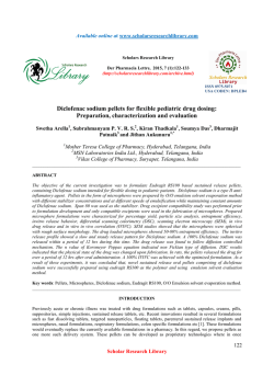
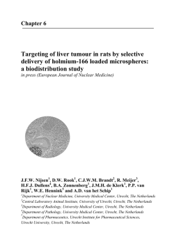
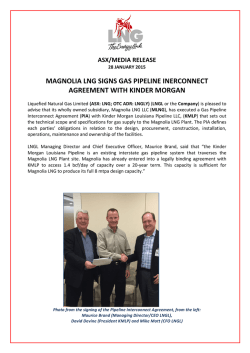
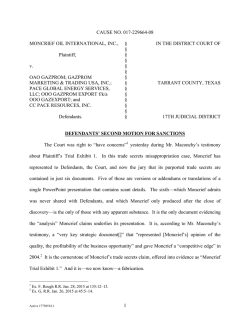
Biodegradation and biocompatibility of contracepti
Contraception 74 (2006) 148 – 156
Original research article
Biodegradation and biocompatibility of contraceptive-steroid-loaded
poly (dl-lactide-co-glycolide) injectable microspheres:
in vitro and in vivo study
Magharla Dasaratha Dhanarajua,b,4, Rajagopalan RajKannanc, Devarajan Selvarajc,
Rajadas Jayakumarc, Chandrasekar Vamsadharab
a
Department of Pharmaceutics, GIET School of Pharmacy, NH-5, Rajahmundry 533 294, India
b
Institute of Pharmacology, Madras Medical College, Chennai 600 003, India
c
Bioorganic and Neurochemistry Laboratory, Central Leather Research Institute, Adyar, Chennai 600 020, India
Received 10 August 2005; revised 14 November 2005; accepted 30 January 2006
Abstract
Purpose: A controlled-release drug delivery of contraceptive steroids levonorgestrel (LNG) and ethinyl estradiol (EE) has been developed by
successful encapsulation of LNG and EE in poly (lactide-co-glycolide) (PLG) microspheres.
Materials and Methods: Smooth, spherical, steroid-loaded PLG microspheres with a mean size of 10–25 Am were prepared by using the
water/oil/water double-emulsion solvent evaporation method.
Results: In vitro release profiles showed an increased burst release of LNG/EE on Week 1; thereafter, the release was sustained. At the end of
Week 7, the release of LNG/EE from 1:5 and 1:10 PLG microspheres was 75.64% and 62.55%. respectively. In vitro degradation studies
showed that the PLG microspheres maintained surface integrity up to Week 8 and then eroded completely by Week 20. In an in vivo study,
the serum concentration of LNG/EE in rats showed a triphasic release response, with an initial burst release of 8 ng/mL LNG and 14 pg/mL
EE on Day 1; thereafter, a controlled release of the drugs to the systemic circulation was maintained until Week 15, maintaining constant drug
levels of 2 ng/mL LNG and 3–4 pg/mL EE in the blood. Histological examination of steroid-loaded PLG microspheres injected
intramuscularly into the thigh muscle of Wistar rats showed minimal inflammatory reaction, demonstrating that contraceptive-steroid-loaded
microspheres were biocompatible.
Conclusion: This controlled-release and biocompatible nature of the PLG microspheres may have potential application in contraceptive therapy.
D 2006 Elsevier Inc. All rights reserved.
Keywords: Poly (lactide-co-glycolide) (PLG) microspheres; Levonorgestrel (LNG); Ethinyl estradiol (EE); Biocompatibility; Biodegradability; Controlled drug delivery
1. Introduction
In recent years, there has been immense interest in using
polymeric microspheres for the sustained or controlled
release of protein and peptide drugs because of their ease
of fabrication, relatively simple administration and versatility. In comparison to conventional dosage forms, biodegradable polymeric matrices provide improved delivery
methods for small molecules, peptides, proteins and nucleic
acids [1]. One of the most commonly used polymers is poly
4 Corresponding author. Department of Pharmaceutics, GIET School
of Pharmacy, NH-5 Rajahmundry 533 294, India. Tel.: +91 44 22362712,
+91 44 22362716; fax: +91 44 22385593.
E-mail address: [email protected] (M.D. Dhanaraju).
0010-7824/$ – see front matter D 2006 Elsevier Inc. All rights reserved.
doi:10.1016/j.contraception.2006.01.015
(lactide-co-glycolide) (PLG) because of its proven nature of
releasing the drug at a relatively slow rate over a prolonged
time. The rate of PLG microsphere degradation in achieving
controlled release affords less frequent administration,
thereby increasing patient compliance, reducing discomfort,
protecting the therapeutic compound and maintaining
constant blood levels of the drug within the body [2–5].
Contraceptive steroids levonorgestrel (LNG) and ethinyl
estradiol (EE) are used in combination to depress the
gonadotrophins follicle-stimulating hormone and luteinizing
hormone, thus preventing ovulation. The oral use of LNG
and EE is limited since they are not tolerated at higher
doses. A long-term, systemic, controlled delivery of
contraceptive steroids appears to be essential in the
regulation of reproductive function, as well as in the case
M.D. Dhanaraju et al. / Contraception 74 (2006) 148 – 156
of postmenopausal therapy [6,7]. However, the pharmacological approach to fertility control is still mainly by oral
administration and transdermal delivery of contraceptive
steroids. The main disadvantage of the oral combined pill is
the requirement of daily ingestion and subsequent daily
variations in blood concentration, leading to blood-druglevel-dependent unwanted side effects and a short biological
half-life of the drug [8]. For transdermal delivery systems,
variations in an individual’s skin permeability and poor
patient compliance can result in insufficient or excessive
mean serum concentrations. Side effects due to variations in
the concentration of the drugs could be avoided by using
a sustained or long-term controlled delivery system to
ensure a longer period of drug availability in the blood at
optimum concentration.
For long-term controlled delivery of contraceptive
steroids, polymeric drug delivery systems have attracted
considerable attention in the past several years [9–15];
currently, there are only a small number of commercially
available products that utilize this technology.
ProgestasertR, a T-shaped device, was designed to provide
a constant release of progesterone through a rate-controlling
membrane of ethylene vinyl acetate, whereas NorplantR is a
silicone-based device for the delivery of LNG. However,
both polymers are nonbiodegradable, and the devices have
to be removed after depletion of the drug [16]. To overcome
this problem, biodegradable PLG microspheres were developed for implantation under the skin without special surgery
[17]. The major advantage of biodegradable PLG microspheres is that they ensure a continuous delivery of the drug
via diffusion to surrounding tissues or by polymer erosion
and they enhance the bioavailability of compounds that are
poorly soluble in body fluids. A constant release prevents
cyclic variations in drug concentrations in the blood with
time and offers maximum pharmacological efficiency at a
minimum drug dose.
Recently, we reported on the development of the
microsphere encapsulation of the lipophilic drugs LNG
and EE using biodegradable PLG and poly (e-caprolactone)
though the water/oil/water (W/O/W) double-emulsion solvent evaporation method [18,19]. The absence of chemical
interactions between the drugs and the polymers and their
size, distribution, surface properties and loading efficiencies
account for the usage of these polymers for the encapsulation of LNG and EE.
In this study, we attempted to examine the feasibility of
formulating contraceptive steroids with PLG microspheres
as an injectable polymeric carrier system for long-term
controlled drug delivery of LNG and EE. We present the
results of our study using this system, with a view of drug
content, surface morphology, in vitro degradation of LNG/
EE-loaded PLG biodegradable microspheres, and in vitro
and in vivo release. The biocompatibility of PLG microspheres is shown by the results of tissue responses to PLG
microspheres injected intramuscularly into the thigh muscle
of rats.
149
2. Materials and methods
2.1. Materials
LNG and EE were obtained as gifts from German
Remedies (Mumbai, India). PLG microspheres
(M W =70,000) were purchased from Birmingham Polymers,
Inc. (Alabama, USA). Polyvinylalcohol (PVA) was obtained
from Sigma (St. Louis, MO, USA); dichloromethane (AR
grade) was from Sisco Research Laboratory Pvt Ltd.
(Mumbai, India); ethanol (AR grade) was from Hayman
Ltd. (England, UK); goat polyclonal interleukin (IL) 1a
(primary antibody) was from Santa Cruz Biotechnology,
Inc. (Santa Cruz, CA, USA); and rabbit antigoat IgG
[secondary antibody conjugated with fluorescein isothiocyanate (FITC)] was obtained from Genei (Bangalore,
India). All other chemicals used were of analytical grade.
2.2. Preparation of microspheres
The PLG microspheres containing both LNG and EE
were prepared by the W/O/W double-emulsion solvent
evaporation method, as described previously [18,19].
Briefly, a saturated solution of LNG (15 mg) and EE
(3 mg) (in a 1:5 ratio) was mixed with ethanol/water (7:3),
which was then emulsified in 10 mL of dichloromethane
containing 180 mg of PLG polymer to form the W/O/W
primary emulsion. The emulsion formed was stirred at
4000 rpm for 10 min and then added to an external phase
containing 1% PVA solution to produce the W/O emulsion.
The formed multiple emulsion was kept under constant
stirring for 4 h at 600 rpm by magnetic spin bar assembly.
Microspheres were separated by centrifugation at 2000 rpm
for 10 min, washed thrice with phosphate buffer (pH 7.4)
and then dried in nitrogen atmosphere.
2.3. Morphology of microspheres
The morphological features of PLG microspheres both
initially and during the degradation process were carried out
using scanning electron microscopy (SEM). The drug
microspheres were sprinkled onto one side of a doublesided adhesive stub. The stub was then coated with
conductive gold with Joel-JFC 1100E sputter coater and
examined under a Joel-JFC 5300 scanning electron microscope (Joel Inc., Peabody, MA, USA) for qualitative
assessment of microsphere morphology.
2.4. Drug content of microspheres
The content of LNG and EE loaded into PLG microspheres was determined by dissolving 100 mg of microspheres in 5 mL of dichloromethane. To do this, 5 mL of
methanol was added, the solution was evaporated under
vacuum to eliminate dichloromethane and the polymer was
allowed to precipitate. The drugs dissolved in methanol
were filtered using a 0.1-Am Millipore [Millipore (India)
Pvt. Ltd., Peenya, Bangalore, India] filter assembly, suitably
diluted and subsequently injected into a Hypersil C18
(250Â4.6-mm) column (Thermo Electron Corp., San Jose,
150
M.D. Dhanaraju et al. / Contraception 74 (2006) 148 – 156
cycle, fed ad libitum with commercial pellet diet (Hindustan
Ltd., Bangalore, India) and given free access to water.
Sterile microspheres containing 5 mg of LNG and 0.69 mg
of EE drug equivalent dose per kilogram of body weight,
and pure contraceptive agents of 5 mg of LNG and 0.69 mg
of EE were injected intramuscularly into the thigh muscle
after reconstitution in a suitable vehicle (2 mL of physiological saline containing 0.1% Tween-80). Blood samples
were collected from orbital venous plexus punctures at
different time intervals up to 5 months. Blood samples were
Fig. 1. Scanning electron micrograph of LNG/EE-loaded PLG microspheres.
CA, USA). Drug content was determined by a previously
reported procedure [20]. The mobile phase used was a
combination of acetonitrile/methanol/water in a ratio of
3.5:1.5:4.5 at a flow rate of 2 mL/min; the eluted sample
was detected at 215 nm using Shimadzu high-performance
liquid chromatography (HPLC) LC 10AT-vp (Shimadzu
Corporation, Kyoto, Japan).
2.5. In vitro degradation studies
One hundred milligrams of PLG microspheres containing steroidal contraceptives LNG and EE was placed
in test tubes containing phosphate-buffered saline (PBS)
buffer (pH 7.4). The test tubes were kept in an incubator
shaker maintained at 37F18C. The buffer medium was
renewed every week. After predetermined periods, samples were taken out by centrifugation of the buffer, then
washed with distilled water and dried under vacuum at
room temperature.
2.6. In vitro drug release studies
Release studies of LNG and EE from PLG microspheres
were carried out under physiological conditions by simulating the in vitro environment. Fifteen milligrams of drug
equivalent microspheres was weighed and added to 50 mL
of PBS in an Erlenmeyer flask. The flask was agitated at
50 rpm at 37F18C in an incubator shaker. A sample (1 mL)
was taken at different intervals up to 5 months and replaced
with fresh medium. The amount of released drug was
estimated from the sample by HPLC [20].
2.7. In vivo drug release study
Colony-inbred female rats of Wistar albino strain were
used for the in vivo drug release study. Twelve rats
weighing between 170 and 200 g were randomized into
two groups of six animals each and were evaluated and used
for the contraceptive efficacy of LNG/EE-loaded PLG
microspheres. The rats were maintained in a room at
25F38C. The animals were exposed to a 12-h dark/light
Fig. 2. Scanning electron micrograph of PLG microspheres retrieved from
phosphate buffer medium (pH 7.4) on: (A) postdegradation Week 2, (B)
postdegradation Week 8 and (C) postdegradation Week 20.
M.D. Dhanaraju et al. / Contraception 74 (2006) 148 – 156
151
Fig. 3. In vitro release profiles of LNG from PLG microspheres prepared
with various (drug/polymer) ratios using the W/O/W method: (E) 1:5 and
( ) 1:10 of PLG. Data are shown as meanFS.E. of three experiments.
!
Fig. 5. Serum LNG concentrations in rats administered intramuscularly with
free drug (x) and drug-loaded PLG microspheres (E). Data are shown as
meanFS.E. of six animals.
centrifuged at 3000 rpm for 10 min, and serum was
collected and stored frozen at À208C until analysis. Drug
concentrations in blood serum were determined after
suitable extraction and dilution with mobile-phase solvent
using the HPLC technique.
body weight). Control tissues were taken from the thigh
muscle of the opposite leg. Retrieved samples were processed
for histological examination.
2.8. In vivo biocompatibility and stability study
2.9. Histological examination
Eighteen rats (n =6) weighing between 170 and 200 g
were used for the in vivo compatibility and stability study.
The in vivo biocompatibility and stability of the LNG/EEloaded PLG microspheres were examined after implanting
the microspheres into the thigh muscle of Wistar rats via
intramuscular injection; 5.69 mg/kg body weight of drug
equivalent dose of sterilized microspheres was suspended in
2 mL of physiological saline containing 0.1% Tween-80 and
injected using an 18-gauge needle. The injected microspheres, along with their surrounding tissues, were excised on
Weeks 1, 8 and 20 postimplantation after anesthetizing the
animals with an overdose of pentothal sodium (80 mg/kg
Tissue samples were fixed in 10% phosphate-buffered
formaldehyde solution and embedded in paraffin. The
samples were then sectioned at a thickness of 7 Am using
an automatic microtome, followed by staining with hematoxylin and eosin (H&E). The stained sections of each test
sample were examined by light microscopy (Polyvar 2
photomicroscope; Leica, Bensheim, Germany) for tissue
inflammatory reaction and were photographed.
Fig. 4. In vitro release profiles of EE from PLG microspheres prepared with
various (drug/polymer) ratios using the W/O/W method: (E) 1:5 and ( )
1:10 of PLG. Data are shown as meanFS.E. of three experiments.
Fig. 6. Serum EE concentrations in rats administered intramuscularly with
free drug (x) and drug-loaded PLG microspheres (E). Data are shown as
meanFS.E. of six animals.
!
2.10. Tissue processing for immunohistochemistry
The tissues were immersed for 24 h at 48C in 10%
phosphate-buffered formaldehyde fixative and rinsed in cold
152
M.D. Dhanaraju et al. / Contraception 74 (2006) 148 – 156
secondary antibody conjugated with FITC at the recommended dilution (1:500) for 1 h at room temperature. The samples
were further washed with PBS, and coverslips were mounted
with bicarbonate-buffered glycerol (pH 8.6) and viewed with
a Zeiss Axioplan 2 fluorescent microscope (NAG f. HBO50;
Carl Zeiss, Jena and Oberkochen, Germany). The following
control procedures were applied to all stainings: tissues from
the control and microsphere-injected area underwent the same
immunohistochemical protocol, but with omission of the
primary antibody and replacement of the primary antibody
with normal goat serum with an additional control [21].
3. Results
3.1. Morphology of microspheres
The PLG microspheres containing LNG and EE, which
were prepared by the W/O/W double-emulsion solvent
evaporation technique, were spherical, individual and nonporous, with mean particle sizes from 10 to 25 Am (Fig. 1).
The microspheres obtained from both 1:5 and 1:10 drug/
polymer ratios were free-flowing and had adequate syringability when mixed with vehicle for in vivo administration.
3.2. In vitro degradation studies
The surface morphology of LNG/EE-loaded PLG microspheres before and after degradation was compared as a
measure of in vitro degradation (Fig. 2). The surface
morphology of PLG microspheres was unchanged up to
the end of Week 1 (Fig. 2A), indicating the crystalline
behavior of the matrix. After Week 8, PLG microspheres
collapsed and were found to be highly porous in nature
(Fig. 2B). Finally, on Week 20, it was observed that the PLG
microspheres had eroded completely (Fig. 2C).
3.3. In vitro release studies
The release profiles of LNG and EE from PLG polymeric
microspheres showed an initial burst release on Week 1,
followed by sustained release of the drugs. The cumulative
release of LNG/EE from 1:5 and 1:10 (drug/polymer) PLG
microspheres at the end of Week 7 was 75 (64%) and 62
(55%), respectively (Figs. 3 and 4).
Fig. 7. Scanning electron micrograph of PLG microspheres retrieved from
an implanted site on: (A) postdegradation Week 1, (B) postdegradation
Week 8 and (C) postdegradation Week 20.
PBS, and specimens were covered with 20% sucrose in PBS
and allowed to stand at 48C overnight. The tissues from all
groups were processed for immunofluorescent localization of
tissue antigens. The specimens were embedded in Optimum
Cutting Temperature (Tissuetek; Sakura Finetek, Torrance,
CA, USA) embedding medium, frozen and sectioned at 16
Am. Serial transverse and longitudinal sections around the
injected muscle area were taken and permeabilized with 0.3%
Triton X-100 in PBS for 2 h and then incubated for 24 h at 48C
with the primary antibody goat polyclonal IL-1a. Slides were
then washed with PBS and incubated with rabbit antigoat IgG
3.4. In vivo drug release studies
After injecting drug-loaded PLG microspheres intramuscularly, the serum concentrations of the LNG and EE in rats
showed a triphasic release response (Figs. 5 and 6). Initially,
8 ng/mL LNG in serum on Day 1 was attributed to the
higher amount of drug release from the microspheres.
Thereafter, the release of LNG in the blood was estimated to
maintain a constant level of 2 ng/mL throughout the study.
The initial release of EE was 14 pg/mL, and the system was
capable of constantly delivering 3 pg/mL EE in the blood.
3.5. In vivo biodegradation of microspheres
After muscular implantation in rats, the morphology of
the microspheres changed progressively with time and
M.D. Dhanaraju et al. / Contraception 74 (2006) 148 – 156
153
Fig. 8. Photomicrograph of tissues implanted with PLG microspheres and stained by H&E (original magnification, Â320) that were retrieved on: (A)
preimplantation, (B) postimplantation Week 2, (C) postimplantation Week 8 and (D) postimplantation Week 20. (Y) Inflammatory cells surrounding the
tissues.
finally disintegrated. On Week 1, PLG microspheres
retained good sphericity, similar to that of the microspheres
before implantation (Fig. 7A). On Week 8, the PLG
microspheres were noticeably degraded into smaller fragments (Fig. 7B). The biodegradation of the PLG microspheres retrieved after Week 20 was more significant
compared to that retrieved on Week 8, indicating that the
microspheres were degraded into fine fragments with
greater size reduction (Fig. 7C).
3.6. Histological examination
The levels of macrophage infiltration were studied
histologically. The tissues injected with LNG/EE-loaded
PLG microspheres stained with H&E and retrieved after
Weeks 1, 8 and 20 showed differential macrophage response
at different time intervals (Fig. 8). Histological analysis of
the normal tissue showed the least macrophage infiltration
(Fig. 8A), whereas the drug-loaded PLG microspheres
injected after Week 1 (Fig. 8B) showed heavy macrophage
infiltration around the muscle at the injection site. These
levels of macrophage infiltration became reduced after
Week 8 (Fig. 8C) and almost disappeared after Week 20
of microsphere injection (Fig. 8D).
3.7. Immunohistochemistry
The level of inflammatory cytokines was determined by
immunostaining for IL-1a (Fig. 9). After Week 1, the
expression of IL-1a was significant in the case of animals
injected with PLG microsphere formulations (Fig. 9B), in
accordance with histological analysis. The presence of
IL-1a confirmed the increased macrophage infiltration
around the injection site. Conversely, after Week 8 of
injection, a moderate amount of IL-1a was observed, which
indicates that the production of inflammatory cytokines at
the injection site declined (Fig. 9C). The immunofluorescent
images taken of tissue samples after Week 20 of PLG
microsphere injection showed little or no reaction to the
antibody against IL-1a. When compared with those of
Weeks 1 and 8, it was clearly observed that the production
of inflammatory cytokines at the injection site had ceased,
as the macrophage infiltration decreased at the injection site
(Fig. 9D).
4. Discussion
The W/O/W double-emulsion solvent evaporation method was used to prepare PLG microspheres in order to obtain
spherical LNG/EE-loaded PLG microspheres with a narrow
size distribution from 10 to 25 Am. The surface morphology
of PLG microspheres in in vitro degradation studies
revealed that, up to Week 1, the microspheres remained
unchanged, indicating the crystalline behavior of PLG
matrices [22]. By Week 8, the microspheres had disintegrated into smaller particles, and the surface of the spheres
had collapsed and were highly porous, signifying that the
PLG polymer was gradually hydrolyzed but had not yet
154
M.D. Dhanaraju et al. / Contraception 74 (2006) 148 – 156
Fig. 9. Immunohistochemical analysis of tissues explanted from the PLG microspheres injection site by fluorescence microscopy: (A) normal thigh muscle, (B)
postimplantation Week 1, (C) postimplantation Week 8 and (D) postimplantation Week 20 (original magnification, Â200). (Y) Expression of IL-1a.
decreased sufficiently in molecular weight to allow an
increased diffusional release of the drug. The PLG microspheres had eroded completely by Week 20, releasing the
remaining drug, because the molecular weight of the
polymer and also the amount of drug present in the polymer
matrix were sufficiently low to allow its solubilization in the
simulated medium (aqueous environment) [23].
The in vitro release profiles of LNG and EE from 1:5 and
1:10 (drug/polymer) PLG microspheres showed an initial
burst release on Week 1 followed by a sustained release of
the drugs. A reason for the observed initial burst release
could be the unstable nature of inner water emulsion
droplets during solvent evaporation, leading to coalescence
and probably causing the drug to locate at the surface of
polymeric microspheres [24]. However, the initial burst
effect of all formulations was well below 20% because of
extensive washings of the microspheres, which removed the
surface-free, poorly entrapped and surface-associated drug
crystals of the PLG microspheres. LNG and EE are
lipophilic in nature, showing lesser tendencies to migrate
toward the aqueous medium; therefore, some of the initial
releases were due to simple partition diffusion of the drugs
through intact polymeric spheres. The drug release rate from
PLG matrices has been controlled by both the diffusion rate
of the drug in the matrices and the degradation rate of
matrices [25]. Discharges of the remaining amount of the
drug from the polymer matrix after Week 1 were dependent
on the rate of polymer erosion [26]. At the end of Week 7,
the cumulative release of LNG/EE from 1:5 and 1:10 (drug/
polymer) PLG microspheres was 75.64% and 62.55%,
respectively. This shows that the degradation rate of PLG
polymers was very slow in an aqueous medium because of
hydrophobicity, thereby assisting a controlled release of the
drug [27].
The serum concentration of LNG and EE in rats showed
a triphasic release response, with an initial burst release of
8 ng/mL LNG and 14 pg/mL EE on Day 1 due to the release
of the steroid adsorbed on the microsphere surface. This
may be attributed to a higher volume of distribution and
to an increased plasma protein binding affinity of the drug.
It was followed by a second sustained release phase from
Week 5, which was initiated because of steroid diffusion
through the pores or channels formed in the polymer matrix,
and a third phase until Week 15 by bulk erosion at the
polymer matrix. This slow advancement in hydrolytic
degradation of the PLG microspheres led to the controlled
release of the drugs to the systemic circulation. These parameters acted as a reservoir and aided in the maintenance
of constant drug levels of 2 ng/mL LNG and 3– 4 pg/mL EE
in the blood until Week 15. After Week 15, the drug
levels were less than the minimum amount for maintaining contraception.
The morphology of the microspheres changed progressively with time and finally disintegrated after muscular
M.D. Dhanaraju et al. / Contraception 74 (2006) 148 – 156
implantation in rats. Once implanted, a biodegradable drug
delivery device should maintain its mechanical property
until it is no longer needed and then be absorbed and
excreted by the body. On Week 1, PLG microspheres
retained good sphericity similar to that of the microspheres
used before implantation. Simple chemical hydrolysis is the
prevailing mechanism for polymer degradation at the initial
stage, after which surface degradation occurs, creating pores
on the surface of the spheres by hydrolysis. Water penetrates
into the bulk of the device, preferentially attacking ester
bonds and converting long polymer chains into shorter
water-soluble fragments, with reduction in molecular weight
followed by metabolism of the fragments, resulting into
their monomers. On Week 8, PLG microspheres were
degraded into smaller fragments; after Week 20, the
microspheres were degraded into fine fragments with
reasonable size reduction. This ensured that the PLG
microspheres had a slow degradation rate and can be used
as a promising device for long-term delivery of contraceptive steroids.
PLG microspheres (average size, 30 Am) generally
induced a mild foreign body reaction and were reported to
be biocompatible [28]. The volume of microspheres injected
into the tissue may be considered as an open porous implant,
which induces an inflammatory response characterized by
the infiltration of macrophages, neutrophils, fibroblasts and
some lymphocytes and by the formation of fibrin, giant cells
and new blood vessels [29–32]. Tissue reaction to the PLG
microsphere injection site after Week 1 showed heavy
macrophage infiltration around the muscle due to a systemic
rise in the level of activated macrophages, which release
cytokines, growth factors and other bioactive agents to
modulate the function of other cell types in the inflammatory milieu [33,34]. The expression of IL-1a was also
significant at the injection site, in accordance with histological analysis. The presence of IL-1a confirmed the
increased macrophage infiltration around the injection site.
The rich neutrophil recruitment in the tissues at this time
point, along with infiltrated macrophages and lymphocytes,
may be due to an increased uptake of particles coated with
immunoglobulin or complement proteins associated with
increased surface Fc and C3 receptors [35]. Thus, the uptake
of opsoninized (and also nonopsoninized) large particles at
inflammatory sites was enhanced.
After Week 8, less macrophage infiltration surrounding
the muscle was observed. Concurrently, a moderate
amount of IL-1a was observed in immunostaining, which
indicated that the production of inflammatory cytokines at
the injection site declined. This minimization in the levels
of macrophage infiltration and IL-1a is due to polymer
degradation as time progresses, with inflammatory reaction
being mild at later time points. This suggests that, during
the early weeks when drug release from the polymers was
in progress, there was decrease in the total volume of the
microspheres, which attracted greater macrophage cell
infiltration due to smaller particles of the microspheres.
155
The macrophage infiltration and the levels of inflammatory
cytokine IL-1a at the microsphere injection site almost
disappeared after Week 20, indicating that the release was
almost complete, resulting in greater degradation of
microspheres and enhanced scavenging by the host
defense mechanism.
The data obtained in this study suggest that the LNG/EEloaded PLG microspheres prepared by the W/O/W doubleemulsion solvent evaporation method can be used as an
intramuscularly injectable drug delivery carrier, in consideration of their biodegradation, biocompatibility and particle
size. The biodegradable property of PLG polymers makes
this delivery system a potential carrier for long-acting
controlled drug delivery. Furthermore, the longer duration
of LNG and EE levels in the blood for contraceptive action
with controlled-release characteristics finds potential application in contraceptive therapy.
Acknowledgments
We are grateful to Dr. T. Ramasami (Director, CLRI) for
granting permission to publish this work. We are also
thankful to Dr. C.V. Gokularathnam (Department of
Metallurgical Engineering, IITM, Chennai) for helping with
SEM analysis.
References
[1] Raman C, Berkland C, Kim K, Pack DW. Modeling small-molecule
release from PLG microspheres: effects of polymer degradation and
nonuniform drug distribution. J Control Release 2005;103:149 – 58.
[2] Berkland C, King M, Cox A, Kim K, Pack DW. Precise control of
PLG microsphere size provides enhanced control of drug release rate.
J Control Release 2002;82:137 – 47.
[3] O’Hagan DT, Rahman D, McGee JP, et al. Biodegradable microparticles as controlled release antigen of pores at the surface of the
microspheres. Immunology 1991;73:239 – 42.
[4] Athanasiou KA, Niederauer GG, Agrawal CM. Sterilization, toxicity,
biocompatibility and clinical applications of polylactic acid/polyglycolic acid copolymers. Biomaterials 1996;17:93 – 102.
[5] Tracy MA, Ward KL, Firouzabadian L, et al. Factors affecting the
degradation rate of poly(lactide-co-glycolide) microspheres in vivo
and in vitro. Biomaterials 1999;20:1057 – 62.
[6] Sagraves R. Estrogen therapy for postmenopausal symptoms and
prevention of osteoporosis. J Clin Pharmacol 1995;35:2S – 10S.
[7] Dupont A, Dupond P, Cusan L, et al. Comparative endocrinological
and clinical effects of percutaneous estradiol and oral conjugated
estrogens as replacement therapy in menopausal women. Maturitas
1991;13:297 – 311.
[8] Petitti DB, Sidney S, Bernstein A, Wolf S, Quesenberry C, Ziel HK.
Stroke in users of low-dose oral contraceptives. N Engl J Med 1996;
335:8 – 15.
[9] Pitt CG, Marks TA, Schindler A. Biodegradable delivery systems based
on aliphatic polyesters: application to contraceptives and narcotic
antagonists. In: Baker R, editor. Controlled release of bioactive
materials. New York7 Academic Press; 1980. p. 19 – 43.
[10] Nash HA. Controlled release systems for contraception. In: Langer RS,
Wise DL, editors. Medical applications of controlled release. Boca
Raton7 CRC Press; 1984. p. 36 – 64.
[11] Lewis DH. Controlled release of bioactive agents from lactide/
glycolide polymers. In: Chasin M, Langer R, editors. Biodegradable
156
[12]
[13]
[14]
[15]
[16]
[17]
[18]
[19]
[20]
[21]
[22]
M.D. Dhanaraju et al. / Contraception 74 (2006) 148 – 156
polymers as drug delivery systems. New York7 Marcel Dekker, 1990.
p. 1 – 42.
Pitt CG. Poly (e-caprolactone) and its copolymers. In: Chasin M,
Langer R, editors. Biodegradable polymers as drug delivery systems.
New York7 Marcel Dekker, 1990. p. 71 – 120.
Gangrade N, Price JC. Poly(hydroxybutyrate–hydroxyvalerate) microspheres containing progesterone: preparation, morphology and release
properties. J Microencapsul 1991;8:185 – 202.
DeLuca PP, Mehta RC, Hausberger AG, Thanoo BC. Biodegradable
polyesters for drug and polypeptide delivery. In: El Nokaly MA,
Piatt DM, Carpentier BA, editors. Polymeric delivery systems:
properties and applications. ACS symposium series. Washington
(DC)7 Am Chem Soc, 1993. p. 53 – 79.
Jameela SR, Kumary TV, Lal AV, Jayakrishnan A. Progesteroneloaded chitosan microspheres: a long-acting controlled delivery
system. J Control Release 1998;52:17 – 24.
Latha MS, Lal AV, Kumary TV, Sreekumar R, Jayakrishnan A.
Progesterone release from glutaraldehyde cross-linked casein microspheres: in vitro studies and in vivo response in rabbits. Contraception
2000;61:329 – 34.
Ogawa Y, Yamamoto M, Takada S, Okada T, Shimamoto T.
Controlled-release of leuprolide acetate from polylactic acid or
copoly(lactic/glycolic) acid microcapsules: influence of molecular
weight and copolymer ratio of polymer. Chem Pharm Bull 1988;
36:1502 – 7.
Dhanaraju MD, Jayakumar R, Vamsadhara C. Influence of manufacturing parameters on development of contraceptive steroid loaded
injectable microspheres. Chem Pharm Bull 2004;52:976 – 9.
Dhanaraju MD, Kiran V, Jayakumar R, Vamsadhara C. Preparation
and characterization of injectable microspheres of contraceptive
hormones. Int J Pharm 2003;268:23 – 9.
Berzas JJ, Rodriguez J, Castaneda G. Simultaneous determination of
ethinylestradiol and levonorgestrel in oral contraceptives by derivative
spectrophotometry. Analyst 1997;122:41 – 4.
Zochodne DW, Cheng C. Neurotrophins and other growth factors in
the regeneration milieu of proximal nerve stumps tips. J Anat 2000;
196:279 – 83.
Porjazoska A, Goracinova K, Mladenovska K, et al. Poly(lactide-coglycolide) microparticles as systems for controlled release of proteins
— preparation and characterization. Acta Pharm 2004;54:215 – 29.
[23] Eppstein DA, Longenecker JP. Alternative delivery systems for
peptides and proteins as drugs. Crit Rev Ther Drug Carr Syst 1988;
5:99 – 139.
[24] Lu W, Park TG. Protein release from poly(lactic-co-glycolic acid)
microspheres: protein stability problems. J Pharm Sci Technol 1995;
49:13 – 9.
[25] Berner B, Kydonieus A. Novel drug delivery systems. In: Welling PG,
Lasagna L, Banakar UV, editors. The drug development process:
increasing efficiency and cost-effectiveness. New York7 Marcel
Dekker; 1996. p. 169 – 201.
[26] Yang YY, Chung TS, Ng NP. Morphology, drug distribution, and in
vitro release profiles of biodegradable polymeric microspheres
containing protein fabricated by double-emulsion solvent extraction/
evaporation method. Biomaterials 2001;22:231 – 41.
[27] Anderson JM. Mechanisms of inflammation and infection with
implanted devices. Cardiovasc Pathol 1993;2:33S – 41S.
[28] Visscher GE, Robinson RL, Maulding HV, Fong JW, Pearsonn JE,
Argentieri GJ. Biodegradation of and tissue reaction to 50:50 poly
(dl-lactide-co-glycolide) microcapsules. J Biomed Mater Res
1985;19:349 – 65.
[29] Anderson JM. In vivo biocompatibility of implantation delivery
systems and biomaterials. Eur J Pharm Biopharm 1994;40:1 – 8.
[30] Anderson JM. Inflammation and the foreign body response. Prob Gen
Surgery 1994;11:147 – 60.
[31] Visscher GE, Pearsonn JE, Fong JW, Argentieri GJ, Robinson RL,
Maulding HV. Effect of particle size on the in vitro and in vivo
degradation rates of poly (dl-lactide-co-glycolide) microcapsules. J
Biomed Mater Res 1988;22:733 – 46.
[32] Visscher GE, Robinson RL, Maulding HV, Fong JW, Pearsonn JE,
Argentieri GJ. Biodegradation of and tissue reaction to poly (dllactide-co-glycolide) microcapsules. J Biomed Mater Res 1986;20:
667 – 76.
[33] Ziats NP, Miller KM, Anderson JM. In vivo and in vitro interaction of
cells with biomaterials. Biomaterials 1988;9:5 – 13.
[34] Ward PA. Recruitment of inflammatory cells into lung: roles of
cytokines, adhesion molecules, and complement. J Lab Clin Med
1997;129:400 – 4.
[35] Bianco C, Griffin FM, Silverstein SC. Studies of the macrophage
complement receptor. Alteration of receptor functions upon macrophage activation. J Exp Med 1975;141:1278 – 90.

© Copyright 2026