
IgE-Independent Interleukin-4 Expression and Induction of a
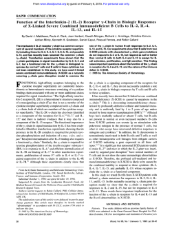
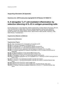
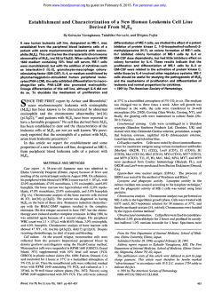
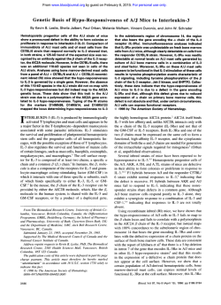
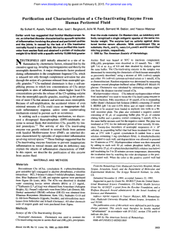
From www.bloodjournal.org by guest on February 6, 2015. For personal use only.
RAPID COMMUNICATION
IgE-Independent Interleukin-4 Expression and Induction of a Late Phase of
Leukotriene C4 Formation in Human Blood Basophils
By Brigitte Ochensberger, Silvia Rihs, Thomas Brunner, and Clemens A. Dahinden
regulated in a similar manner. However, C5ainduces a rapid,
T-helper cells can differentiate into at least two subtypes
transient burst of leukotriene formation only if added after
secreting distinct profiles of cytokines, Thl and Th2, regulatIL-3. Interestingly, upon prolonged culture, a late phase of
ing immunoprotection and different immunopathologies.Incontinuous LTC, production is observed, which also requires
terleukin-4 (IL-4) is both the product and the inducer of Th2
two signals (IL-3 andC5a). but rather depends on their concells, raising the question whether IL-4 can be produced in
tinuous presence than on their sequence of action. These
responset o antigen-independentstimuli. Here we show that
data describe an antigen-independent pathway of very rehuman basophils produce IL-4on stimulation with IL-3 and
stricted IL-4 expression.Thus, basophils must be considered
in similar amounts as induced by IgE-recepC5a or =a,-,
as central immunoregulatory cells of the innate immune systor-cross-linking. C5a-induced IL-4production requires the
tem. Furthermore, the results show that LTC, can also be
presence of IL-3,with little effect of the sequence of stimuli
generated more continuously for many hours, a phenomeaddition. No ’7hl-cytokines“ (interferon-y andIL-2)and
non that may beof particular importance in chronic allergic
even no “Th2-cytokines” (IL-3, IL-5, IL-10. and granulocyteinflammation, such as asthma.
macrophage colony-stimulating factor) are produced by basophils in response t o either IgE-dependent or IgE-indepen- 0 1995 by The American Societyof Hematology.
dent activation. The generation of leukotriene C, (LTC,) is
I
NTERLEUKIN-4 (IL-4) plays an important role in the
regulation of differentiation of T and B lymphocytes. In
B cells IL-4 provides an essential signal for isotype switching
and the production of IgE and IgGl.1-3More importantly,
the presence of IL-4 is critical in the differentiation of CD4+
T cells toward a phenotype, T helper 2 (Th2), producing a
restricted and distinctive pattern of cytokines (eg, IL-4,
IL-5, but no interferon-y [IlW-y]). More recently IL-4 has
also been shown to influence CD8+ cells leading to a Tcell subset of reduced cytotoxicity and a change in cytokine
secretion (eg, reduced IFN-y and increased IL-4 and
IL-5).8,9This key immunoregulatory role of IL-4 has been
demonstrated in vitro as well as in vivo in the murine system,
in diverse experimental models.4”2 For IL-4 to have this
effect, it must be present early in a immune response and
early during T-cell activation. However, the cellular source
of IL-4 in this process is still unclear and has been the subject
of considerable debate, because IL-4 is both a product of
Th2 cells, but is also needed for the induction of a Th2 type
immune re~ponse.~”
In contrast to most other cytokines, the expression of
IL-4 appears to be restricted to a few specialized cell types.3
Beside its established production by T lymphocytes, alternative IL-4 sources by non-T cells have received increasing
attention. In the murine system, mast cell lines and certain
IgE receptor (1gER)-positive non-B non-T cells have been
shown to transcribe IL-4 message and, in some cases, also
to produce IL-4 upon IgER activation.I3-l5Weand others
have shown that mature human basophils are capable of
producing IL-4 after activation of the IgER in synergy with
the hematopoietic growth factor IL-3.16-20
However, the induction of IL-4 in IgER+ cells depends on cross-linking of
cell bound IgE by antigen. No antigen-independent activation pathway for IL-4 production has yet been described in
any cell type. Antigen-independent induction of IL-4 synthesis, in the absence of IFN-y expression, by cells of the
innate immune system at an early stage of an inflammatory
response may be crucial for the initiation of a Th2 response.
The most potent IgE-independent basophil agonist for mediator release are the complement fragment C5a21-24
and its
Blood, Vol86, No 11 (December 1), 1995: pp 4039-4049
degradation product C5a,,,.25
We now examined whether
and under what conditions C5a/C5a,,esarg
are also capable of
inducing the expression of IL-4. Indeed, our results indicate
that basophils maynot only have an amplifying (by IgEdependent IL-4 production) but also an initiating role in the
development of a Th2 response.
Leukotrienes (LTs) are important inflammatory lipid mediators, formed by 5-lipoxygenase-catalyzed oxidation of
free arachidonic acid. The epoxid leukotriene & is further
metabolized to leukotriene B4 or C4 depending on the cell
type and the enzymes they express. In contrast to prostaglandins, leukotrienes are only generated by myeloid leukocytes
and mast cells. Neutrophils and monocytes produce LTB4,
a chemoattractant, whereas mast cells, basophils, and eosinophils generate LTC.,, a mediator inducing smooth muscle
contraction and an increase in vascular permeability.26The
production of leukotrienes can be induced by cross-linking
of Ig receptors, particularly IgE receptors of mast cells and
However, leukotrienes are involved in many
inflammatory conditions without the participation of Ig-antigen complexes. Particular high levels of leukotriene C, are
found in the exudate of allergic late phase reactions and at
chronic inflammatory sites in allergic or “intrinsic” types
of asthma.26329
Thus, antigen-independent pathways for leukotriene formation in response to soluble endogenous stimuli
From the Institute of Immunology and Allergology, Inselspital,
Bern, Switzerland.
Submitted September 1, 1995; accepted September 14, 1995.
Supported in part by the Swiss National Science Foundation
(Grant No. 3100-041906.94).
Address reprint requests to Clemens A. Dahinden, MD, Institute
of Immunology and Allergology, University Hospital, Inselspital,
CH-3010 Bern, Switzerland.
The publication costs of this article were defrayed in part by page
charge payment. This article must therefore be hereby marked
“advertisement” in accordance with 18 U.S.C.section 1734 solely to
indicate this fact.
0 I995 by The American Society of Hematology.
0006-4971/95/8611-0045$3.00/0
4039
From www.bloodjournal.org by guest on February 6, 2015. For personal use only.
4040
OCHENSBERGER ET AL
must exist in vivo. Yet,
a physiologic mode of leukocyte
activation could not be found for years, until it was discovered that two sequential signals are required for leukotriene
However, leukotrienes are always formed in
a veryrapidandtransientburstthat
is rather unlikely to
occur in chronicinflammatory processes. In ourstudyof
IgE-independent IL-4 expression we observed that LTC4can
also be produced more slowly and continuously for many
hours, reaching very high levels of this important lipid mediator.
MATERIALS ANDMETHODS
Reagents and media. Reagents used were HEPES (CalbiochemBehring Corp, La Jolla, CA); EDTA (Fluka AG, Buchs, Switzerland); Percoll and dextran (Pharmacia, Uppsala, Sweden); and bovine serum albumin fatty acid-free (BSA; Boehringer Mannheim
Inc, Mannheim, Germany). All other reagents were of the highest
purity available. HA buffer contained 20 mmol/L HEPES, 125
mmol/L NaCl, 5 mmol/L KCI,and 0.5 mmol/L glucose and 0.25
mg/mL BSA. Culture medium was RPM1 1640 supplemented with
10% heat-inactivated fetal calf serum (FCS), 25 mmol/L HEPES,
100 U/mL penicillin, and 100 mg/mL streptomycin, 2 mmol/L
nonessential amino acids, and 2 mmol/L L-glutamine (GIBCO, Paisley, Scotland).
Preparation of basophils and mononuclear cells. Highly purified basophils and control mononuclear cells were prepared as previously described.“ Briefly, blood of unselected healthy volunteers
was anticoagulated with 10 mmol/L EDTA and mixed with 0.25 v01
of 6% dextran in 0.9% NaCI,and erythrocytes were allowed to
sediment atroom temperature (RT). Leukocytes were pelleted by
centrifugation (l50g, 20 minutes RT) and separated over three-step
discontinuous Percoll gradients (1.0795/1.070/1.065 g/mL isotonic
Percoll solution, respectively) at 40013 for 30 minutes at RT. Basophil-enriched interphases between the densities 1.0795 and 1.070,
and for control experiments the upper cell layers, containing lymphocytes, monocytes, and generally less than 0.5% basophils (MNC),
were obtained and used in parallel under identical experimental conditions. Basophil-enriched fractions, containing 10% to 40% basophils (with lymphocytes and variable proportions of neutrophils),
were washedtwice in HA buffer and thenfurther purified by negative
selection using antibody-coated paramagnetic beads (MACS system;
Miltenyi Biotec, Bergisch Gladbach, Germany). Basophil purity was
examined by Giemsa-stained cytospin smears, and generally ranged
between 70% and 95%, the contaminating cells consisting mainly
of small lymphocytes and occasionally few monocytes.
In some experiments basophils enriched to 30% to 40%by Percoll
gradients, the contaminating cell population consisting almost exclusively of lymphocytes ( < l % monocytes, <5% neutrophils), was
directly used for culture without negative selection. Basophils were
also purified to near homogeneity ( ~ 0 . 5 %lymphocytes, monocytes,
neutrophils, eosinophils) by incubation with a cocktail of IgGl
monoclonal antibodies (MoAbs) (aCD2,aCD3,aCD4,aCD8,
aCDI4, aCD16, aCD19, aCD20, aCD21,
and aCD56) and negative
selection with rat-antimouse IgGl paramagnetic beads.
Culture conditions. Basophil preparations, andMNC from the
same blood specimen (cultured in parallel for comparison), were
resuspended at a cell density of 1.0 X 10‘ cells/mL in culture medium, incubated in sterile round-bottom 96-well microtiter plates
(100 pL/well) (Becton Dickinson, Lincoln Park, NJ) at 37°C in a
humidified atmosphere with5% CO2. Reagents were added at a
1 : 1 0 0 vol:vol ratio. After the time indicated, cell-free supernatants
were procured and stored at -70°C until measurements of cytokine
production by enzyme-linked immunosorbent assay (ELISA) and
sulfidoleukotriene synthesis by radioimmunoassay (RIA).
Measurement of cytokines and leukotriene C4/D4/E4. IL-4 and
IL-3 was measured with an ELISA-assay (kindly provided by Sandoz, Vienna, Austria) as described.” Both assays had a sensitivity
of 10 pg/mL with a dynamic range up to 1 ng/mL. In some experiments, IL-4 was also measured using the kit supplied by Genzyme
Corp (Cambridge, MA), according to manufacturer’s protocols or
by an ELISA using anti-IL-4 MoAb pairs obtained by Pharmingen
(San Diego, CA) with identical results. IL-2, IL-5, IL-10, and granulocyte-macrophage colony-stimulating factor (GM-CSF) were measured using antibody pairs from Pharmingen (San Diego). IFN-y
was measured with ELISA (detection limit 30 pg/mL) as previously
described?’ Sulfidoleukotrienes were determined inanRIA-assay
as described.*’
Cell stimuli. rhIL-3 was a kind gift of Dr M. Schreier (Sandoz,
Basel, Switzerland).*’ The complement products C5a and C ~ Q ~ \ ~ ~ ~
were purified from yeast-activated human serum as previously desCribed,21,25.3)and were found to be homogenous as determined by
amino acid analysis, sodium dodecyl sulfate-polyacrylamide gel
electrophoresis (SDS-PAGE), and microzone paper electrophoresis
at pH 8.6. Purified MoAb 29C6 (aIgER), directed against the nonIgE-binding epitope of the high-affinity IgER a-chain (FccRI), was
a generous gift from Drs J. Hakimi and R. Chizzonite (HoffmannLa Roche, Nutley, NJ).’4
Stutistical analysis. Statistical analysis was performed using linear regression analysis. Differences associated with probability values of P < .05 were considered significant.
RESULTS
C5a o r C5ade.,,,,,induce IL-4 synthesis in mature human
basophilsculturedwith
IL-3. Itisnowwellestablished
that human basophils produceIL-4 in response to IgER activation in synergy with 1L-3.’6-20
We now examined whether
the complement product C5a is also capable of inducing
IL-4 expression, under the experimental conditions used in
our previous study.16 Figure 1 shows that the IgE-independent chemotactic agonist C5a promotes IL-4 production in
cells cultured with IL-3 in amounts overall similar to that
induced by IgER cross-linking, but to variable degrees depending on the donor. Stimulation of basophils cultured in
medium alone with C5a does not lead to IL-4 production.
Lipid mediator formation is regulated in
a similar manner
with an absolute requirement of IL-3 for C5a-induced LTCJ
generation (Fig 1). A logarithmic scale was used to better
visualize the fact thatC5a strongly enhancedIL-4 generation
consistently over that induced by IL-3 alone and that IL-4
was always detected after C5a stimulation of cells cultured
with IL-3, even in experiments in which IL-3 alone was an
ineffective stimulus. In vivo the half-life and, therefore, the
radius of action of C5a within an inflammatory site is very
short due to rapid cleavage of the C-terminal arginine
by
carboxypeptidase(s) resulting in the generation of
C5&,,,,.
is still a potent neutrophil chemotaxin, but has lost
most of the anaphylactic and cell-activating properties of
C5a. Consistent with our recent finding that C5%,,,, retains
its capacity to induce basophil mediator
release,”
also promotes IL-4 generation with an efficacy identical to
C5a (Fig l), but with somewhat lower potency at
a molar
basis (Fig 2). Priming for C5a-induced IL-4 expression occurs over the same concentration range of IL-3 as reported
for the enhancement of the IgE-dependent response, reaching
optimal effects at 1 to 10 ng/mL (data not shown). Contami-
From www.bloodjournal.org by guest on February 6, 2015. For personal use only.
4041
IgE-INDEPENDENT IL-4 PRODUCTION IN BASOPHILS
o
l
0
0
lr
10n!
1 1° i
Cuit~~re: 0
Stimulation: C5a
IL-3
IL3
0 a-lgER
IL3
C5a
C5a
IL-3
da
Fig 1. IL-4 and LTC, production of human basophils cultured with
or without IL-3. Purified basophils from different donors were cultured during 18 hours with or without 10 ng/mL IL-3 before addition
(100
of buffer control, algER (100 ng/mL), C5a (10 nmol/L) or C5ah,
nmol/L) and further cuttured for 10 hours. The top panel shows 11-4
synthesis, the bottomshows LTC, generation. Values obtained with
cells from the same donor are connected by lines. Mean values of
duplicates from nine different experiments are shown. Note thelogarithmic scale.
nating lymphocytes do not appear to influence IL-4 release
by basophils, because (1) there is no correlation between
IL-4 release and the degree of lymphocyte contamination,
and ( 2 ) basophils purified to near homogeneity ( < O S lymphocytes, monocytes, neutrophils, eosinophils) and basophils
purified (30% to 40%) without negative selection produce
identical amounts of IL-4 in response to IL-3 and C5a (data
not shown). No IL-4 is detected in parallel experiments with
control MNC depleted of basophils (data not shown).
Effect of time interval between IL-3 and C5a forIL-4 and
LTC, production. To investigate to what extent the time
of exposure to IL-3 influences the responsiveness of the
cells to C5a, the time interval between maximally effective
concentrations of IL-3 and C5a was variedbetween 10
minutes and 18 hours. Figure 3 shows that neither LTC4
generation nor IL-4 expression is influenced in an important
manner by the preincubation time with IL-3.
Two signals are required for antigen-independent IL-4
production. Basophils are capable of expressing IL-4 in
response to IgER activation alone, although IL-4 production
is
more
consistent and pronounced after culture with
IL-3.I6Our further studies showed that, under optimal isolation conditions, freshly isolated unprimed basophils can produce IL-4 in response to IgER cross-linking in variable
amounts, depending on the blood donor. Although undetectable in cells of some donors, IL-4 release can be quite sub-
stantial and reach up to 800 pg IL-4 per million basophils
(unpublished observations, January 1994), consistent with
data from a study with basophil enriched mixed leukocyte
c ~ l t u r e s .Therefore,
'~
we examined the response of freshly
isolated basophils to C5a stimulation with and without pretreatment with IL-3 for 15 minutes in cells isolated from
several donors differing in their responsiveness to IgER stimulation. From the data shown in Fig 4 it is evident that even
cells from donors which produce relatively high levels of
IL-4 in response to cYIgER alone, IL-4 release upon stimulation with C5a is undetectable or minimal. However, after
preincubation with IL-3 for 15 minutes, large amounts of
IL-4 are produced in response to both IgE-dependent and
IgE-independent stimulation. These data also demonstrate
that after a short preincubation with IL-3, cYIgER-induced
IL-4 generation is enhanced in nearly all experiments, with
a mean enhancement of approximately twofold. The formation of LTC4 measured in the same cell supernatants follows
a similar pattern.
Correlations betweenIgE-dependent and IgE-independent
induction of IL-4 and of LTC, release. The data shown in
Fig 4 indicate that basophils from donors with a particularly
high responsiveness for the production of IL-4 upon combined stimulation with IL-3 and C5a do not necessarily produce high levels of IL-4 in response to d g E R stimulation,
and vice versa. To investigate whether the marked donor
variability of IL-4 and LTC, release is due to a difference
400
1"-
c5a
0
0.1
l
10
o
o
l
"nM C
Fig 2. Dose response of C5a- and &--induced
114 synthesis.
Basophils were incubated for 18 hours with 10 ng/mL IL-3 before
exposure to either control buffer (effect of 11-3alone) or to increasing
concentrationsof C5a (0)and C5aWm (m),respectively. for 10 hours.
IL-4, top panel; LTC,, bottom panel. Mean values f SEM; n = 6.
From www.bloodjournal.org by guest on February 6, 2015. For personal use only.
OCHENSBERGER ET AL
200"
40
0
0.2
0.5
1
2
10
lime Interval IL3/C5a (h)
Fig 3. Effect of the time of preexposure with 11-3 on the C5ainduced 114.Purified basophils were primed with 11-3 (10 ng/mL) for
different times indicated in the X-axis, andthen stimulated with C5a
(lo-* mol/L) for 18 hours. Basophils pretreated with IL-3 during 18
hours before C5a addition were cultured during 28 hours. 0, Basophils
stimulated with C5a alone. Kinetic studies showed that under all
conditions 11-4 release was complete and optimal. The top panel
shows the I L 4 synthesis(picograms/106basophils), the bottom
shows LTC, production (nanograms/106 basophils). Three independent experiments (meanof duplicate determination) are shown.
a component of the FCS used in the culture medium may
provide a necessarysecondsignal,suchassomebovine
C5%,, formed
during clotting.
Correlation between IL-4 expression and LTC4 generation. StimulationconditionsleadingtoIL-4expression
consistently promote LTC4 generation. However, although
the correlation between IL-4 and LTC4 release upon stimulation with IL-3 and C5a reached statistical significance, the
association is weak and the cells from donors secreting large
amounts of IL-4 are not necessary the most efficientin producing LTC, and vice versa (Table 1, see also Fig 6). Similarly, no correlation between cytokine expression and lipid
mediator formation is found after stimulating the cells
by
IgER cross-linking regardless of whether or not cells have
beenpretreatedwithIL-3.Thus,thedonorvariabilityof
IL-4release in responsetoeitherIgE-dependent or IgEindependent activation cannot be simply explained
by a variability of the general responsiveness of the cells toward the
stimuli used.
Requirement for the persktence of the stimuli inducing IL-4
and LTC,production.
Mediatorreleaseis a veryrapid process2 1.2527
whereas IL4production occuls more slowly,'"2o regardless of the mode of activation. Thus, we examined whether
one of thetwo signals, or both, have to
be present for prolonged
periods of time to promote IgE-independent IL4 expression.
For this purpose,basophilsexposedto
L 3 andC5awere
washed after an incubation period of 30 minutes, and recultured
withmediumwith or without addition of one or of the
two
stimuli for further 18 hours. Figure 6 shows, that both L 3 and
C5amust be continuously present for optimal IL-4secretion.
During the initial stimulation of 30 minutes, large amounts of
in the general capacity of the basophils to express this cytokine or to produce lipid mediator, respectively, or rather to
a different responsivenessof the basophils toward the stimuli
used, we examined the correlations of product generation
after different modes of activation.A significant correlation
is found for both IL-4 and LTC4 generation in response to
IgERcross-linkingofuntreatedversusIL-3primed
cells.
The correlation is particularly strong for LTC, release but
weaker for IL-4 expression (Fig 5). By contrast, IL-4 produced in response to IgE-dependent versus IgE-independent
stimulation does not correlate at all (Fig
5 , Table 1). Correlations ofLTC4 generation between the different modes of
activation are generally higher than those
of IL-4 expression.
Our previous study and the present study have shown that
IL-3, at leastin the basophils of certain donors, canby itself
promote IL-4 production. It was most interesting to find that
J
v
C5a
IL-3/C5a
algER lL-3/a-lgER
IL-4 release in response to IL-3 alone is highly correlated
tothatinducedbystimulationofIL-3primedcellswith
4. 114and LTC, synthesis by freshly isolated human basophils
C5a, but not to that observed after IgE-dependent activation inFig
response to IgE-dependent or IgE-independent activation. Baso(aIgER alone or aIgER after IL-3 priming) (Table1). This
phils purified from nine different donors were preincubated with
may indicate that the donor variability of IgE-independent
buffer or IL-3 (10 ng/mLl for 15 minutes followed by a stimulation
IL-4 expression( L 3 or IL-3K5a) could be caused by differ- with either C5a (lo-* mol/L) or algER (100 nglmL) for18 hours. The
top panel shows 114synthesis, the bottom shows the LTC. generaences in the cellular responsiveness to IL-3, although the
tion. Each data point represents the mean value of duplicates or
lackof correlation for IL-4 production between IL-3/C5a
triplicates. The data from each of nine different experiments with
versus IL-3hIgER and the strong correlation ofaIgER vercells from distinct donors are connected by lines. Columns represent
sus IL-3hIgER argues against this hypothesis. Alternatively, the mean values ofall experiments.
From www.bloodjournal.org by guest on February 6, 2015. For personal use only.
IgE-INDEPENDENTIL-4PRODUCTION
4043
IN BASOPHILS
IL-4 (pg/l 0 Basophils)
I
-
Fig 5. Correlationsof 11-4 and
LTC, formation in response to
different modes of activation.
The figure shows the correlations of the products releasedby
basophils in response to algER
versus 11-3 algER (leftpanels)
+ C5a
or to algERversusIL-3
(right panels). (Top) Correlations
of the IL-4 expression. (Bottom)
LTC, formation. Curve fit was
done by linear regression analysis.Eachsymbolrepresentsa
datapair of the mean of duplicates ortriplicates from one separate experiment. All experiments were performed with
basophils purified from different
donors. The experimental conditions were as in Fig 4.
I
A
0'
0
0200
100
300
m
A A
210
.o
y
20
A
0'
I
0
IO
20
a-lgER
LTC4
n
P
r
n
P
A.
IL-3
<.0001
ND v25IL-3/C5a
.91
ND ,015 13
IL-3 .65
v algER
IL-3
ND,386
v IL-3/algER
16 .23
,217
IL-3/C5a
12 .38
,85712
v algER
-.06
IL-3/C5a vIL-3IalgER 15 .36
algER vIL-3/algER
,000112 .89,005 12 .75
.l84
,00615 .67
IL-4 versus LTC,
r
n
P
B.
IL-3/C5a
O/algER
IL-3/algER
300
;,/-------
IL-4
Stimulation
200
a-lgER
LTC4 (ng/lOe Basophils)
Table 1. Correlationsof IL-4 and of LTC, Formation Between
Different Modes of Activation (A) and Correlations Between IL-4
and LTC, Production (B)
r
I
100
a-lgER
+
Stimulation
0
.45,023 25
,0013
.g97 12
. l 1,698 16
Individual data pairs are the mean of duplicate or triplicatedeterminations and were derived from seperate experiments. Experimental
conditions were as described in Figure legends 4 and 5.
Abbreviations: r, correlation coefficient, calculated by linear regression analysis; n, number of experiments with basophils from different
unselected donors; P, probability; ND, not determined because in
many experiments LTC, release in response to IL-3 alone was not
detected.
30
0
10
20
30
a-lgER
consistentwith our previous ~tudies,2'.*~
LTC4areformed,
whereas IL-4 release is minimal. Most inte&ingly,during further culture for 18 hours, a second phase of pronounced lipid
mediatorreleasewasobservedunderconditionsofoptimal
IL-4 expression. For this late phase of LTC, generation the readdition of both stimuli is required. For comparison, a similar
experimental set-up was adapted using
CuIgER as a trigger. Again
LTC, is formed very rapidly as compared to IL-4 expression.
In contrast to antigen-independent activation, the basophils efficiently produce IL4 after washing and culturing the cells with
medium alone, possibly because of irreversible IgER cross-linkingbytheantibody.However,
L - 3 has to be present for an
optimal response. No second phase of LTC, generation during
the time of IL-4release is observed, in contrast to cells cultured
with L 3 and C5a.
Importance of the sequence of the two signals. Several
previous studies have clearly shown that the rapid burst of
antigen-independent lipid mediator formation is critically dependent on the sequential action of an appropriate priming
cytokine followed by triggering witha chemotactic agoniSt.z~-z5.3n
The need for a more prolonged presence of IL-3
and C5a for cytokine expression could indicate that IL-4
production is regulated differently. Indeed, Fig 7 shows that
IL-4 is also produced when IL-3 and C5a are added ina
reversed order, in amounts that are even significantly higher.
During culture of 18 hours the basophils also produce large
amounts of LTCI even if C5a is added 15 minutes before
IL-3, but not in response to C5a alone.
Time course of IL-4 and LTC, production. IL-4 release
From www.bloodjournal.org by guest on February 6, 2015. For personal use only.
OCHENSBERGER ET AL
1201
1501
1
I
d
,
e
dJ
i3
'P
Wash
=!
Immediate Response
Restimulation
:l
Immediate Response
Restimulation
I
I
l
l0
0
Fig 7. 11-4 and LTC, production by basophils in response to C5a
and 11-3. Effect of the sequence of stimuli addition. Freshly purified
basophils from five different donors were stimulated with C5a (10"
mollLI, IL-3 (10 ng/mL), or both. IL-3 was added 15 minutes before
llL-3lC5a) or after (C5allL-3) C5a. 11-4 (top) and LTC, (bottom) were
measured in the cell-free supernatant after 18 hours of culture. The
data from the same donor (mean of duplicates indicated by circles)
are connectedby lines. The mean value five
of experiments is shown
in columns.
Fig6.Requirement
for the
persistence ofthe stimuli. Basophils were stimulated with 11-3
( l 0 nglmL) and C5a (lo-' mollL)
or dgER (100 nglmL), respectively. After 30 minutes the supernatant was obtained (Imma
diate Response)and the cells
were washed with medium and
exposed to the stimuli indicated
in the figure for further 18 hours
(Restimulation). Each column
representsthe mean value of duplicates, three experiments are
shown (presentedwithdifferent
shadings).
is induced rapidly and continues during 16 hours after stimulation, with identical kinetics regardless of the sequence of
IL-3 and C5a addition (Fig 8). However, marked differences
in the kinetics of LTC, production are observed depending
on whether C5a is added before or after IL-3. In contrast to
the rapid burst of LTC, formation triggered by C5a in IL-3
primed cells?'.25 no LTC, is formed early after stimulation
if the order of the stimuli is inversed. After 2 hours, however,
and in parallel with IL-4 release, large amounts of LTC, are
continuously generated, resulting in levels comparable to
those after C5a stimulation of primed cells (Fig 8). The
kinetics of LTC, accumulation in the supernatant of IL-3
primed basophils exposed to C5a may be explained by some
degradation of sulfido-leukotrienes during the first 4 hours
after the rapid burst which lasts only a few minutes.2'*2'The
second phase of LTC, increase may reflecta balance between
leukotriene production and degradation, a hypothesis consistent with the data shown in Fig 6.
Effect of the sequence of the two signals upon the dose
response of C%. Figure 9 shows that about one order of
magnitude higher concentrations of C5a are needed for LTC,
production if C5a is added before IL-3, as determined by
measurements of LTC4 after culture of 18 hours. This difference is almost certainly due to the fact that lower amounts
of C5a are needed for the rapid burst of mediator formation
by IL-3 primed ~ e l l s ~ ' ~compared
~ " ~ ' with the second, late
phase of LTC4 generation accompanying IL-4 expression.
The dose response of IL-4 release was affected to a lesser
extent by the order of the stimuli (Fig 9).
Restricted expression of IL-4 by human basophils. We
have previously shown that basophils cultured withIL-3
and stimulated by IgER cross-linking produce IL-4 without
detectable amounts of the "Thl cytokines," IL-2 and
IFN-y. Thus, human basophils may represent a kind of innate Th2-like cells. We now determined whether basophils
can also produce other cytokines expressed by T-helper 2
From www.bloodjournal.org by guest on February 6, 2015. For personal use only.
IgE-INDEPENDENT IL-4 PRODUCTION IN BASOPHILS
le01
4045
l
I
400-A-
lL-3/C5a
300- -e C5allL-3
200-
-o- C5a
I
160
4
12
8
16
40
houn
m
10
--l
C5a (nM)
200
Fig 9. Dose response of C5a-induced 114 and LTC, synthesis
added before or after
IL-3. Purified human basophils were stimulated
with different concentrationsof C5a with (closed symbols) or without
(open symbols) IL-3 ( l 0 ng/mL) given either 15 minutes before (0)
or after (A)C5a. Each point represents the meanvalue of triplicates
from one representative experiment out ofthree.
100
0
60t
404
10
.
e
e
l hour
.I
or IgE-independent mechanism are unable to produce any
detectable amounts of GM-CSF, IL-3, IL-5, and IL- 10 (Table
2). Therefore, it appears that basophils express IL-4 in a
very restricted manner and that the nature of the stimuli does
not influence the cytokine profile produced, at least under
the experimental conditions of this study.
4 hours
18 hours
Fig 8. l i m e course of IgE-independent 11-4 and LTC. production.
Table 2. Cytokine Profileof Stimulated HumanBasophils
Effect of the sequence of IL-3 and C5a. (A) Basophils were stimulated
Th 1
with C5a (lo-* mol/L) added 15 minutes before (0)or after (A)11-3
Th 2 Cytokines
Cytokines
(10 nglmL) and the supernatant was obtained at different times indicated in the X-axis. One representative experiment out of three is
11-4
GM-CSF
11-3
IL-5
IL-10
IL-2
ylFN
shown. In all experiments, the time course of product release was
C5a
15
<30 < l 0 < l 0
<30
<20
<30
virtually identical, but the amountof IL-4 and LTC, differed markedly.
10
<30
< l<0l 0
<30
<20
<30
(B) Basophils were exposed t o IL-3 (10 ng/mL) C5a (lo-' mol/L), or
both. C5a was added either 15 minutes before (C5allL-3) or after
0
<30 1 3<0l<0l 0
<20
<30
(IL-3/C5a) IL-3. Supernatants were obtained 1, 4, and 18 hours after
IL-3/C5a
506
<30
ND
<l0
<30
<20
c30
stimulation. Four experiments (IO1mean of triplicates) and the mean
<30
520
<30
ND
<l0
<30
<20
of all thedata (columns) are shown. No IL-4 and LTC. were detected
322
<30
ND
<l0
<30
<20
<30
in basophils cultured in medium alone.
<l0
<30
<20
<30
594
ulgER
<30
<l0
440
<30
<l0
<l0
<30
<20
130
0
<30
<l0
<l0
<30
<20
<30
clones,4" and whether the cytokine profile may depend on
IL-3/ulgER
<30850
ND
<l0
<30
<20
130
the mode of activation of the cells. For this purpose, larger
ND
<20
<30
604
<30
<l0
<30
amounts of supernatants from basophils stimulated with C5a
<20
<30
372
<30
ND
<l0
c30
or aIgER with or without pretreatment with IL-3 were produced in several experiments with cells from different donors. Table 2 shows that no Thl cytokines L 2 and 1FN-y
can be detected, regardless of the mode of activation and
the amount of IL-4 produced. Even more interesting is the
finding that basophils stimulated by either an IgE-dependent
Purified basophils stimulated withC5a (lo-*mmol/L) oralgER (100
ng/mL) with or without a preincubation with IL-3 (10 ng/mL) for 15
minutes were cultured for 18 hours. The different cytokines were measured in thecell-free supernatants. The data (mean of duplicates) of
three representative experiments are shown. All data are in pglml.
Abbreviation: ND, not determined.
From www.bloodjournal.org by guest on February 6, 2015. For personal use only.
4046
OCHENSBERGER ET AL
DISCUSSION
Protective immunity of the host to different pathogens
and immune-mediated diseases does not only depend on the
recognition of antigens but also on the activation of distinct
effector systems. The differentiation of T-helper cells, and
possibly also CD8’ cells, into at least two subsets, “Thl”
and “Th2,” producing distinct sets of cytokines, leads to the
different types of immune response^.^-^ There is increasing
evidence that cytokines themselves determine whether an
immune response will be dominated by Thl or Th2 cells.“
12.75.36
The importance of the innate immune system in this
process has been clearly demonstrated for the induction of
a Thl response (IL-12 production by macrophages with the
participation of natural killer [NK] cells, IFN-y, and tumor
necrosis factor-cu [TNF-(Y]).~.”~”~”
How a Th2 response is
initiated is more controversial because IL-4 is both the inducer and the product.”I’ The capacity of basophils to produce large amounts of IL-4 in response to IgE-independent
activation, as shown here, now provides a potential mechanism for the induction of a Th2 type immune response. Other
mechanisms regulating T-cell differentiation into Th2 that
have recently been proposed in the murine system are the
type of costimulatory molecules on antigen-presenting cells3’
andthe
activation of a T-cell subpopulation ofCD4’,
N K I . l + phenotype.3xHowever, it is rather unlikely that the
skewing of the immune response toward Thl or Th2 is under
all conditions controlled by the same mechanism or cell type.
Thepresentstudy
shows that basophils produce large
amounts of IL-4 in response to the combination of low concentrations of IL-3 and CSa or CSadeaarg.
The time interval
between IL-3 and C5a addition is not critical for the degree
of1L-4 expression. However, the amount of the product
released varies strongly, raising the question of whether the
isolation procedure of this study may have affected the function of this rare cell type. We do not believe this to be the
case because the variability wasonlyseenbetween
cells
from different donors and not between separate experiments
with cells from the same donor (data not shown). The lack of
correlation for IL-4 formation in response to IgE-dependent
versus IgE-independent stimulation shown here also argues
against an isolation-artefact, because this is expected to affect the basophil responsiveness in general. Nevertheless,
the isolation of basophils to high purity and with preserved
functionality remains a problem, and this may explain some
discrepant findings in the literature with regard tothe optimal
conditions for IgE-dependent IL-4 production.’6.‘X~2”
In marked contrast to IgE receptor activation, the presence
of IL-3 is essential for CSa-induced IL-4 production. Thus,
IL-4 production seems to be regulated in a manner similar
to lipid mediator formation. Although IgER activation is by
itself sufficient to promote LTC, formation, two sequential
signals are necessary for all IgE-independent endogenous
agonists.*”” The first is provided by a growth factor interacting with a tyrosine kinase associated re~eptor’“’~.~~
and
the second by a chemotactic agonist interacting with a Gprotein coupled receptor leading to phospholipase C (PLC)
activation and a transient elevation of intracellular calc i ~ m . ’ ” ~This has been found to be a basic principle for
triggering leukotriene formation not only in basophils but in
other myeloid cells as
Although
the rapid and transient burst of LTC, formation strictly depends on the priming
cytokine preceding the triggering agent,” nosuch dependence of the sequence of the stimuli was found with regard
to IL-4 expression. In fact, even somewhat larger amounts
of IL-4 are produced when the sequence of the two stimuli
is reversed, at least at higher C5a concentrations. Furthermore, both stimuli need to be present for longer periods of
time, as shown by removing the agonists from the medium.
IL-4 release is induced rapidly uponC5a addition and continues for further 16 hours, in contrast tothe response after
IgER activation, which is complete within 4 hours under all
experimental conditions.”.” This difference in the duration
of IL-4 release may be one of the reasons why in a previous
study XL-4 could in general not be detected in response to
IgE-independent stimuli, because IL-4 was measured4 hours
after cell activation.”
In some experiments, basophils also produced IL-4 upon
culture with IL-3 without further stimulation. At present it
is unclear whether IL-3 alone is sufficient or whether two
signals are always required to induce IL-4 release for an
IgE-independent response. The very strong correlation between IL-3 versus IL-3 + CSa induced IL-4 release may
indicate that a second signal acting similarly to C5a is provided by the serum in the medium. Indeed, we found that
under serum-free culture only the combination of IL-3 and
C5a, but not IL-3 alone, promoted IL-4 expression, even in
cells of donors responding well to IL-3. However, these
results are preliminary because we could not yet find serumfree culture conditions allowing IL-4 expression in amounts
comparable to those induced in media containing FCS.
Most interesting and unexpected was the finding that IgEindependent IL-4 production was accompanied by a sustained generation of LTs that accumulate in large amounts
after 18 hours of culture. In contrast to the rapid burst observed after triggering primed cells, which is completed in
a few minutes, this late phase is, like the IL-4 expression,
independent of the sequence of the stimuli but requires their
more continuous presence. This observation represents a
novel pathway of LT production by endogenous agonists. In
contrast to prostaglandins, which can be generated slowly
and continuously,46 only a rapid and transient burst of LT
release hasbeen described up to nowin all cell types in
response to any effective s t i r n ~ l i . ’ ~ ~ ’ The
~ ~ ’ slow
” ~ ’ ~phase
~~~
of LTC, formation described here may be of much greater
pathophysiologic importance than the rapid burst requiring
stimulation of primed myeloid effector cells by a bolus of
chemotactic agonist, hitherto theonly available model for
lipid mediator formation in response to endogenous agonists.
LTs are found to accumulate in allergic late-phase reactions
in amounts even exceeding that formedduring the immediate
reaction, presumably through the action of host-derived factors on infiltrating leukocytes.*‘.’’ The importance of LTs in
this process is now supported by the increasing evidence for
the efficacy of LT antagonists in the treatment of chronic
allergic inflammation such as asthma.29However, it is unlikely that in vivo LTC,, is formed only in a rapid and transient burst. Furthermore, both the chemotactic agonist C5a
From www.bloodjournal.org by guest on February 6, 2015. For personal use only.
IgE-INDEPENDENT IL-4 PRODUCTION IN BASOPHILS
4047
The role of complement as an important humoral effector
and the cytokine IL-3 may rather be generated continuously
arm inhost defense of the innate immune system and in
and concomitantly at inflammatory sites.
Although LTC4 can be produced in the absence of L- inflammation is well established?6 However, whether complement is also involved in immunoregulation ofthe adaptive
4 expression (unpublished observation, October 1993), all
immune system is less clear. This study indicates that, deconditions leading to IL-4 release also induce lipid mediator
pending on the microenvironment in the presence of IL-3
formation. Mitogen activated protein [MAP) kinase was reand basophils, complement activation can lead to IL-4 syncently proposed to regulate cytosolic PLA, activity, and thus
thesis with all its possible implications for the regulation of
activation of MAP kinase may be a necessary common step
in regulating gene expression and mediator f ~ r m a t i o n . ~It’ . ~ ~ the immune system. The requirement of IL-3 for antigenindependent IL-4 expression in response to C5dC5adesarg
sugwill also be interesting to examine whether the signaling
gests that this cytokine may play a yet unexplored important
mechanisms differ for the rapid and the slow phase of LTC4
role in the development of a Th2 response. In addition, ILgeneration. In this respect it is intriguing that both cPLA,
3 is a growth and differentiation factor of basophils in huand the 5-iipoxygenase, in contrast to the cyclooxygenase,
mans and promotes mast cell hyperplasia in the murine sysrequire high concentrations of calcium for activity and that
tem, expanding the pool of IgER+ cells capable of expressing
chemotactic agonists are thought to induce only a rapid and
IL-4. Thus, L - 3 may act very early in a Th2 response,
transient elevation of intracellular free cal~ium.4~
similar to the recently proposed role of IL-12 in initiating
The marked donor variability of IL-4 release cannot be
cell-mediated irnm~nity.’~
explained solely by differences in the general capacity of
In conclusion, we findthat basophils secrete large amounts
basophils to express IL-4. This observation may be related
of IL-4 in a very restricted manner under a variety of experito the complexity in the genetic predisposition for allergic
mental conditions by antigen-dependent as well as antigendisease. Differences in the responsiveness of basophils to ILindependent activation, implying a key immunoregulatory
3, C5a, IgER cross-linking, and in the corresponding signal
function of this effector cell of the innate immune system.
transduction pathways may be separately inherited compoThus, basophils may represent a kind of counterpart of NK
nents. The lack of correlation of IL-4 release induced by
cells that generate IFN-y but no IL-4. Furthermore, the data
IgE-independent and IgE-dependent activation could also
provide a novel IgE-independent pathway of continuous and
indicate that initiation and amplification of Th2 type immune
prolonged lipid mediator formation that may be of particular
responses are separately regulated. In this regard it is interesting that up to now two gene loci involved in atopy have
relevance in the pathogenesis of chronic allergic processes.
been proposed: one was localized to the cytokine gene cluster
ACKNOWLEDGMENT
on chromosome 5 and the other to the of the P-chain gene
We
express
our
sincerest
thanksto J. Engeli-Zingg and A. Urwyler
of the high affinity IgER.49-5’
for technical assistance. We also thank Drs F. Kalthoff and E. Liehl
Basophils appear to produce IL-4 in a very exclusive man(Sandoz, Vienna, Austria),Dr H. Galati (Hoffmann-La Roche,
Basel,
ner, because no IL-2, IFN-y, IL-3, L-5, IL-10, and GMJ
.
Hakimi
and
R.
Chizzonite
(Hoffmann-La
Switzerland),
and
Drs
CSF could be detected in supernatants containing large
Roche, Nutley, NJ) for providing us with MoAbs.
amounts of IL-4, regardless of the mode of activation. Thus,
the cytokine profile of basophils is even more restricted than
REFERENCES
that of Th2 lymphocytes, which produce also IL-3, L-5,
1. Del Prete G, Maggi E, Parronchi P, Chretien I,Tin A, Macchia
IL-10, and GM-CSF.’” Furthermore, L-5,
for example, is
D, RicciM,Banchereau J, de Vries JE, Romagnani S: L - 4 is an
generally secreted in much larger amounts than IL-4 by Th2
essential factor in the IgE synthesis inducedinvitrobyhumanT
cells. “Th2 type cytokines” such as IL-5, E-10,and, in
cell clones and their supernatants. J Immunol 140:4193, 1988
2. Wne J, Chrktien I, Rousset F, Britre F, Bonnefoy YF, Spits
particular, GM-CSF, are also produced by a number of cell
H, Yakota Y, Arai N, Banchereau J, de Vries J: I g E production by
types incapable of producing IL-4, whereas IL-4 expression
normalhumanlymphocytes is inducedbyinterleukin 4 andsupappears to be more restricted (T cells and IgER’ cells). Thus,
pressed by interferon y and a and prostaglandin E2. Proc Natl Acad
IL-4 expression in the absence of GM-CSF and IL-5 was a
Sci USA 85:6880, 1988
rather unexpected finding, indicating that IL-4 expression in
3. Paul WE: Interleukin-4: A prototypic irnmunoregulatory lymbasophils and T cells is regulated d i f f e r e n t l ~ . ~ ~
phokine. Blood 77:1859, 1991
Bythe capacity to produce large amounts of IL-4 in a
4. MosmannTR,Coffman RL: TH1andTH2
cells: Different
very restricted manner, basophils could play a prominent
patterns of lymphokine secretion lead to different functional properimmunoregulatory role not shared by other cell types. Basoties. Annu Rev Immunol 7:145, 1989
phils differ from human mast cells, which express other
5. Kapsenberg ML, Jansen HM, Bos JD, Wierenga EA: Role of
type 1 andtype2Thelper
cells inallergic diseases. Cum Opin
cytokines such as TNF-a, IL-5, and IL-6.53.54
To which exImmunol 4:788, 1992
tent human mast cells are capable of secreting IL-4 is, how6. Romagnani S: Lymphokineproductionbyhuman
T cells in
ever, still c o n t r ~ v e r s i ain
l ~contrast
~
to the capacity of basodisease
states.
Annu
Rev
Immunol
12:227,
1994
phils to produce large amounts of this cytokine, which has
7. Seder R A , Paul WE: Acquisition of lymphokine-producing
now been confirmed by independent groups.’”20Therefore,
phenotype by CD4’ T cells. Annu Rev Immunol 12:635, 1994
we propose the hypothesis that in the human system mast
8. Croft M, CarterL, Swain SL, Dutton RW: Generationof polarcells play mainly a proinflammatory role by secreting cytoised antigen-specific CD8 effector populations: reciprocal action of
kines such as TNF-a, IL-5, and chemokines, whereas basointerleukin(1L)-4andIL-12inpromotingtype2versustype
1
phils are the primary IgER+ immunoregulatory cells.
cytokine profiles. J Exp Med 180:1715, 1994
From www.bloodjournal.org by guest on February 6, 2015. For personal use only.
4048
9. Seder RA, Le Gros GG: The functional role of CD8+ T helper
type 2 cells. J Exp Med 1815, 1995
10. Swain SL, Weinberg AD, English M, Huston G: L - 4 directs
the development of different subsets of helper T cells. J Immunol
145:3796, 1990
11. Maggi E, Parronchi P, Manetti R, Simonelli C, Piccinni MP,
Santoni Rugiu F, de Carli M, Ricci M, Romagnani S: Reciprocal
regulatory effects of IFN-y and IL-4 on the in vitro development of
human Thl and Th2 clones. J Immunol 148:2142, 1992
12. Kopf M, Le Gros G, Bachmann M, Lamers MC, Bluethmann
H, Kohler G: Disruption of the murine IL-4 gene blocks Th2 cytokine responses. Nature 362:245, 1993
13. Brown MA, Pierce JH, Watson CJ, Falco J, Ihle JN, Paul
WE: B cell stimulatory factor-lhnterleukin-4 mRNA is expressed
by normal and transformed mast cells. Cell 50809, 1987
14. Ben-Sasson SZ, Le Gros G, Conrad DH, Finkelman FD, Paul
WE: Crosslinking Fc receptors stimulates splenic non-B, non-T cells
to secrete IL-4 and other lymphokines. ProcNatl Acad Sci USA
88:8656, 1990
15. Galli SJ, Gordon JR, Wershil BK: Cytokine production by
mast cells and basophils. Cum Opin Immunol 3:865, 1991
16. Brunner T, Heusser CH, Dahinden CA: Human peripheral
blood basophils primed by interleukin 3 (IL-3) produce IL-4in
response to immunoglobulin E receptor stimulation. J Exp Med
177:605, 1993
17. Arock M,Merle-BCral H, Dugas B, Ouaaz F, Le Goff L,
Vouldoukis I, Mencia-Huerta JM, Schmitt C, Leblond-Missenard V,
Debre P, Mossalayi MD: IL-4 release by human leukemic and activated normal basophils. J Immunol 151:1441, 1993
18. MacGlashan DW Jr, White JM, Huang SK, Ono SJ, Schroeder
JT, Lichtenstein LM: Secretion of IL-4 from human basophils. J
Immunol 152:3006, 1994
19. Schroeder JT, MacGlashan DW Jr, Kagey-Sobotka A, White
JM, Lichtenstein LM: IgE-dependent IL-4 secretion by human basophils. J Immunol 153:1808, 1994
20. Mueller R, Heusser CH, Rihs S, Brunner T, Bullock GR,
Dahinden CA: Immunolocalisation of intracellular interleukin-4 in
normal human peripheral blood basophils. Eur J Immunol 24:2935,
1994
21. Kurimoto Y, de Weck AL, Dahinden CA: Interleukin-3 dependent mediator release in basophils triggered by C5a. J Exp Med
170:467, 1989
22.Bischoff SC, Brunner T, de WeckAL, Dahinden CA: Interleukin 5 modifies histamine release and leukotriene generation by
human basophils in response to diverse agonists. J Exp Med
172:1577, 1990
23. Bischoff SC, Dahinden CA: Effect of nerve growth factor on
the release of inflammatory mediators by mature human basophils.
Blood 79:2662, 1992
24. MacGlashan DWJr. Hubbard WC: IL-3 alters free arachidonic acid generation in C5a stimulated human basophils. J Immunol
151:6358, 1993
25. Biirgi B, Brunner T, Dahinden CA: The degradation product
of the anaphylatoxin CS&,,, retains basophil-activating properties.
Eur J Immunol 24:1583, 1994
26. Lewis RA, Austen KF, Soberman RJ: Leukotrienes and other
products of the 5-lipoxygenase pathway. Biochemistry and relation
to pathobiology in human diseases. N Engl J Med 323645, 1990
27. Kurimoto Y, de Weck AL, Dahinden CA: The effect of interleukin 3 upon IgE-dependent and IgE-independent basophil degranulation and leukotriene generation. Eur J Immunol21:361, 1991
28. Bischoff SC, Dahinden CA: C-kit ligand: A unique potentiator
of mediator release by human lung mast cells. J Exp Med 175:237,
1992
29. Israel E: Moderating the inflammation of asthma: Inhibiting
OCHENSBERGER ET AL
the production or action of products of the 5-lipoxygenase pathway.
Ann Allergy 72:279, 1994
30. Dahinden CA, Bischoff SC, Brunner T, Krieger M, Taktfuji
S, de Weck AL:Regulation of mediator release by human basophils:
Importance of the sequence and time of addition in the combined
action of different agonists. Int Arch Allergy Appl Immunol 94:161,
1991
31. Van Reijsen FC, Bruijnzeel-Koomen CAFM, Kalthoff FS,
Maggi E, Romagnani S, Westland JKT Skin-derived aeroallergenspecific T-cell clones of Th2 phenotype in patients with atopic dermatitis. J Allergy Clin Immunol 90184, 1992
32. Gallati H: Interferon. Wesentlich vereinfachte, enzymimmunologische Bestimmung mit zwei monoklonalen Antikorpem. J Clin
Chem Clin Biochem 20:907, 1982
33. Hugli TE, Gerard C, Kawahara M, Scheetz ME 11, Barton R,
Briggs S, Koppel G, Russell S: Isolation of three separate anaphylatoxins from complement-activated human serum. Mol Cell Biochem
41:59, 1981
34. Riske F, Hakimi J, Mallamaci M, Griffin M, Pilson B, Tobkes
N, Lin P, Danho W, Kochan J, Chizzonite R: High affinity human
IgE receptor (FceRI). J Biol Chem 266:11245, 1991
35. Manetti R, Gerosa F, Giudizi MG, Biagiotti R, Parronchi P,
Piccini MP, Sampognaro S, Maggi E, Romagnani S, Trinchieri G:
Interleukin 12 induces interferon y production during differentiation
of human T helper cells and transient IFN-y production in established Th2 cells clones. J Exp Med 179:1273, 1994
36. Trinchieri G: Interleukin-l2 and its role in the generation of
TH1 cells. Immunol Today 14:335, 1993
37. Kuchroo VK, Das MP, Brown JA, Ranger AM, Zamvil SS,
Sobel RA, Weiner HL,Nabavi N, Glimcher H: B7-1and B7-2
costimulatory molecules activate differentially the ThlRh2 developmental pathways: application to autoimmune disease therapy. Cell
80:707, 1995
38. Yoshimoto T, Paul WE: CD4POS,NK1.lPOsT cells promptly
produce interleukin 4 in response toin vivo challenge with antiCD3. J Exp Med 179:1285, 1994
39. Krieger M, von Tschamer V, Dahinden CA: Signal transduction for interleukin 3 dependent leukotriene synthesis in normal human basophils: Opposing role of tyrosine kinase and protein kinase
C. Eur J Immunol22:2907, 1992
40. Bischoff SC, Krieger M, Brunner T, Dahinden CA: Monocyte
chemotactic protein-l is a potent activator of human basophils. J
Exp Med 175:1271, 1992
41. Bischoff SC, Krieger M, Brunner T, Rot A,von Tscharner
V, Baggiolini M, Dahinden CA: RANTES and related chemokines
activate human basophil granulocytes through different G protein
coupled receptors. J Exp Med 177505, 1993
42. Dahinden CA, Geisser T, Brunner T, von Tschamer V, Caput
D, Ferrara P, Minty A, Baggiolini M: Monocyte chemotactic protein
3 is a most effective basophil- and eosinophil-activating chemokine.
J Exp Med 179:751, 1994
43. Dahinden CA, Zingg J, Maly FE, de Weck AL: Leukotriene
production in human neutrophils primed by recombinant human
granulocyte/macrophage colony-stimulating factor and stimulated
with the complement component C5a and fMLP as second signals.
J Exp Med 167:1281, 1988
44. Takafuji S, Bischoff SC, de WeckAL, Dahinden CA: Interleukin 3 and interleukin 5 prime normal human eosinophils to
produce leukotriene C4 in response to soluble agonists. J Immunol
147:3855, 1991
45. Dahinden CA: Regulation of leukotriene production by cytokines, in Dahldn SE ( 4 ) : Advances in Prostaglandin, Thromboxane,
and Leukotriene Research. New York, NY, Raven, 1994, p 327
46. Murakami M, Matsumoto R, Austen KF, Arm J P Prostaglandin endeoperoxide synthase-l and -2 couple to different transmem-
From www.bloodjournal.org by guest on February 6, 2015. For personal use only.
IQE-INDEPENDENT lL-4 PRODUCTION IN BASOPHILS
brane stimuli to generate prostaglandin D2 in mouse bone marrowderived mast cells. J Biol Chem 269:22269, 1994
47. Nemenoff, RA, Winitz S, Qian NX, Van Putten V, Johnson
GL, Heasley LE: Phosphorylation and activation of a high molecular weight form of phospholipase A2 by p42 microtubule-associated protein 2 kinase and protein kinaseC. J Biol Chem 268:1960,
1993
48. Lin LL, Wartmann M, Lin AY, Knopf JL, Seth A, Davis RJ:
cPLA2 is phosphorylated and activated by MAP kinase. Cell 72:269,
1993
49. Standford AJ, Shirakawa T, Moffatt MF, Daniels SE, Ra C,
Faux JA, Young RP, Nakamura Y, Lanthrop GM, Cookson WOCM,
Hopkin JM: Localisation of atopy and p subunit of high-affinity IgE
receptor (FccRI) on chromosome llq. Lancet 341:332, 1993
50. Hizawa N, Yamaguchi E, Ohe M, Itoh K, Furuya K, Ohnuma
N. Kawakami Y Lack of linkage between atopy and locus 1lq13.
Clin Exp Allergy 22:1065, 1992
5 1. Marsh DG, Neely JD, Breazeale DR, Ghosh B, Friedhoff LR,
Ehrlich-Kautzky E, Schou C, Krishnaswamy G, Beaty TH: Linkage
4049
analysis of L - 4 and other chromosomal 5q 31.1 markers and total
serum immunoglobulin E concentrations. Science 264:1152, 1994
52. Henkel G, Brown MA: PU.1 and GATA: Components of a
mast cell-specific interleukin 4 intronic enhancer. Proc Natl Acad
Sci USA 91:7737, 1994
53. Bradding P, Roberts JA, Britten K M , Montefort S, Djukanovic R, Mueller R, Heusser CH, Howarth PH, Holgate S T Interleukin-4, -5, -6 and tumor necrosis factor-alpha in normal and asthmatic
airways: evidence for the human mast cell asa source of these
cytokines. Am J Respir Cell Mol Biol 10471, 1994
54. Church MK, Okayama Y, Bradding P: The role of the mast
cell in acute and chronic allergic inflammation. Ann NY Acad Sci
725:69, 1994
55. Bradding P, Feather IH, Howarth PH, Mueller R, Roberts JA,
Britten K, Bews JPA. Hunt TG, Okayama Y, Heusser CH: Interleukin 4 is localized to and released by human mast cells. J Exp Med
176:1381, 1992
56. Frank MM, Fries L F The role of complement in inflammation
and phagocytosis. Immunol Today 12:322, 1991
From www.bloodjournal.org by guest on February 6, 2015. For personal use only.
1995 86: 4039-4049
IgE-independent interleukin-4 expression and induction of a late
phase of leukotriene C4 formation in human blood basophils
B Ochensberger, S Rihs, T Brunner and CA Dahinden
Updated information and services can be found at:
http://www.bloodjournal.org/content/86/11/4039.full.html
Articles on similar topics can be found in the following Blood collections
Information about reproducing this article in parts or in its entirety may be found online at:
http://www.bloodjournal.org/site/misc/rights.xhtml#repub_requests
Information about ordering reprints may be found online at:
http://www.bloodjournal.org/site/misc/rights.xhtml#reprints
Information about subscriptions and ASH membership may be found online at:
http://www.bloodjournal.org/site/subscriptions/index.xhtml
Blood (print ISSN 0006-4971, online ISSN 1528-0020), is published weekly by the American
Society of Hematology, 2021 L St, NW, Suite 900, Washington DC 20036.
Copyright 2011 by The American Society of Hematology; all rights reserved.

© Copyright 2026