
Recent Progress in Poxvirus Research
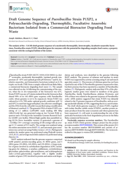
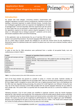
BACrERIOLOGICAL REVIEWS, June 1968, p. 127-137
Copyright © 1968 American Society for Microbiology
Vol. 32, No. 2
Printed in U.S.A.
Recent Progress in Poxvirus Research
Department
of
BRUCE WOODSON
Microbiology, University of California Medical Center, San Francisco, California 94122
INTRODUCTION ................................ 127
STRUCTURE
AND
CHEMICAL
CoMPosmON
.........................................
127
General Features ........................................................
127
Disassembly of the Virus In Vitro
Structural Proteins of Poxviruses
Isolation of the DNA as a Single Molecule
128
.............................................
..............................................
UNCOATING
OF THE
TRANSCRIPTION
EARLY
OF THE
130
VIRAL GENOME
RNA POLYMERASE
AND
129
.........................................
ACID
131
........................
LATE FUNCTIONS ...................................................
CONCLUSION ......................
.........................................
LITERATURE CITED ............................................................
INTRODUCTION
This review will deal primarily with studies
reported within the past 2-year period. Except to
place recent works in their proper perspective, no
attempt has been made to consider previous
advances; for a review of these, the reader should
consult Joklik (27).
Recent advances include the development of
techniques for the disassembly of poxvirus
particles in vitro and for the isolation of the viral
deoxyribonucleic acid (DNA) as a single molecule. In addition, a "late" enzyme has been discovered. Recent studies indicate, moreover, that
a number of the structural viral proteins are
coded for by the "parental" genome. Perhaps the
most exciting discovery is the finding that poxviruses contain a DNA-dependent ribonucleic
acid (RNA) polymerase. Transcription of the
viral genome has been demonstrated in vitro by
investigators using intact virus particles and
particles of subviral dimensions known as
"cores." Recent studies have also forced a complete re-evaluation of the mechanism by which
poxvirus particles are disassembled (uncoated) in
vivo, and it now appears likely that "derepression" of the host genome is not required to
initiate infection.
STRUCTURE
CHEMICAL CoMPosITION
General Features
Before proceeding with recent advances, it will
be instructive to consider certain aspects of viral
architecture and composition. For convenience, I
have illustrated some of these features in Fig. 1.
Three structures are clearly recognizable in
sections of the virus particle-the outer coat, the
lateral bodies, and the dense central structure
AND
127
133
133
135
135
known as the "core" or nucleoid (9). It is this
last structure, the core, which contains the poxvirus DNA. The addition of trypsin to cores prepared in vitro (15) causes them to rupture, releasing the DNA. Once the DNA has been released, it is sensitive to deoxyribonuclease. The
suggestion that a special enzyme, the "uncoating
protein" (25), is required to release the DNA
during the course of infection will be dealt with
in the section on uncoating.
Recent studies indicate that the core consists at
least partially of components which are coded for
by the parental genome (20, 21). In addition, the
recently discovered DNA-dependent RNA polymerase of poxviruses (35) is associated with this
structure. At the moment, it is impossible to tell
whether the enzyme resides within the core, or
whether it is a component of the core structure
itself. Chemical composition data have been obtained for vaccinia virus (60) and for fowlpox
virus (49). Vaccinia is about 89% protein and
5 to 6% DNA; the rest is lipid. Presumably, the
lipid components (phospholipid, cholesterol, and
neutral fat) are constituents of the membrane
structures (see Fig. 1). Copper, biotin, and riboflavine were also detected in these preparations
(60). However, at this time it is not known
whether these components are constituents or
contaminants, as no metabolic role can be
demonstrated.
Vaccinia virus, rabbitpox, cowpox, and ectromelia have the same DNA content and base
composition. The guanine plus cytosine (GC)
content is about 36%, and the base ratio [adenosine plus thymine (AT)/GC] is approximately
1.7. The Tm of cowpox DNA is 87 C, and its
density in CsCl is 1.695.
It is only recently that the DNA of poxviruses
Downloaded from http://mmbr.asm.org/ on February 6, 2015 by guest
POXVIRUS
VIRAL NUCLEIC
128
......................................
128
WOODSON
OUTER COAT
CORE
LATERAL BODIES
DNA
MEMBRANES
had been isolated in an apparently intact form.
Recent studies (22, 55) indicate that the DNA is
a linear duplex, 87 to 100 Mi in length. This would
put the molecular weight in the range of 160 X
106 to 200 X 106 daltons, in excellent agreement
with previous estimates based on chemical composition (23, 49).
Provided that the entire genome is transcribed,
sufficient DNA is present to code for several
hundred proteins. Only a few of these are known.
Seventeen to twenty have been implicated in virus
structure (see below), and five (thymidine kinase,
DNA polymerase, and three deoxyribonucleases),
which are presumably virus-coded, have been
detected by enzyme assay (7, 33, 38, 39, 40, 42).
Disassembly of the Virus In Vitro
A simple chemical procedure which can be
utilized to dissect the virus in vitro has been described by Easterbrook (15). The method is as
follows. The virus particles are treated first with
a nonionic detergent, NP40, and then with 2mercaptoethanol. This treatment alone results in
rupture and release of the outer coat. Lateral
bodies, which are still attached to the cores at this
point, can be removed enzymatically by the
addition of a small quantity of trypsin. This
results in a preparation which consists almost
entirely of viral cores. Sonic treatment or excess
trypsin will cause the cores to rupture, yielding
"ghosts" and naked viral DNA.
Treatment of virus particles with NP40 alone
or 2-mercaptoethanol alone produces the following result. With 2-mercaptoethanol, the outer coat
appears to loosen or swell. Easterbrook sug-
gested that this is due to the presence of disulfide
bonds which are involved in maintaining the
integrity of the outer coat. NP40 was thought to
attack the lipoprotein of the membrane layer,
resulting in increased permeability to phosphotungstic acid.
The beauty of the procedure is that it can be
applied to milligram quantities of purified virus,
each step appears to be reasonably quantitative,
and the intermediate structures can be purified by
sedimentation in sucrose or tartrate gradients. I
suspect that the procedure will eventually have
widespread application, especially in the determination of virus composition. The method has
already been put to use by Fenner and Sambrook
(17) and by Holowczak and Joklik (20, 21).
Structural Proteins of Poxviruses
Adequate studies on the structural proteins
have not been undertaken, and, as a result, information is limited. The primary difficulty is that
the virus particles are extremely insoluble in all
but the most drastic reagents (60). The attempts
to solubilize the proteins of the virus by mechanical disintegration and extraction with alkaline
buffers have been discussed (27). Although the
percentage of the total viral protein solubilized by
these techniques is small (as little as 20%), as
many as eight antigens have been recognized.
The ultimate description of the number of
components present in the outer coat, the lateral
bodies, and the core, may well depend upon the
development of special techniques such as those
described by Easterbrook (15). To acquire such
knowledge would indeed be a major achievement.
Nevertheless, it should be kept in mind that the
ultimate objective of the work is the description
of the chemical and physical properties of the
constituents. The technical problems involved in
elucidating the manner in which each component
is associated with the lipid, nucleic acid, and other
protein components of the virus are severe, and at
the moment it is safe to say the solutions are not
in sight.
One procedure is available which results in
complete solubilization of the virus particletreatment with sodium dodecyl sulfate (SDS),
urea, and 2-mercaptoethanol. However, in terms
of the objectives outlined above, the procedure is
clearly of limited value. These reagents not only
destroy the secondary and tertiary structure of
the proteins but they also produce a complex
mixture of polypeptides which is difficult to
resolve.
The recent studies by Holowczak and Joklik
(20, 21) demonstrate the difficulties. These investigators analyzed the polypeptides of the virus
by acrylamide gel electrophoresis. In spite of the
length of the gel columns employed and the reproducibility of the electropherograms, the number of polypeptides, though clearly in excess of 17,
Downloaded from http://mmbr.asm.org/ on February 6, 2015 by guest
FIG. 1. Features of poxvirus architecture.
BACrERIOL. REV.
VOL. 32, 1968
PROGRESS IN POXVIRUS RESEARCH
Becker (55) for isolating intact molecules of poxvirus DNA. In spite of these accomplishments, the
reasons for previous failures are not at all clear.
It has generally appeared to make little difference
whether phenol and SDS were used separately
or together, or in conjunction with proteolytic
enzymes, or with previously defatted virus. The
result in most cases was the same-extensive
fragmentation of the DNA (27). Nor is it known
whether the major damage to the DNA results
during its release from the core or during subsequent manipulations. The latter possibility is a
very real one, however, as the DNA is extremely
large (larger, in fact, than that of any other known
virus).
The DNA of fowlpox virus has been released
successfully with SDS by Gafford and Randall
(19). These investigators concluded that phenol
results in extensive fragmentation, even to purified DNA, and that the extent of the damage is
proportional to the exposure to phenol. The possible involvement of protein linkers was investigated, and was ruled out since trypsin had no
effect. Purification of the DNA was accomplished
by extraction in chloroform-n-butyl alcohol
followed by methylated albumin kieselguhr
(MAK) column chromatography.
In addition to the problems encountered with
phenol, the authors discuss the problems involved
in converting sedimentation coefficients to
molecular weights-a problem in formula selection. The molecular weight of fowlpox DNA by
sedimentation techniques was judged to be 200 x
106 to 240 X 106 daltons. The contour length of
fowlpox DNA has also been determined (22).
Forty-one molecules of DNA were measured;
the average length was 100 ,u, equivalent to 192 X
106 daltons.
A second procedure for releasing poxvirus
DNA using sodium deoxycholate (DOC) and
Pronase has been described recently by Sarov
and Becker (55). The special virtue of this technique is that it protects the DNA by releasing it in
the presence of sucrose. The authors imply that
once the DNA has been released in this fashion
it can be deproteinized with phenol; however, no
data on this point were actually given. The contour lengths of four molecules of vaccinia DNA
have been measured; the average length, 87 ,u, is
equivalent to a molecular weight of approximately 167 X 106 daltons.
In the past 2 years, electron microscopy has
been the technique most frequently employed to
determine molecular weight. The studies of
Isolation of the DNA as a Single Molecule
McCrea and Lipman (41) indicate that the oneMethods have been described recently by step osmotic shock method is to be avoided beGafford and Randall (19) and by Sarov and cause it results in extensive fragmentation. Even
Downloaded from http://mmbr.asm.org/ on February 6, 2015 by guest
could not be ascertained. The location within the
virus of certain polypeptide components was
determined by application of the Easterbrook
(15) technique.
Treatment of vaccinia virus with NP40 alone
resulted in the release of a single (multiple polypeptide) component (VSP-6). Holowczak and
Joklik suggested that this component, which
accounted for 18.7% of the total virus protein,
is situated at the surface of the virus particle.
Treatment of the virus particle with NP40 followed by 2 ,2'-dithiodiethanol liberated all of the
polypeptides except those associated with the
viral core. The polypeptides of the core, which
accounted for 36.7% of the total viral protein,
consisted of two components believed to be single
polypeptides (VSP-1 and VSP-2) and a third
component (VSP4) which was a mixture of
polypeptides. No information was provided on
the constituents of the lateral bodies.
An approach which circumvents the problems
of solubilization is that of examining the proteins
present after infection-i.e., the soluble antigens.
At the present time, they represent the best source
of virus coded proteins which are suitable for
purification and characterization. As many as
17 components have been observed in rabbit skin
infected with vaccinia (64), 17 components also
have been observed in chick chorioallantoic membranes infected with vaccinia (51), and up to 20
components have been observed in HeLa
cells infected with rabbit pox (4). It has not been
established yet whether all of the components
capable of reacting with hyperimmune serum are
structural viral proteins. Nevertheless, it is clear
that many are identical to the antigens which
can be extracted from virus particles (8, 64).
Two of these antigens have been isolated and
purified-an [S-antigen which has a molecular
weight of approximately 240,000 (61) and an
antigen (molecular weight 100,000 to 200,000)
which will combine with virus-neutralizing antibody (5). (The LS antigen has two immunogenic
sites: one which is labile and is inactivated at 60 C,
and a second which is stable even at higher temperatures.) Physical and chemical methods have
also been applied recently to the soluble viral
antigens of KB cells (8). Three antigens have
molecular weights of 200,000 or greater, and the
remaining antigens are in the 50,000 to 100,000
range. Of the high molecular weight components,
at least one stimulated the synthesis of virusneutralizing antibody.
129
130
WOODSON
when released from cores (16), only 1 of 91 molecules appeared unbroken by this technique. The
suggestion that this surviving molecule was at
one time circular is, in my opinion, completely
unwarranted. No good evidence has yet been obtained to show that animal viruses other than
those of the papova group contain circular molecules.
A technique has also been reported for increasing the density difference (normally about 0.004
g/cc in CsCl) between viral DNA and host-cell
DNA, based on the selective ability of viral
DNA to renature at 60 C. The new value for the
density difference was 0.017 g/cc (32).
stitute the first stage of disassembly, result in the
appearance of viral cores within the cytoplasm.
At this stage, the genome is insensitive to deoxyribonuclease.
The second stage of uncoating proceeds more
slowly than the first. Generally, there is a lag of
30 min to 1 hr before the release of the viral
DNA begins. The exact timing depends upon the
multiplicity of infection: the higher the multiplicity, the shorter the lag. Judging from its
susceptibility to deoxyribonuclease, all of the
DNA which can be released from cores will be
released by 3 to 4 hr. This may be as little as 10%
or as much as 60 to 70% of the total input DNA
(24).
For second-stage uncoating to occur, there
is an absolute requirement for both RNA and
protein synthesis. This point is very secure, and
it has been demonstrated many times by the use
of inhibitors which result in the accumulation of
cores (10, 25, 34). The result suggests that
information encoded in the base sequence of
DNA is required for liberation of the viral DNA
from the core.
Several years ago, a scheme was proposed by
Joklik (25) to account for these results. He
predicted that first-stage uncoating proceeded
spontaneously but that second-stage uncoating
required information encoded within the host
genome. [It was presumed by Joklik (25) on a
priori grounds that a viral genome which was
insensitive to deoxyribonuclease, and, therefore,
presumably still surrounded by protein, was not
yet ready to assume its genetic function. In view
of the requirement for protein synthesis, this
necessitated the involvement of the host genome
in the uncoating mechanism.] Joklik believes
that the host genome codes for the synthesis of
an "uncoating protein," a proteolytic enzyme
which is essential in facilitating the release of the
viral nucleic acid from the core. Two pieces of
information-the lag required before secondstage uncoating commenced and the effect of
inhibitors in preventing the release of the viral
DNA-suggested to him that the protein was
not present in uninfected cells. To complete the
theory, it was proposed that a "viral inducer"
protein released during the first stage of uncoating "derepressed" the host genome, thus allowing synthesis of the uncoating protein.
Although the theory requires several assumptions, the crucial one is that the viral genome,
still within the core and inaccessible to deoxyribonuclease, is an unlikely template for messenger RNA (mRNA). Recent studies (see the
following section) indicate that this assumption
is unwarranted. Viral cores, blocked in their
uncoating by inhibitors, are extremely active in
Downloaded from http://mmbr.asm.org/ on February 6, 2015 by guest
UNCOATING OF THE VIRAL NUCLEIC ACID
The intracellular uncoating of poxviruses is a
two-stage process-the first uncoating step removes the outer coat and the lateral bodies, and
the second uncoating step liberates the DNA from
the core, rendering it sensitive to deoxyribonuclease (11, 24, 25). Until recently, it was thought
that both uncoating steps were required in order
for the viral DNA to assume its genetic function.
As it turns out, this concept is wrong. Recent
experiments show that the poxvirus genome can
be transcribed even though it resides within the
viral core and is completely resistant to deoxyribonuclease (34, 44, 66). This is apparently due
to the fact that poxviruses contain a DNAdependent RNA polymerase (35, 45). The net
result of recent studies is that they discredit, to a
large extent, a theory proposed recently to account for the molecular basis of the uncoating
phenomenon (25). Before discussing the nature of
this proposal, it will be instructive to examine the
uncoating process itself. Only the essentials of the
process need concern us here.
Knowledge of the uncoating process derives
from studies with the electron microscope (11)
and from studies employing radiocative isotopes
(24, 25). Briefly, the following concept has
emerged. Poxviruses are taken into their hosts by
a process akin to phagocytosis and eventually
reside within phagocytic vesicles within the cell
(11, 12). Once within these vesicles, first-stage
uncoating commences, resulting in the degradation of the viral coat and, presumably, the lateral
bodies. Dales (11) believes that the walls of the
phagocytic vesicles collapse simultaneously with
the rupture of the viral coat and has suggested
that a concerted mechanism is involved. Whether
the process is primarily an enzymatic one, or
mechanical, or some combination of these factors,
is not known. Whatever the mechanism, it accounts for the release of nearly 100% of the viral
phospholipid and as much as 50 to 60% of the
viral protein (20, 24). These events, which con-
BACTERIOL. REV.
VOL. 32, 1 968
PROGRESS IN POXVIRUS RESEARCH
TRANscRIPTION OF THE VIRAL GENOME
Before discussing the problems of transcription, it will be profitable to outline the events
C. RM.
I
B/
TIME (HOURS)
FIG. 2. Pattern of vaccinia virus specific mRNA
synthesis in HeLa cells (A) in the presence of cycloheximide or with ultraviolet-inactivated infecting virus,
(B) no additions, and (C) in the presence of FUDR (66).
which are associated with the replicative cycle.
The exact timing of these events depends largely
upon the system employed and upon the multiplicity of infection. At high input multiplicities
(several hundred particles per cell), transcription
of the parental genome begins immediately upon
infection; uncoating of the viral nucleic acid and
the "takeover" of host cell functions begin by 30
min to 1 hr, induced enzymes and viral antigens
appear between 1 and 2 hr; DNA replication
begins at 1 to 1.5 hr and is essentially complete
by 4 to 5 hr; and progeny particles, which may
number 104 per cell at 24 hr, begin to appear as
early as 4 to 5 hr after infection. These events
take place exclusively within the cytoplasm (27).
The problems of transcription can be handled
in a variety of ways. The simplest is to discuss
them in relation to my own studies, shown in
Fig. 2. This figure describes the relative rate at
which transcription of the vaccinia genome proceeds in HeLa cells under three different conditions. In all cases, the pattern of viral mRNA
synthesis was determined by administering 10min pulses of '4C-uridine as described elsewhere
(66). Curve C represents transcription of the
parental genome only, since viral DNA synthesis
has been prevented with fluorodeoxyuridine
(FUDR). Curve B shows the pattern of RNA
synthesis under normal conditions-i.e., transcription of both parental and progeny genomes.
Curve A also represents transcription of the
parental genome, but under conditions such that
second-stage uncoating (release of the viral
genome from the core) does not occur. It is this
last result which has prompted the re-evaluation
Downloaded from http://mmbr.asm.org/ on February 6, 2015 by guest
the synthesis of viral mRNA. As a result, it must
be assumed that the DNA of the virus particle is
capable of directing its own release and replication.
In his efforts to obtain evidence for the viral
inducer concept, Joklik (26) described the
properties of a viral protein component which
was essential for infectivity. He concluded that
the viral inducer protein was sensitive to denaturation by heat, urea, and ultraviolet irradiation. In each case, the virus particles were
rendered noninfectious by these treatments, and
they failed to undergo second-stage uncoating.
Ultraviolet irradiation was presumed to damage
the histidine residues of the inducer protein (26).
Recent studies in this laboratory suggest that
the inducer protein postulated by Joklik (25, 26)
and the DNA-dependent RNA polymerase described by Kates and McAuslan (35) are one and
the same. As might be expected, the activity of
this enzyme is readily destroyed by heat and urea,
but not by ultraviolet irradiation (Woodson,
unpublished data). The recent findings of Kim
and Sharp (37, 57) also support this contention.
These investigators concluded that virus particles
rendered noninfectious by ultraviolet irradiation
and by nitrogen mustard show multiplicity
reactivation, whereas those inactivated by heat
and urea do not.
The requirement for protein synthesis is indeed
real. Inhibitors such as cycloheximide and streptovitacin inhibit second-stage uncoating completely
(10, 25, 34). Although no good evidence has been
obtained to show that a specific protein is
required, this has generally been assumed to be
the case. As indicated previously by Joklik (27),
attempts to demonstrate the activity of the
postulated protein failed. Abel (1) claimed,
however, that she could detect uncoating enzymes
in extracts of infected chick embryo fibroblasts.
Furthermore, she and Trautner (2) reported that
infectious DNA, prepared by treating virus
particles with these enzymes, could initiate a
round of virus replication in Bacillus subtilis.
Claims of a similar nature have been made
recently by Babbar et al. (6). Furthermore,
infectious DNA has been isolated from fowlpox
(50), and infectious particles (of subviral dimensions) have been obtained from cultures
infected with myxoma and fibroma viruses (63).
At the present time, these findings are not well
understood, and a conclusion must await further
studies.
131
132
WOODSON
which case presumably no viral mRNA is translated.
These results suggest that, when uncoating does
not occur, transcription is uncontrolled. An explanation for this behavior will be offered in the
following section. When uncoating does occur,
transcription of the parental genome appears to
subside. The possibility that this behavior results
from the dissociation of the RNA polymeraseDNA complex is being investigated.
Transcription of the viral genome has also
been studied by Oda and Joklik (46). These
investigators examined the behavior of the
vaccinia genome in L cells and HeLa cells and
found that the two patterns were entirely different.
In HeLa cells, the pattern was essentially that
which I have shown in Fig. 2; by 2.5 hr, transcription of the parental genome was barely
detectable. In L cells, however, the parental
genome was transcribed at a much greater
velocity, and by 4 hr was still being transcribed
at a substantial rate. Competition hybridization
experiments were undertaken to determine the
nature of the sequences being transcribed in the
two cell lines. In HeLa cells, parental genomes
were transcribed exclusively from 0 to 2 hr after
infection. From this time on, "early" and "late"
message sequences were transcribed at nearly
equivalent rates. (Late sequences were defined as
those which could not be transcribed from
parental genomes.) In L cells, parental genomes
appeared to be transcribed almost exclusively at
all times. In spite of this finding, progeny particles
were produced in roughly equivalent numbers in
the two cell lines. From these experiments, it
may be concluded that there is little or no
correlation between the yield of progeny particles
and the quantity of late sequences transcribed.
Similar observations have been made in regard to
protein synthesis (53) and will be discussed later.
The experiments also suggest that it is the host
which determines the pattern of viral transcription. Additional studies, in a variety of cell lines,
will be required to determine whether this is
literally true. Finally, there is the possibility that
the observed result is due to the efficiency of uncoating. That uncoating can vary, not only among
cell lines but among strains of the same cell line,
has been shown previously by Joklik (24).
Clearly the problems of transcription are many.
The factors which may influence and control
transcription are, nevertheless, becoming more
apparent. One of these factors is an enzyme involved in the transcription process itself. Evidence that it is a component of the virus particle
will be presented next.
Downloaded from http://mmbr.asm.org/ on February 6, 2015 by guest
of the uncoating theory proposed by Joklik (25),
since it shows that the virus, though not yet fully
uncoated, is capable of synthesizing mRNA. This
result is obtained with ultraviolet-treated virus
and, provided that protein synthesis is inhibited
at zero-time, with normal virus. Evidence has
been obtained that the RNA transcribed under
the conditions of curve A is viral mRNA. This
material associates rapidly with polyribosomes,
and, when extracted from these structures, it had
the base composition of legitimate viral mRNA
(66).
Additional proof that transcription of the
viral genome can occur, though not yet fully uncoated, comes from the experiments of other
workers. Although Munyon and Kit (44) were
the first investigators to come to this conclusion,
they made no attempt to identify the RNA transcribed in the presence of inhibitors as being
viral. Convincing data have been supplied by
Kates and McAuslan (34). These investigators
showed that the RNA transcribed under the
conditions of curve A will hybridize with poxvirus DNA, but not with host-cell DNA. In fact,
on the basis of competition hybridization experiments, these workers concluded that the majority
of the genes which can be transcribed from the
parental genome can also be transcribed from
cores. Additional experiments indicated that
certain genes (those controlling the synthesis of
DNA polymerase, for example) could be transcribed only after the release of the viral genome
from the core.
The factors which regulate transcription of the
viral genome are not at all clear. Nevertheless,
the following points can be made in reference to
Fig. 2. First, in order to study the transcription
of parental genomes, one must include either
FUDR or cytosine arabinoside in the medium;
otherwise, this event is completely obscured by
the transcription of progeny genomes. In the
presence of FUDR, the initial rate of viral mRNA
synthesis is directly proportional to the input
multiplicity, over a fairly wide range (66). We
ascribe this behavior to the fact that each particle
brings into the cell its own DNA-dependent
RNA polymerase. Second, transcription of the
parental genome under conditions of high
multiplicity and maximal synchronization (as in
curve C) is tightly controlled and occurs in a
burst-like fashion. Finally, under a variety of
conditions, transcription of the parental genome
occurs in an "uncontrolled" or extended fashion,
as in curve A. Kinetics of this nature have been
observed with inhibitors of protein synthesis and
with ultraviolet-treated virus (34, 66), in studies
performed in vitro with virus particles and cores
(35, 45), and in studies on interferon (31), in
BACTERIOL. REV.
VOL. 32, 1968
PROGRESS IN POXVIRUS RESEARCH
sensitive to ribonuclease. However, after extended
periods of transcription (8 to 10 min is sufficient),
the majority of the material was then ribonuclease-sensitive (Hedgpeth and Woodson,
unpublished data). Further evidence for the
release of the product from the core has been obtained by sedimentation techniques (35, 45). In
addition, Kates and McAuslan (35) have shown
that a portion of the product is the same size as
that which is transcribed in vivo, and that it can
be annealed to poxvirus DNA.
A comparison of the properties of the poxvirus
system with those of the DNA-dependent RNA
polymerase from Escherichia coli reveals several
interesting features. When the E. coil enzyme is
primed by native DNA, chains are first initiated,
followed by chain growth. At the end of the
reaction, nascent RNA chains remain bound to
the polymerase which, in turn, remains firmly
bound to the DNA (39). The poxvirus system
behaves quite differently. First, overall transcription proceeds in a linear fashion for a period
of several hours (35, 45). During this time,
quantities of RNA equivalent to the weight of the
poxvirus genome are transcribed and released
(35). The results imply that portions of the poxvirus genome are being transcribed in a repetitive
fashion. Experiments which should confirm or
deny this result are presently in progress.
EARLY AND LATE FuNcrIoNs
The multiplication of certain bacteriophages
(phage x, phage 2C, phage T4, etc.) requires the
formation of virus-specific enzymes which appear
"early" or "late" in the replicative cycle. The
synthesis of these enzymes, and the expression
of the genome generally, is under strict control
(18, 47, 59). For poxviruses, a similar regulation
of gene action has been postulated (30). Induced
enzymes (7, 33, 38, 39, 42) appear shortly after
infection which are subject to switchoff control
(38, 39) when progeny genomes appear. In addition, certain of the structural viral proteins are
synthesized early and others are synthesized late
(21, 53, 65).
Available evidence suggests that expression of
the poxvirus genome is controlled largely at the
level of transcription. Thus, a portion of the
parental genome is transcribed at the core level,
and additional genes are presumably transcribed
once the genome is free (34). These sequences of
mRNA presumably code for the early enzymes
and for the early structural viral proteins. Late
sequences of mRNA are transcribed after the
appearance of progeny genomes, and presumably
these result in the activation of the switchoff
Downloaded from http://mmbr.asm.org/ on February 6, 2015 by guest
PoxviRus RNA POLYMERASE
Until recently, there was no reason to think
that the poxvirus system would behave differently
from any other; the input DNA, after its release
from the core, would be transcribed by a hostcell enzyme. The first indication that this was not
the case came from the experiments (34, 44, 66)
which demanded the re-evaluation of the uncoating theory (25). These said, in essence, that
the DNA was transcribed prior to its release from
the core. Although it is conceivable that a host
enzyme could enter the core (though deoxyribonuclease cannot), or that a host enzyme could
act upon a few genomes released in some anomalous fashion, the evidence clearly indicates
that the enzyme responsible resides within the
core, and that it is a component of the virus
particle. This was demonstrated by the experiments of Kates and McAuslan (35). These
investigators examined the cytoplasmic extracts
of uninfected cells and of infected cells containing
viral cores for polymerase activity. No activity
was found in the cytoplasm of uninfected cells
in spite of the addition of rabbitpox DNA (100
fig, either native or denatured) in amounts
equivalent to the combined genomes of 3.6 x 10¹¹
virus particles. In contrast, the extracts containing
viral cores rapidly catalyzed the incorporation of
labeled uridine triphosphate (UTP) into an acidinsoluble product. The activity required the
presence of all four ribonucleoside triphosphates,
but it was not stimulated by the addition of
primer DNA, nor was it inhibited by the addition of deoxyribonuclease. Transcription was
inhibited by actinomycin D, and the product was
sensitive to ribonuclease.
Further evidence that the enzyme is a component of the virus particle was obtained by
showing that highly purified preparations of
cores will catalyze RNA synthesis in vitro. The
reaction requires only three additional components-Mg+, triphosphates, and buffer.
Virus particles are also active in vitro, provided
that the outer coat has been loosened or made
permeable by exposure to mercaptoethanol or
trypsin. Similar observations have now been
made in this laboratory (Woodson, unpublished
data) and by Munyon et al. (45). Although no
one has yet been successful in obtaining from
poxviruses an enzyme which will respond to exogenous primer, efforts with this goal in mind are
presently being made in several laboratories.
The nature of the product which is transcribed
in vitro has been investigated to some extent.
When the product was examined after a period of
limited transcription (30 sec to 2 min), the bulk
of it was found to be core-associated and in-
133
134
WOODSON
arise which result in misreading of early (stable)
mRNA sequences (18). Two studies have been
reported recently which bear upon the problem.
The studies by Sebring and Salzman (56)
indicate that a sizable fraction of vaccinia virus
mRNA synthesized late sediments in the 30 to
74S region. In contrast to the results obtained by
Joklik and Becker (29), this RNA was judged to
be nonfunctional in that it did not decay in the
presence of actinomycin and it could not be
chased into the polysome region. Additional
studies will be required to determine the nature
of the binding of this RNA to other cell constituents, and to determine the sequence composition of the RNA-i.e., whether it is predominantly early or late, or both. Oda and Joklik
(46) found that early sequences transcribed at a
late time enter the polyribosome region. A
similar observation has been reported in the case
of bacteriophage T4 (18).
In the remainder of this section, I would like
to discuss (i) the synthesis and assembly of the
structural viral proteins and (ii) the function of
several nonstructural proteins which arise during
the course of infection.
(i) Recent studies show that three to five of
the structural viral proteins are coded for by the
parental genome, and that these proteins are
synthesized early. This is a rather unusual situation, and, as far as I know, it has not been observed in any other systems.
The early structural proteins begin to appear
at about 1 to 2 hr after infection and, like the
induced enzymes, are subject to switch-off control
at 4 to 6 hr. They constitute about 15 to 25% of
the total protein of the virus particle (20, 21, 53).
The studies by Shatkin (58) were probably the
first to indicate that structural viral proteins were
produced early. He showed that a sizable fraction of the total viral antigen was produced in
the presence of FUDR. Five early antigens have
now been recognized by Appleyard (3) and by
Salzman and Sebring (53), four by Wilcox and
Cohen (65), and three polypeptide components
have been detected by Holowczak and Joklik
(21).
The location of the early components within
the virus particle has been examined, and it is
clear that some are the structural proteins of
cores. This was first suggested by Wilcox and
Cohen (65) on the basis of the fact that the low
molecular weight antigens of vaccinia virus which
were synthesized early did not elicit the formation of virus-neutralizing antibody. The studies
by Holowczak and Joklik (21) offer a more direct
proof. These investigators have utilized a combination of techniques to investigate the location
and time of synthesis of various polypeptide
Downloaded from http://mmbr.asm.org/ on February 6, 2015 by guest
mechanism and the synthesis of the late structural
viral proteins (46).
At the present time, there is no indication of the
mechanism by which the information is selected.
Presumably this would occur in a fashion analogous to that which has been predicted for other
systems-i.e., different portions of parental and
progeny genomes are available for transcription
owing to physical and chemical differences, or
enzymes of different specificity recognize one
portion of the genome or another (47). In addition, repressor proteins may be involved in
regulating the expression of the DNA (48). At
the moment, there is no information on whether
the poxvirus genome is transcribed symmetrically
or asymmetrically (62), or whether early and late
message sequences derive from distant segments
of the poxvirus genome, as in the case of phage
x (59).
Once a particular mRNA molecule has been
transcribed, or while it is in the process of being
transcribed, it will associate with ribosomes (29)
in such a fashion as to initiate polypeptide synthesis. As a result of this act, or owing to the
presence of soluble degradative enzymes within
the cell, or both, it will be degraded in some
fashion rendering it nonfunctional. The observation that vaccinia mRNA transcribed early in
HeLa cells decays slowly and that late message
sequences decay more rapidly has led to speculation that protein synthesis is controlled additionally at the level of translation.
In recent studies by Sebring and Salzman (56),
it was found that at early times mRNA synthesis
proceeded at substantial rates and that the halflife of the RNA was in excess of 2 hr. At late
times, the quantity of mRNA which could be
extracted from polyribosomes was sharply
reduced, and the half-life of this material was
about 13 min. In spite of these events, protein
synthesis was observed to continue at a substantial rate until late in infection. Similar studies
were undertaken by Oda and Joklik (46). These
investigators found that, in HeLa cells, less than
20% of early vaccinia mRNA was degraded in a
period of 5.5 hr. Late sequences had a half-life
of less than 1 hr. The situation in L cells was
quite different-vaccinia mRNA transcribed at
early and late times had identical half-lives (about
2 to 3 hr).
The fate of the messenger molecules which are
subject to switchoff control has also been touched
upon to some extent. The molecular basis of this
phenomenon is still completely unknown, and at
the present time there is insufficient evidence to
support strongly either the notion that a specific
protein is coded for by the progeny genome (18,
39) or that new transfer RNA (tRNA) molecules
BACrERIOL. REV.
VOL. 32, 1968
PROGRESS IN POXVIRUS RESEARCH
coding for its synthesis. The specific function of
the protein is not known.
The synthesis and regulation of virus-induced
deoxyribonucleases has also been investigated by
these workers. Recent studies by McAuslan and
Kates (40) indicate that, of three nucleases which
are induced by cowpox in HeLa cells, one acts
optimally and almost exclusively on denatured
DNA at pH 4.5, a pH at which the enzyme is
insoluble. The enzyme has been purified (3,000fold), and its chemical and physical properties
have been assessed. It is synthesized continuously
from about 3 to 18 hr postinfection, and during
this time undergoes a sharp transition from the
soluble to the bound state. No enzyme was synthesized in the presence of FUDR, and the rate
of enzyme synthesis in the absence of FUDR was
strictly proportional to the quantity of DNA
synthesis allowed. In all respects, the enzyme
appears to be a "late" function.
CONCLUSION
The major accomplishments of the past 2 years
may be summarized as follows. The viral DNA
has been isolated in a form which, for the first
time, appears to be reasonably intact. In addition, a method has been described for disassembling the virus in vitro. Two enzymes have been discovered, a DNA-dependent RNA polymerase
which resides within the virus particle, and a
deoxyribonuclease which appears to be a late
function. Recent experiments have indicated,
moreover, that a number of the structural viral
proteins are coded for by the parental genome.
Our concept of uncoating has been revised
completely, and our understanding of the factors
which regulate transcription and the expression of
early and late functions has increased substantially. Although many problems remain to be
solved, most of these are presently under attack.
If the progress of the past 2 years can be taken
as a measure of what is yet to come, then prospects for the next 2 years should be excellent
indeed.
ACKNOWLEDGMENT
supported by Public Health Service
grant Al 06862 from the National Institute of Allergy and Infectious Diseases.
This work was
LITERATURE CITED
1. Abel, P. 1963. Reactivation of heated vaccinia
virus in vitro. Z. Vererbungslehre 94:249-252.
2. Abel, P., and T. A. Trautner. 1964. Formation of
an animal virus within a bacterium. Z. Verer-
bungslehre 95:66-72.
3. Appleyard, G., V. B. M. Hume, and J. C. N.
Westwood. 1965. The effect of thiosemicar-
Downloaded from http://mmbr.asm.org/ on February 6, 2015 by guest
components. Two of the polypeptides synthesized
early are components of cores. A third component synthesized early apparently resides at the
surface of the particle.
Studies with the electron microscope suggest
that the structural components of cores, which
are coded for by the parental genome, can be
assembled into structures which have the appearance of cores, but which lack DNA. These
structures, which are known as "immature
forms," accumulate in the presence of FUDR
(14), hydroxyurea (52), and IBT (isatin-3-thiosemicarbazone) (13). Unlike FUDR and hydroxyurea, IBT does not inhibit viral DNA
synthesis (13, 67). It inhibits protein synthesis,
but only after the appearance of progeny genomes (presumably by interfering with the functioning of "late" mRNA molecules). Apparently
the packaging of DNA into the core requires the
synthesis of late proteins. This conclusion is
supported by the finding that one of the polypeptide components of cores (VSP-4) is synthesized late (21), and by the studies with puromycin (28) which showed that proteins synthesized late were necessary to convert progeny
DNA to deoxyribonuclease-resistant subvirion
forms.
As I have just indicated, structures (presumably
intermediates in the assembly process) can be
accumulated by the use of inhibitors. More than
likely, structures of a similar nature would be
accumulated in cells infected by conditional
lethal mutants. Now that such mutants have been
isolated (54), it is probably safe to assume that a
serious study of poxvirus assembly will soon get
underway. Fenner and Sambrook (17) have
analyzed certain of these mutants already. Many
of the host cell-dependent mutants are capable of
synthesizing DNA, all four antigens of the core,
and all but a few of the remaining proteins. [In
view of the studies suggesting that some of the
structural viral proteins are coded for by the
parental genome (21, 53, 65), it is clear that DNA
synthesis alone is no longer a sufficient criterion
for classifying a mutant as either "early" or
"late."] It would be interesting indeed to determine whether these components are assembled
into cores or lateral bodies or both.
(ii) The relationship between protein synthesis
and poxvirus DNA synthesis has been investigated by Kates and McAuslan (36). These
investigators have provided evidence that an
early protein is required both to initiate and to
sustain viral DNA synthesis. The protein, which
was itself stable, was required in stoichiometric
rather than catalytic amounts and could be
distinguished from DNA polymerase and thymidine kinase because of the instability of mRNA
135
136
WOODSON
virus. II. Kinetics of the synthesis of individual
groups of structural proteins. Virology 33:726739.
22. Hyde, J. M., L. G. Gafford, and C. C. Randall.
1967. Molecular weight determination of
fowlpox DNA by electron microscopy. Virology 33:112-120.
23. Joklik, W. K. 1962. The purification of four
strains of poxvirus. Virology 18:9-18.
24. Joklik, W. K. 1964. The intracellular uncoating
of poxvirus DNA. I. The fate of radioactively
labeled rabbitpox virus. J. Mol. Biol. 8:263276.
25. Joklik, W. K. 1964. The intracellular uncoating of
poxvirus DNA. II. The molecular basis of the
uncoating process. J. Mol. Biol. 8:277-288.
26. Joklik, W. K. 1964. The intracellular fate of
rabbitpox virus rendered noninfectious by
various reagents. Virology 22:620-633.
27. Joklik, W. K. 1966. The poxviruses. Bacteriol.
Rev. 30:33-66.
28. Joklik, W. K., and Y. Becker. 1964. The replication and coating of vaccinia DNA. J. Mol.
Biol. 10:452-474.
29. Joklik, W. K., and Y. Becker. 1965. Studies on
the genesis of polyribosomes. II. The association of nascent messenger RNA with the 40S
subribosomal particles. J. Mol. Biol. 13:511520.
30. Joklik, W. K., C. Jungwirth, K. Oda, and B.
Woodson. 1967. Early and late functions during the vaccinia virus multiplication cycle, p.
473-494. In J. S. Colter and W. Paranchych
[ed.], The molecular biology of viruses. Academic Press, Inc., New York.
31. Joklik, W. K., and T. C. Merigan. 1966. Concerning the mechanism of action of interferon.
Proc. Natl. Acad. Sci. U.S. 56:558-565.
32. Jungwirth, C., and I. B. Dawid. 1967. Vaccinia
DNA: separation of viral from host cell DNA.
Arch. Ges. Virusforsch. 20:464-468.
33. Jungwirth, C., and W. K. Joklik. 1965. Studies
on "early" enzymes in HeLa cells infected with
vaccinia virus. Virology 27:80-93.
34. Kates, J. R., and B. R. McAuslan. 1967. Messenger RNA synthesis by a "coated" viral genome.
Proc. Natl. Acad. Sci. U.S. 57:314-320.
35. Kates, J. R. and B. R. McAuslan. 1967. Poxvirus
DNA dependent RNA polymerase. Proc.
Natl. Acad. Sci. U.S. 58:134-141.
36. Kates, J. R., and B. R. McAuslan. 1967. Relationship between protein synthesis and viral
deoxyribonucleic acid synthesis. J. Virol. 1:
110-114.
37. Kim, K. S., and D. G. Sharp. 1967. Multiplicity
reactivation of vaccinia virus particles treated
with nitrogen mustard. J. Virol. 1:45-49.
38. McAuslan, B. R. 1963. Control of induced thymidine kinase activity in the poxvirus-infected
cell. Virology 20:162-168.
39. McAuslan, B. R. 1963. The induction and repression of thymidine kinase in the poxvirusinfected HeLa cell. Virology 21:383-389.
40. McAuslan, B. R., and J. R. Kates. 1967. Pox-
Downloaded from http://mmbr.asm.org/ on February 6, 2015 by guest
bazones on the growth of rabbitpox virus in
tissue culture. Ann. N.Y. Acad. Sci. 130:92-104.
4. Appleyard, G., and J. C. N. Westwood. 1964. The
growth of rabbitpox virus in tissue culture. J.
Gen. Microbiol. 37:391-401.
5. Appleyard, G., H. T. Zwartouw, and J. C. N.
Westwood. 1964. A protective antigen from
the poxviruses. I. Reaction with neutralizing
antibody. Brit. J. Exptl. Pathol. 45:150-161.
6. Babbar, 0. P., B. L. Chowdhury, and G. Rani.
1966. Pock formation by vaccinia virus deoxyribonucleic acid after passage through Escherichia coli speroplats. Acta Virol. 10:15-19.
7. Chang, L. M. S., and M. E. Hodes. 1967. Induction of DNA nucleotidyltransferase by the
Shope fibroma virus. Virology 32:258-266.
8. Cohen, G. H., and W. C. Wilcox. 1966. Soluble
antigens of vaccinia-infected mammalian cells.
I. Separation of virus-induced soluble antigens
into two classes on the basis of physical characteristics. J. Bacteriol. 92:676-686.
9. Dales, S. 1963. The uptake and development of
vaccinia virus in strain L cells followed with
labeled viral deoxyribonucleic acid. J. Cell
Biol. 18:51-72.
10. Dales, S. 1965. Effects of streptovitacin A on the
initial events in the replication of vaccinia and
reoviruses. Proc. Natl. Acad. Sci. U.S. 54:
462-468.
11. Dales, S. 1965. Penetration of animal viruses into
cells. Progr. Med. Virol. 7:1-43.
12. Dales, S., and R. Kajioka. 1964. The cycle of
multiplication of vaccinia virus in Earle's
strain L cells. I. Uptake and penetration.
Virology 24:278-294.
13. Easterbrook, K. B. 1962. Interference with the
maturation of vaccinia virus by isatin-betathiosemicarbazone. Virology 17:245-251.
14. Easterbrook, K. B. 1963. Conservation of vaccinial DNA during an abortive cycle of multiplication. Virology 21:508-510.
15. Easterbrook, K. B. 1966. Controlled degradation
of vaccinia virions in vitro: an electron microscope study. J. Ultrastruct. Res. 14:484-496.
16. Easterbrook, K. B. 1967. Morphology of deoxyribonucleic acid extracted from cores of vaccinia virus. J. Virol. 1:643-645.
17. Fenner, F., and J. F. Sambrook. 1966. Condiditional lethal mutants of rabbitpox virus. II.
Murants (p) that fail to multiply in PK-2a
cells. Virology 28:600-609.
18. Friesen, J. D., B. Dale, and W. Bode. 1967.
Presence of T4 "early" messenger RNA on
polysomes late in infection. J. Mol. Biol.
28:413-422.
19. Gafford, L. G., and C. C. Randall. 1967. The
high molecular weight of the fowlpox virus
genome. J. Mol. Biol. 26:303-310.
20. Holowczak, J. A., and W. K. Joklik. 1967.
Studies on the structural protein of vaccinia
virus. I. Structural protein of virions and cores.
Virology 33:717-725.
21. Holowczak, J. A., and W. K. Joklik. 1967.
Studies on the structural protein of vaccinia
BACrERIOL. REV.
VOL. 32, 1968
41.
42.
43.
45.
46.
47.
48.
49.
50.
51.
52.
53.
virus-induced acid deoxyribonuclease: Regulation of synthesis; control of activity in vivo;
purification and properties of the enzyme.
Virology 33:709-716.
McCrea, J. F., and M. B. Lipman. 1967. Strandlength measurements of normal and 5-iodo2'-deoxyuridine-treated vaccinia virus deoxyribonucleic acid released by the Kleinschmidt
method. J. Virol. 1:1037-1044.
Magee, W. E., and 0. V. Miller. 1967. Immunological evidence for the appearance of a new
DNA polymerase in cells infected with vaccinia virus. Virology 31:64-69.
Maitra, U., S. N. Cohen, and J. Hurowitz. 1966.
Specificity of initiation and synthesis of RNA
from DNA templates. Cold Spring Harbor
Symp. Quant. Biol. 31:113-122.
Munyon, W. H., and S. Kit. 1966. Induction of
cytoplasmic ribonucleic acid (RNA) synthesis
in vaccinia-infected LM cells during inhibition
of protein synthesis. Virology 29:303-309.
Munyon, W., E. Paoletti, and J. T. Grace, Jr.
1967. RNA polymerase activity in purified
infectious vaccinia virus. Proc. Natl. Acad.
Sci. U.S. 58:2280-2288.
Oda, K., and W. K. Joklik. 1967. Hybridization
and sedimentation studies on "early" and "late"
vaccinia messenger RNA. J. Mol. Biol. 27:
395-419.
Pene, J. J., and J. Marmur. 1967. Deoxyribonucleic acid replication and expression of
early and late bacteriphage functions in Bacillus
subtilis. J. Virol. 1:86-91.
Ptashne, M. 1967. Isolation of the X phage
repressor. Proc. Natl. Acad. Sci. U.S. 57:306313.
Randall, C. C., L. G. Gafford, R. W. Darlington, and J. Hyde. 1964. Composition of fowlpox virus and inclusion matrix. J. Bacteriol.
87:939-944.
Randall, C. C., L. G. Gafford, R. L. Soehner,
and J. M. Hyde. 1966. Physicochemical properties of fowlpox virus deoxyribonucleic acid
and its anomalous infectious behavior. J.
Bacteriol. 91:95-100.
Rodriquez-Burgos, A., A. Chordi, R. Diaz, and
J. Tormo. 1966. Immunoelectrophoretic analysis of vaccinia virus. Virology 30:569-572.
Rosenkranz, H. S., H. M. Rose, C. Morgan,
and K. C. Hsu. 1966. The effect of hydroxyurea on virus development. II. Vaccinia virus.
Virology 28:510-519.
Salzman, N. P., and E. D. Sebring. 1967. Sequential formation of vaccinia virus proteins and
viral deoxyribonucleic acid replication. J.
Virol. 1:16-23.
137
54. Sambrook, J. F., B. L. Padgett, and J. K. N.
Tomkins. 1966. Conditional lethal mutants of
rabbitpox virus. I. Isolation of host celldependent and temperature-dependent mutants. Virology 28:592-599.
55. Sarov. I., and Y. Becker. 1967. Studies on vaccinia virus DNA. Virology 33:369-375.
56. Sebring, E. D., and N. P. Salzman. 1967. Metabolic properties of early and late vaccinia
virus messenger ribonucleic acid. J. Virol.
1:550-558.
57. Sharp, D. G., and K. S. Kim. 1966. Multiplicity
reactivation and radiation survival of aggregated vaccinia virus. Calculation of plaque
titer based on MR and particle aggregation
seen in the electron microscope. Virology
29:359-366.
58. Shatkin, A. J. 1963. The formation of vaccinia
virus protein in the presence of 5-fluorodeoxyuridine. Virology 20:292-301.
59. Skalka, A., B. Butler, and H. Echols. 1967.
Genetic control of transcription during development of phage X. Proc. Natl. Acad. Sci.
U.S. 58:576-583.
60. Smadel, J. E., and C. L. Hoagland. 1942. Elementary bodies of vaccinia. Bacteriol. Rev.
6:79-110.
61. Smadel, J. E., and T. Shedlovsky. 1942. Antigens
of vaccinia. Ann. N.Y. Acad. Sci. 43:35-46.
62. Summers, W. C., and W. Szybalski. 1968. Totally
asymmetric transcription of coliphage T7 in
vivo: correlation with poly G binding sites.
Virology 34:9-16.
63. Takehara, M., and C. E. Schwerdt. 1967. Infective subviral particles from cell cultures infected with myxoma and fibroma viruses.
Virology 31:163-166.
64. Westwood, J. C. N., H. T. Zwartouw, G. Appleyard, and D. H. J. Titmuss. 1965. Comparison
of the soluble antigens and virus particle antigens of vaccinia virus. J. Gen. Microbiol.
38:47-53.
65. Wilcox, W. C., and G. H. Cohen. 1967. Soluble
antigens of vaccinia-infected mammalian cells.
II. Time course of synthesis of soluble antigens
and virus structural proteins. J. Virol. 1:500508.
66. Woodson, B. 1967. Vaccinia mRNA synthesis
under conditions which prevent uncoating.
Biochem. Biophys. Res. Commun. 27:169175.
67. Woodson, B., and W. K. Joklik. 1965. The inhibition of vaccinia virus multiplication by isatinbeta-thiosemicarbazone. Proc. Natl. Acad.
Sci. U.S. 54:946-953.
Downloaded from http://mmbr.asm.org/ on February 6, 2015 by guest
44.
PROGRESS IN POX VIRUS RESEARCH

© Copyright 2026