
Characterization of an Interleukin-6-Mediated Autocrine
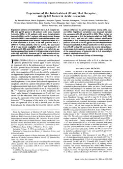
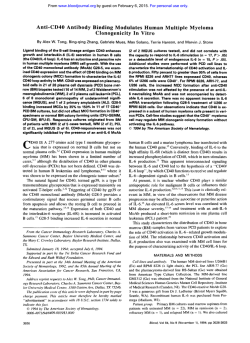
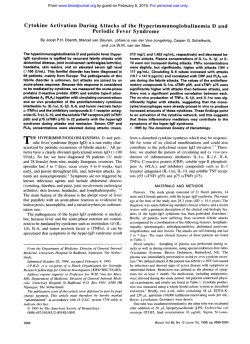
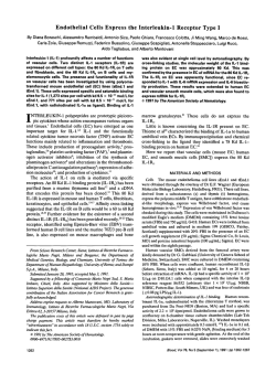
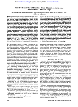
From www.bloodjournal.org by guest on February 6, 2015. For personal use only.
Characterization of an Interleukin-6-Mediated Autocrine Growth Loop in the
Human Multiple Myeloma Cell Line, U266
By Gisela Schwab, Clay E. Siegall, Lucien A. Aarden, Leonard M. Neckers, and Richard P. Nordan
It has been reported recently that freshly isolated human
myeloma cell cultures proliferate in response to added
interleukin-6 (IL-6). Endogenous levels of IL-6 found in the
same cultures suggested that an autocrine growth loop may
contribute to cell growth. However, the lack of homogenous
cell populations in primary myeloma cultures has made it
difficult to distinguish between paracrine and autocrine
growth mechanisms. To precisely address the autocrine
growth issue we have evaluated the growth of the human
myeloma cell line, U266. We have found that a neutralizing
anti-IL-6 monoclonal antibody can inhibit U266 proliferation.
Furthermore, the addition of IL-6 antisense oligonucleotides
also inhibits U266 proliferation.These effects are reversed by
adding IL-6, suggesting the presence of an autocrine loop.
Using bioassays with two different IL-Mependent cell lines,
we were able to detect IL-6 in concentrated U266 supernatants. IL-6 mRNA was detected by polymerase chain reaction
amplification of cDNA. Cell cycle parameter analysis shows
that IL-6 acts to release a block in G1. Taken together these
results present conclusive evidence for I L - h e d i a t e d autocrine growth in the U266 human myeloma cell line.
This is a US government work. There are no restrictions on is
use.
I
directed against human IL-2, was purchased from Genzyme
(Boston, MA). Plasmid pIL-6 was purchased from British Biotechnologies Limited (Oxford, UK) and carries a synthetic gene for
IL-6 inserted between the Hind111 and EcoRI sites in the polylinker of pUC 18.
Cell culture. U266pl (referred to as U266), a human myeloma
cell line, and T24, a human bladder carcinoma line, were obtained
from the American Type Culture Collection (Rockville, MD).
CHP100, a human neuroepithelioma line, was kindly provided by
Dr Angelo Rosolen (National Cancer Institute, Bethesda, MD).
B9, an IL-&dependent B-cell hybridoma, has been described
previously.I67TD1, another IL-&dependent B-cell hybridoma cell
line, was a kind gift from J. Van Snick.I7All cells were maintained
in complete medium that consisted of RPMI 1640, 10% heatinactivated fetal calf serum (FCS), 50 mmol/L 2-mercaptoethanol,
and 50 KgImL gentamycin. In addition, the IL-6-dependent B9 and
7TD1 cells were maintained in medium supplemented with rIL-6.
IL-6 bioassays. Culture supernatants were evaluated for IL-6
activity using the B9 and 7TD1 hybridoma growth factor assay^.'".'^
Briefly, 2,000 B9 or 7TD1 cells in a final volume of 200 pL of
complete medium (no IL-6) were cultured with serial dilutions of
the test sample. After 85 hours the B9 assays were pulsed with 0.5
pCi 'H-thymidine per well for 4 hours, harvested over glass fiber
filters, and counted in a liquid scintillation spectrophotometer. In
the 7TD1 assays the wells were pulsed with 20 KL of 3-4.5dimethylthiazol-2.5-diphenyltetrazoliumbromide (MTT) at a concentration of 5 mg/mL for 4 hours at 37"C, and then further
processed as previously described.'* Briefly, the cells were pelleted
in the wells and resuspended in 200 pL of dimethyl sulfoxide. The
samples were then read on an enzyme-linked immunosorbent assay
reader at 540-nm and 690-nm wavelengths. The absorbance values
in this assay closely correlated with the growth of the 7TDl cells. In
these assays one hybridoma growth factor (HGF) unit represents
NTERLEUKIN-6 (IL-6), also known as interferon p2,
B-cell stimulatory factor-2, or plasmacytomabybridoma growth factor, has multiple biologic activities. It has
been shown to induce B-cell maturation,'.' growth and
differentiation of T cell^^^ and has multi-colony-stimulating factor activity on hematopoietic progenitor cells: IL-6
has also been shown to induce the differentiation of
myeloid cells' and nerve cells,' and is responsible for the
production of acute-phase proteins in
In
addition to its activity on normal cells, IL-6 is necessary to
sustain the in vitro growth of many murine plasmacytomasll.'z
and has been shown to enhance the proliferation of
freshly explanted human myeloma cells." The hypothesis
that IL-6 can also support myeloma cell growth in vivo is
supported by the observation that myelomas arise in or are
found in environments that contain cells which produce
IL-6 in
Recently, Kawano et all3 found measurable concentrations of IL-6 in cultures of freshly explanted myeloma cells
as well as cell surface expression of the p80 chain of the
IL-6 receptor. They also showed that inhibition of the
endogenously produced IL-6 with polyclonal antibodies
could inhibit the proliferation of some freshly cultured
myelomas, and suggested that an autocrine mechanism may
account for the myeloma cell proliferation. However, in a
similar study, Klein et all4 were unable to verify the
autocrine hypothesis in human myeloma and assigned the
in vitro production of IL-6 to contaminating adherent cells
from the bone marrow rather than to the myeloma cells
themselves. Thus, according to Klein et al,I4human multiple myeloma growth is regulated in a paracrine rather than
an autocrine manner. Although paracrine growth remains a
likely mechanism for many myelomas, in this report we
present data on the human multiple myeloma cell line,
U266, that clearly show the existence of IL-&driven autocrine growth.
MATERIALS AND METHODS
Reagents. Moloney murine leukemia virus (M-MLV) reverse
transcriptase was obtained from Bethesda Research Laboratories
(Gaithersburg, MD). Taq DNA polymerase was purchased from
Perkin Elmer-Cetus (Norwalk, a).
Recombinant human IL-6
(rIL-6) was obtained from R&D Systems (Minneapolis, MN).
CLB.IL618 is a murine monoclonal antibody (MoAb) (IgG,, K)
directed against human IL-6.I5 DMS 1, a murine MoAb (IgG,, K)
Blood, Vol77, No 3 (February 1). 1991: pp 587-593
From the Tumor Cell Biology Section, Medicine Branch and
Laboratory of Molecular Biology, National Cancer Institute, National
Institutes of Health, Bethesda, MD; and the Central Laboratory of the
Red Cross Blood Transfusion Service, Amsterdam, The Netherlands.
Submitted March 23, 1990; accepted October 3, 1990.
Address reprint requests to Richard P. Nordan, PhD, Medicine
Branch, Bldg 10, Room 12N234, National Cancer Institute, NIH,
Bethesda, MD 20892.
The publication costs of this article were defrayed in part by page
charge payment. This article must therefore be hereby marked
"advertisement" in accordance with 18 U.S.C. section 1734 solely to
indicate this fact.
This is a USgovemment work. There are no restrictions on its use.
0006-4971I9117703-0015$0.0010
587
From www.bloodjournal.org by guest on February 6, 2015. For personal use only.
588
SCHWAB ET AL
the half maximal growth response of the IL-Mependent cells and
is equivalent to approximately 1pg/mL of native IL-6.
Cell cycle analysis. Test cells were harvested, washed in phosphate-buffered saline (PBS), and futed in 50% cold ethanol for 1
hour on ice. After pelleting, the cells were resuspended in RNase
A solution (580 U/mL PBS) and incubated for 30 minutes at 37°C.
Equal volumes of 0.005% propidium iodine were added. After a
20-minute incubation at room temperature, cell cycle analysis was
performed using a FACSstar flow cytometer (B-D Immunocytometry Systems, Mountain View, CA) as previously described.I9
Oligodeoynucleofidesfor in vitro assays. Fifteen-base sequences
from three different regions of the IL-6 cDNA’ were chosen for the
production of antisense, sense, or random oligodeoxynucleotide
(Fig 1). Random oligonucleotide sequences of the same base
composition as the antisense oligomers were used as controls.
Region I spanned the transcriptional start site. Region I1 was
downstream and immediately adjacent to region I, and region 111
corresponded to an area in the second exon of the IL-6 gene that
had previously been described as an effective antisense sequence in
reducing growth and IL-6 production in Kaposi sarcoma cells.”
The oligonucleotides were synthesized by cyanoethyl phosphoramidite methodology using an Applied Biosystems Model 380 B
DNAsynthesizer (Foster City, CA). An aliquot of each sample was
analyzed for homogeneity on a 15% polyacrylamide denaturing gel.
The oligomers were ethanol precipitated, washed with 70% ethanol, and subjected to three sequential cycles of sterile water
resuspension and lyophilization.
cDNA amplijicafion using the polymerase chain reaction (PCR).
Poly A selected RNA was prepared using the guanidine isothiocyanatekesium chloride method,21.” followed by poly-A-selection on
an oligo-dT column. The first strand of cDNA was synthesized
using random hexamers and M-MLV reverse transcriptase. 24mer
oligodeoxynucleotide primers which flanked 639 bases of the
mature IL-6 message spanning five exons and four introns in the
IL-6 gene were synthesized by the cyanoethyl phosphoroamidite
methodology. cDNA was amplified using taq polymerasez3 on a
Perkin Elmer-Cetus thermocycler for 30 cycles with cycles designed
as follows: denaturation at 94°C for 1minute, annealing at 42°C for
2 minutes, and polymerization at 72°C for 3 minutes with 10
seconds of extension time in each cycle. An aliquot of each sample
was analyzed on a 1% agarose gel. Amplified DNA could be
detected at the expected size by ethidium bromide staining of the
gel. Subsequently, the samples were transferred to nitrocellulose
and hybridized with a 3zP-labeled IL-6 probe (HindIII-EcoRI
insert of pIL-6) prepared by nick translation. The signal was
detected by autoradiography.
RESULTS
Production of IL-6 by U266 cells. In searching for
IL-6-mediated autocrine growth we initially asked if U266
myeloma cells produced biologically active IL-6. Conditioned medium was collected at various times from U266
cells growing at densities ranging from 0.2 to 1.5 x lo6
cells/mL and analyzed in the B9/HGF assay. IL-6 was not
detected in any of the neat supernatants. However, after a
20-fold concentration, IL-6 could be detected at concentrations of 5 to 20 HGF U/mL in bioassays using either of the
IL-&dependent cell lines, B9 or 7TD1. In the neat supernatants this corresponds to approximately 0.5 to 2 HGF
U/mL, which corresponds to 0.5 to 2 pg/mL of IL-6.
Specificity of the IL-&dependent support of B9 or 7TD1
cells was proven by inhibition of the supernatant-mediated
growth with CLB.IL6/8, a neutralizing anti-IL-6 MoAb
(Table 1).
Inhibition of U266 proliferation with anti-IL-6. To test
whether the endogenously produced IL-6 was acting as an
autocrine growth factor, the neutralizing MoAb CLB.IL6/8
was added directly to U266 cultures. As measured by
’H-thymidine incorporation, a 70% inhibition of U266
proliferation was obtained with 10 g / m L CLB.IL6/8 (Fig
2). Partial inhibition of U266 proliferation was observed
with antibody concentrations as low as 0.1 p.g/mL. This
effect was reversible by adding excess IL-6. The addition of
CLB.IL6/8 also prevented increases in cell numbers but did
not result in cell death, even after 5 days of incubation at 50
pg/mL (data not shown), suggesting that IL-6 is necessary
for U266 proliferation but not for the maintenance of
viability. In our hands 1 pg/mL of CLB.IL6/8 completely
neutralized 20 HGF U of IL-6 in the B9 assay. The
monoclonal murine antibody DMSl that was used as a
control did not affect ’H-thymidine uptake of U266 cells.
We also evaluated the effect of CLB.IL6/8 inhibition on
U266 cell cycle parameters. As shown in Fig 3, anti-IL-6treated U266 cells displayed a 50% reduction of cells in the
S phase of the cell cycle (P < .001). This was accompanied
by an accumulation of cells in GI (P < .Ol), indicating that
U266 cells encounter a G, block in the absence of IL-6.
Detection of IL-6 message in U266 cells. We attempted
to evaluate the level of IL-6 mRNA using Northern blot
and RNase protection assays. Although others have reported the presence of IL-6 message in U266 cell^,^'^^^ we
were unable to detect IL-6 message in poly A-selected
U266 RNA (data not shown) using these methods. PCR
amplification of cDNA, a more sensitive assay for the
detection of specific mRNAs, was performed. First-strand
IL6 messenger RNA
ATG
Region:
Antisense:
I
GGAGlTCATAGCTGG
Sense:
CCAGCTATGAACTCC
Random:
TGTCGGAGTCGTGAA
II
111
CTTACTTGTGGAGAA
TCCTGGGGGTACTGG
ND
TCTCATGTTAGGAGA
ND
CTCGTGTGGACGGGT
Fig 1. Schematic illustration of location and sequence of the oligodeoxynucleotides used.
From www.bloodjournal.org by guest on February 6, 2015. For personal use only.
589
IL-MEDIATED AUTOCRINE GROWTH IN U266 MYELOMA
Table 1. EndogenousProduction of 11-6 by U266
Sample
69 assay
Expt 1
Expt 2
7TDl assay Expt 1
Expt 2
Expt 3
Expt 4
U266
Medium
RPMl
1640 +
10% FCS
u266
Medium +
Anti-lL-6
2
1
0.5
0.5
0.5
0.25
0
0
0
0
0
0
ND
0
ND
0
0
ND
Values are HGF units per milliliter. Productionof IL-6by U266 cells. 69
or 7TDl cells were cultured in triplicate with serial dilutions of 20-fold
concentrated U266 conditioned medium in presence or absence of 10
pg/mL of the monoclonal anti-IL-6 antibody CLB.IL6/8. Twentyfold
concentrated RPMl 1640 with 10% FCS served as control. Results were
corrected to reflect the IL-6activity present in the neat supernatant.
Abbreviations: Expt, experiment; ND, not done.
cDNAs were synthesized from 1 pg of poly A-selected
RNA using reverse transcriptase. Specific primers corresponding to the 5' and 3' ends of the mature IL-6 message
were used in the PCR reaction to amplify the IL-6 cDNA.
This reaction produced the 639-bp fragment predicted for
the IL-6 cDNA using these primers (Fig 4). The same
fragment was generated using T24 bladder carcinoma cells,
which have been shown to express IL-6 message,= whereas
no detectable fragment was generated using CHP100, a
neuroepithelioma.
Effect of treatment with IL-6 antisense oligodeoxynucleotides. To establish whether IL-6 antisense oligodeoxynucleotides can be used to inhibit the growth of autocrine
myeloma cells, we cultured U266 with an antisense IL-6
oligomer that spanned the transcriptional start site of the
IL-6 message (Fig 1, region I). After 48 hours the growth of
mm
0
c
n
0)
0
%
0
-m
C
0
0
c
0
L.
C
0
2
0
n
Cell Cycle Phase
Fig 3. Effect of anti-lL-6 on U266 cell cycle parameters. U266 cells
were cuttured for 2 days with CLB.IL6/8 (monoclonal anti-hll-6) and
then analyzed for DNA content using a flow cytometer. Data represent the mean and standard error of three experiments. (m), Control;
anti-lL-6.1 pg; (0).
anti-ll-6.10 pg.
(a),
639 Base Pairs
Fig 2. Inhibition of U266 proliferation with anti-ll-6 MoAb. U266
cells were cultured at 5 x l0'lmL in triplicate with 10,OOO HGF U of
11-6 or the indicated concentrations of CLB.IL6/8, a neutralizing
monoclonal anti-lL-6 antibody. On days 1through 5 the cultures were
pulsed with 3H-thymldineand harvested. Data represent one of four
similar experiments. Monoclonal anti-IL-2 antibody of the same
10 pg/mL
isotype (DMS1) sewed as a control. (-m-), Control; (.-.O.-.),
anti-116; I-.-),
10 pg/mL anti-ll-2; (.-.A,-.), 10 pg/mL anti-ll-6 +
IL-6.
4
Fig 4. Detection of lL-6 mRNA First-strand cDNA generated from
poly A selected RNA was amplified by PCR using 24mer primers
corresponding to amino acids 1through 8 and 206 through 213 of the
mature IL-6 protein. PCR products were electrophoresed on a 1%
agarose gel, transferred to nitrocellulose, and hybridized with a
UP-labeled11-6 probe. Lane 1, control cDNA; lane 2, CHP100; lane 3,
U266; lane 4, T24.
From www.bloodjournal.org by guest on February 6, 2015. For personal use only.
SCHWAB ET AL
590
antisense-treated cells was diminished by 40% when compared with sense-treated or control cells (P < .01) (Fig 5).
Analysis of cell cycle parameters showed that treatment
with antisense oligodeoxynucleotidesalso resulted in a 40%
reduction of cells in S phase (P < .05), whereas sense
treatment had no effect (P = .63) (Fig 5). The antisense
effect on cell growth and cell cycle parameters could be
reversed by adding IL-6 to the cultures. Similar results were
obtained in experiments in which the antisense oligomer
was added every day for 6 days (data not shown). As with
the addition of anti-IL-6, no loss of cell viability was
observed. In titration studies of IL-6 antisense oligodeoxynucleotides we saw growth inhibition at concentrations as
low as 10 pmol/L oligomer (data not shown). A 15mer of
random configuration, but the same base composition as
the antisense oligomer to the transcriptional start region,
also had no effect on cell growth or distribution of the cells
in the cell cycle. We further evaluated the effect of
antisense oligonucleotides directed to other regions of the
IL-6 cDNA (Fig 6). An antisense 15mer directed against a
region of the second exon of the IL-6 gene (Fig 1, region
111) exhibited similar effects on the distribution of cells in S
phase and GI phase to those obtained with antisense
oligonucleotide against the transcriptional start site of the
IL-6 gene (region I). After treatment with this oligomer,
U266 cells in S phase decreased by 45% (P < .001) and
cells in G, increased by 45% as compared with untreated
U266 cells (P < .001). The corresponding random base
oligomer did not affect either parameter significantly. An
antisense oligonucleotide directed to the region immediately adjacent to and downstream from the transcriptional
start site oligomer (Fig 1, region 11) did not exert any effect
on cell growth or cell cycle distribution of U266 cells.
DISCUSSION
The ability of IL-6 to serve as an in vitro growth factor for
myelomas has been well documented. Both murine and
human iL-Mependent myeloma cell lines have been
established in vitro,".11z6and studies with freshly explanted
human myeloma cells have shown that IL-6 enhances, in a
4
Cell
Count
Percenl in S Phase
Fig 5. Inhibition of U266 with 11-6 antisense oligodeoxynucleotides. U266 cells were cultured at lO*/mL. Cell counts and percent of
cells in S phase of the cell cycle were determined at 2 days after
treatment with 100 pmol/L sense or antisense IL-6 oligodeoxynucleotides at 0 and 24 hours. Where indicated, rlL-6 was added at a
concentration of 10.000 HGF UlmL. Data represent the mean and
standard error of four experiments. (m), Control; (B), sense; (0).
antisense; (0).
antisense + IL-6.
paracrine manner, the in vitro proliferation of some tumor~.".'~
The issue of autocrine growth in myeloma is less
clear. Using freshly explanted myeloma cells, Kawano et all3
detected exogenous IL-6 production and mRNA expression
and could inhibit the proliferation of some myelomas with
anti-IL-6 antibodies. In a similar study Klein et all4were
unable to confirm the autocrine hypothesis and attributed
the endogenous in vitro IL-6 expression to the presence of
contaminating cells. The concept of an autocrine mechanism in tumor development is supported by the studies of
Freeman et al? who found IL-6 message expressed in some
B-cell lymphomas, but because of the presence of contaminating cells were unable to determine if freshly isolated
myeloma cells expressed IL-6 message. In our studies we
avoid the issue of contaminating cells by analyzing the
human myeloma cell line, U266. In this report we demonstrate the existence of an IL-&mediated autocrine growth
loop in U266. Evidence of an autocrine growth loop is
provided by the specific inhibition of U266 growth by the
monoclonal anti-IL-6 antibody, CLB.IL6/8. When U266 is
inhibited with anti-IL-6 the cells accumulate in GI and the
proportion of cells in S phase is reduced by 40% to 50%,
indicating that these cells encounter a GI block in the
absence of IL-6. This observation agrees with experiments
in which IL-6 has also been shown to relieve a GIblock
in IL-Mependent murine plasmacytomas'' and B-cell hybridoma~.~'
It is interesting that unlike the IL-64ependent
murine plasmacytomas and hybridomas, which die in the
absence of IL-6, the inhibition of U266 does not lead to cell
death even with a 100-fold excess of antibody. This suggests
either that IL-6 is not required to maintain the viability of
U266 or that IL-6 can interact with its receptor in regions
that cannot be reached by the antibody, perhaps internally.
In either case, if the inhibition of IL-6 does not result in the
death of autocrine myeloma cells, the potential therapeutic
effectiveness of such reagents against in vivo autocrine
tumors will be reduced.
The production of IL-6 by U266 was documented using
two different highly sensitive IL-Mependent hybridoma
cell lines, and this response was specifically inhibited with
the monoclonal anti-IL-6 antibody. The accumulated IL-6
reached levels of about 0.5 pg/mL to 2 pg/mL, and could be
detected only after concentration of the U266 conditioned
medium. These exceedingly low levels of IL-6 would be
unable to support most IL-6 responses and raise questions
about the level of sensitivity of the U266 myeloma to IL-6.
Most cells that exhibit a response to IL-6, eg, IL-6dependent murine plasmacytomas, require levels of native
IL-6 that are 100-fold higher than levels required by B-cell
hybridomas, such as the B9 and 7TD1 cell lines used in this
study (half maximum = 100 v 1 pg/mL). The low levels of
IL-6 found in U266 medium might imply that, like B9 and
7TD1, the stimulation of U266 growth occurs at very low
concentrations (1 pg/mL) of IL-6. Alternatively, U266 may
be similar in sensitivity to IL-6-dependent murine plasmacytomas (> 100 pg/mL) and may produce a concentration
gradient of secreted IL-6 in the immediate vicinity that is
high enough to meet this requirement. It can also be argued
From www.bloodjournal.org by guest on February 6, 2015. For personal use only.
591
IL-6-MEDIATED AUTOCRINE GROWTH IN U266 MYELOMA
W Control
Fig 6. Comparison of the effect of three different antisense
IL-6 oligodeoxynucleotides and
the corresponding random base
sequences on the distribution of
U266 cells in the cell cycle. Cells
were cultured and processed as
described in the legend to Fig 5.
Regions 1,2, and 3 refer to oligonucleotide sequences described
in the Fig 1 legend and in the
text. Data represent one of two
similar experiments performedin
quadruplicate. An asterisk (*)
above a column indicates a statistically significant response
when compared with the control
(P < ,001):
0
Region1 Random
Region 1 Antisense
Region2 Random
[7 Region 2: Antisense
Region 3: Random
Region 3: Antisense
60
40
20
0
% Gl
% S
receptors on U266 cells, and we have repeated this observathat U266 rapidly internalizes the secreted IL-6, thus
depleting it from the medium and complicating the detion for the subline used in this
In addition, the
tection of this molecule. Our results do not allow us to
expression of IL-6 message in U266 has been reported by
distinguish between these possibilities.
Kawano et all3 and Freeman et alZ4;however, Klein et all4
Antisense oligodeoxynucleotides to p r o t o - o n ~ o g e n e s ~ ~did
~ ~ not detect IL-6 message in their studies of U266.
and growth factor^^^^^^ have previously been used to inhibit
Likewise, Kawano et all3detected a low level of IL-6 activity
cell growth. In our studies a specific inhibition of U266 cell
produced by U266, whereas Klein et a1 found no IL-6
growth is seen after treatment with an antisense oligomer to
produced by U266. As suggested by Klein et al,I4 these
the transcriptional start site of the IL-6 cDNA, whereas the
discrepancies are possibly due to the use of different U266
sense and random sequence oligomers do not affect growth.
sublines.
Cell cycle analysis of antisense-treated U266 cells yields
The presence of IL-6 mRNA is a conditio sine qua non of
essentially identical results to treatment with anti-IL-6
IL-6
antisense oligomer action. In our hands Northern blot
antibody, ie, reduction of cells in S phase and accumulation
analysis
was not sensitive enough to detect IL-6 mRNA in
of cells in GI. Recently, an antisense IL-6 15mer against a
U266
cells.
We find that IL-6 mRNA can be detected in
region in the second exon of the IL-6 gene has been shown
U266 cells, but only after amplification of the reverse
to inhibit IL-6 production and growth of Kaposi sarcoma
transcribed mRNA by PCR. The need to use the PCR
cells.20Using this same antisense oligonucleotide on U266
technique indicates that the level of IL-6 message is
also yielded the same growth inhibition and cell cycle
exceedingly low and correlates with the low levels of IL-6
effects obtained with the transcriptional start site sequence.
The inhibition of U266 with two distinct IL-6 antisense
detected in the conditioned medium. Thus, our results
sequences and their reversal with exogenous IL-6 confirms
indicate that vanishingly low amounts of IL-6 are sufficient
the specificity of the antisense effect on the IL-6 message.
for allowing autocrine growth of this U266 subline. FurtherThe failure of a third sequence to inhibit is not surprising
more, these studies show that the failure to detect message
because the most effective region for antisense inhibition
in Northern blots should not be taken as evidence for the
spans the transcriptional start site,32whereas inhibition in
absence of an autocrine mechanism. Taking all results
other regions of the mRNA is unpredictable. IL-6 antisense
together, we conclude that in the U266 cell line there is
oligomers presumably exert their effect by annealing with
clear evidence of an IL-&mediated autocrine growth loop
the IL-6 mRNA in the region of complementarity, thus
in which IL-6 acts to relieve a GI block in the cell cycle.
producing a DNNRNA hybrid. At least two potential
Monoclonal anti-IL-6 antibody or antisense IL-6 oligodemechanisms exist that may mediate the inhibitory effect. In
oxynucleotides interrupt this loop at different stages and
the first, the hybrid becomes a substrate for RNAse H and
may be useful for further understanding the role of this
is degraded, allowing the oligomer to hybridize to another
cytokine in human myeloma. Interestingly, in the sera of
mRNA m ~ l e c u l e . ~In~ .the
~ ~ second, if the antisense
patients
with advanced disease of multiple myeloma inoligomer is complementary to the initiation codon, the
of IL-6 have been described, whereas in early
creased
levels
attachment of the ribosome is inhibited and translation is
stages IL-6 is usually not detected." It is possible that, in
arrested."
some tumors, high serum levels of IL-6 might reflect a
Other investigators have conducted studies that address
progression from paracrine to autocrine growth in myeloma
various aspects of IL-6 and U266 growth; however, none
have specifically demonstrated an IL-&mediated autocrine
cells. U266 might represent a window in an advanced stage
loop. Taga et aP8 initially described the expression of IL-6
of the disease.
From www.bloodjournal.org by guest on February 6, 2015. For personal use only.
592
SCHWAB ET AL
REFERENCES
1. Hirano T, Yasukawa K, Harada H, Taga T, Watanabe Y,
Matsuda T, Kashiwamura S, Najajima K, Koyama K, Iwamutsa A,
Tsurasawa S, Sakiyama F, Matsui H, Takahara Y, Taniguchi T,
Kishimoto T Complementary DNA for a novel human interleukin
(BSF-2) that induces B lymphocytes to produce immunoglobulin.
Nature 324:73,1986
2. Kishimoto T T cell stimulatory factors (BSFs). Molecular
structure, biological function and regulation of expression. J Clin
Immunol7:343,1987
3. Vink A, Coulie PG, Wauters P, Nordan RP, VanSnick J: B
cell growth and differentiation activity of interleukin HPI and
related plasmacytoma growth factors. Synergy with IL1. Eur J
Immunol18:607,1988
4. Uyttenhove C, Coulic PG, VanSnick J: T cell growth and
differentiation induced by interleukin HPI/IL6, the murine hybridoma/plasmacytoma growth factor. J Exp Med 167:1417,1988
5. Takai Y, Wong GG, Clard SC, Burehoff SJ, Hemnann SH: B
cell stimulatory factor 2 is involved in the differentiation of
cytotoxic T lymphocytes. J Immunol140:508,1988
6. Igebuchi K, Wong GG, Clark SC, Ihle JN, Hirai Y, Ogawa M:
Interleukin 6 enhancement of interleukin 3 dependent proliferation of multipotential hematopoietic progenitors. Proc Natl Acad
Sci USA 84:9035,1987
7. Shabo Y, Lotem J, Sachs L: Autoregulation of interleukin 6
and granulocyte-macrophage colony stimulating factor in the
differentiation of myeloid leukemic cells. Mol Cell Biol 9:4109,
1989
8. Satoh T, Nakamura S, Taga T, Mabuda T, Hirano T,
Kishimoto T, Kaziro T Induction of neural differentiation in PC12
cells by the B cell stimulatory factor 2/interleukin 6. Mol Cell Biol
8:3546, 1988
9. Gauldie J, Richards C, Harnish D, Landorp D, Baumann H:
Interferon p2/B cell stimulatory factor type 2 shares identity with
monocyte-derived hepatocyte-stimulating factor and regulates the
major acute phase protein response in liver cells. Proc Natl Acad
Sci USA 84:7251,1987
10. Andus T, Geiger T, Hirano T, Northoff H, Ganter U, Bauer
J, Kishimoto T, Heinrich PC: Recombinant human B cell stimulatory factor (BSF-2/IFNP,) regulates P-fibrinogen and albumin
mRNA levels in. FEBS Lett 221:18,1987
11. Nordan RP, Potter M: A macrophage-derived growth factor
required by plasmacytomas for survival and proliferation in vitro.
Science 233:566,1986
12. Van Snick J, Vink A, Cayphas S, Uyttenhove C InterleukinHP-1, a T cell-derived hybridoma growth factor that supports the
in vitro growth of murine plasmacytomas. J Exp Med 165:641, 1987
13. Kawano M, Hirano T, Makuda T, Taga T, Horii Y, Iwato K,
Asaoku H, Tang B, Tanabe 0,Tanaka H, Kuramoto A, Kishimoto
T Autocrine generation and requirement of BSF-2/IL-6 for human
multiple myelomas. Nature 332233,1988
14. Klein B, Zhang X-G, Jourdan M, Content J, Houssiau F,
Aarden L, Piechaczyk M, Bataille R: Paracrine rather than
autocrine regulation of myeloma-cell growth and differentiation by
interleukin-6. Blood 73:517,1989
15. Brakenhoff JPJ, Hart M, DeGrot ER, Di Padova F, Aarden
LA: Structure-function analysis of human IL-6: Epitope mapping
of neutralizing monoclonal antibodies with amino- and carboxylterminal deletion mutants. J Immunol145:561,1990
16. Aarden LA, deGroot ER, Schaop OL, Lansdorp PM:
Production of hybridoma growth factor by human monocytes. Eur J
Immunol17:1411,1987
17. VanSnick J, Cayphas S, Vink A, Uyttenhove C, Coulie PG,
Rubira MR, Simpson RJ: Purification and NH,-terminal amino
acid sequence of a T-cell derived lymphokine with growth factor
activity for B-cell hybridomas. Proc Natl Acad Sci USA 83:9679,
1986
18. Twentyman PR, Fox NE, Rees JKH: Chemosensitivity
testing of fresh leukemia cells using the MTT colorimetric assay. Br
J Hematol71:19, 1989
19. Neckers LM, Nordan R P Regulation of murine plasmacytoma transferrin receptor expression and G1 traversal by plasmacytoma cell growth factor. J Cell Physiol135:495, 1988
20. Miles SA, Rezai AR, Salazar-Gonzalez JF, VanDer Meyden
M, Stevens RH, Logan DM, Mitsuyashu RT, Taga T, Hirano T,
Kishimoto T, Martinez-Maza 0: AIDS Kaposi sarcoma-derived
cells produce and respond to interleukin 6. Proc Natl Acad Sci
USA 874068,1990
21. Chirgwin JM, Przybala AE, MacDonald RJ, Rutter WJ:
Isolation of biologically active ribonucleic acid from sources
enriched in ribonuclease. Biochemistry 18:5294,1979
22. Aviv H, Leder P: Purification of biologically active globin
messenger RNA by chromatography on oligothymidylic acidcellulose. Proc Natl Acad Sci USA 69:1408,1972
23. Saki R, Gelfand DH, Stoffel S, Scharf SJ, Higuchi R, Horn
GT, Mullis KB, Ehrlich HA:Primer-directed enzymatic amplification of DNA with a thermostable DNA polymerase. Science
239:487, 1988
24. Freeman GJ, Freedman AS, Rabinowe SN, Segil JM,
Horowitz J, Rosen K, Whitman JF, Nadler LM: Interleukin 6 gene
expression in normal and neoplastic B cells. J Clin Invest 83:1512,
1989
25. Tanabe 0, Akira S, Kamiya T, Wong GG, Hirano T,
Kishimoto T: Genomic structure of the murine IL-6 gene. High
degree conservation of potential regulatory sequences between
mouse and human. J Immunol141:3875,1988
26. Shimizu S, Yoshioka R, Hirose Y, Sugai S, Tachibana J,
Konda S: Establishment of two interleukin 6 (B cell stimulatory
factor 2hnterferon P2)-dependent human bone marrow-derived
myeloma cell lines. J Exp Med 169:339, 1989
27. Landsdorp PM, Aarden LA, Calafat J, Zeiljemaker WP: A
growth factor dependent B cell hybridoma. Curr Top Immunol
Microbiol132:105,1986
28. Heikkila R, Schwab G, Wickstrom E, Loke SL, Pluznik DH,
Watt R, Neckers LM: A c-myc antisense oligodeoxynucleotide
inhibits entry into S phase but not progress from Go to G,. Nature
328:445,1987
29. Gewirtz AM, Calabretta B: A c-myb antisense oligodeoxynucleotide inhibits human hematopoiesis in vitro. Science 242:1303,
1988
30. Schaller HC, Druffel-Augustin S, Duebel S: Head activator
acts as autocrine growth factor for NH15-CA2 cells in the G2/
mitosis transition. EMBO J 8:3311, 1989
31. Becker D, Meier CB, Herlyn M: Proliferation of human
malignant melanomas is inhibited by antisense oligodeoxynucleotides targeted against basic fibroblast growth factor. EMBO J
8:3685,1989
32. Goodchild J: Inhibition of gene expression by oligonucleotides, in Cohen J (ed): Oligodeoxynucleotides. Antisense Inhibitors of Gene expression. Topics in Molecular and Structural
Biology. London, UK, MacMillan, 1989, p 53
33. Minshull J, Hunt T: The use of single-stranded DNA and
RNAse H to promote quantitative “hybrid arrest of translation” of
mRNA/DNA hybrids in reticulocyte lysate cell free translation.
Nucleic Acids Res 14:6433, 1986
From www.bloodjournal.org by guest on February 6, 2015. For personal use only.
IL-&MEDIATED AUTOCRINE GROWTH IN U266 MYELOMA
34. Dash P, Lotan J, Knapp M,Kandel ER, Goelet P: Selective
elimination of mRNAs in vitro: Complementaryoligodeoxynucleotides promote RNA degradation by an RNAse H like activity.
Proc Natl Acad Sci USA 84:7896,1987
35. Cazanove C, Loreau N, Thuong N-T, Toulme JJ, Helene C:
Enzymatic amplificationof translation inhibition of rabbit P-globin
mRNA mediated by antimessenger oligodeoxynucleotides covalently linked to intercalating agents. Nucleic Acids Res 15:4717,
1987
36. Walder RY, Walder J A Role of RNAse H in hybrid
arrested antisense oligonucleotides. Proc Natl Acad Sci USA
85:5011,1988
37. Kawasaki ES: Quantitative hybridization-arrestof mRNA in
593
xenopous oocytes using single-stranded complementary DNA or
oligonucleotides. Nucleic Acids Res 13:4991, 1985
38. Taga T, Kawanishi K, Hardy RR, Hirano T, Kishimoto T
Receptors for B cell stimulatory factor 2 (BSF-2): Quantitation,
specificity, distribution and regulation of the expression. J Ekp
Med 166:967,1987
39. Siegall CB, Nordan RP, FitzGerald DJ, Pastan I: Cellspecific toxicity of a chimeric protein composed of interleukin 6
and pseudomonas exotoxin (IL6-PE40) on tumor cells. Mol Cell
Biol10:2443,1990
40. Bataille R, Jourdan M,Zhang X-G, Klein B: Serum levels of
interleukin 6, a potent myeloma cell growth factor, as a reflect of
disease severity in plasma cell dyscrasia.J Clin Invest 84:2008,1989
From www.bloodjournal.org by guest on February 6, 2015. For personal use only.
1991 77: 587-593
Characterization of an interleukin-6-mediated autocrine growth loop in
the human multiple myeloma cell line, U266
G Schwab, CB Siegall, LA Aarden, LM Neckers and RP Nordan
Updated information and services can be found at:
http://www.bloodjournal.org/content/77/3/587.full.html
Articles on similar topics can be found in the following Blood collections
Information about reproducing this article in parts or in its entirety may be found online at:
http://www.bloodjournal.org/site/misc/rights.xhtml#repub_requests
Information about ordering reprints may be found online at:
http://www.bloodjournal.org/site/misc/rights.xhtml#reprints
Information about subscriptions and ASH membership may be found online at:
http://www.bloodjournal.org/site/subscriptions/index.xhtml
Blood (print ISSN 0006-4971, online ISSN 1528-0020), is published weekly by the American
Society of Hematology, 2021 L St, NW, Suite 900, Washington DC 20036.
Copyright 2011 by The American Society of Hematology; all rights reserved.


© Copyright 2026