
Adrenergic Modulation of Human Polymorphonuclear
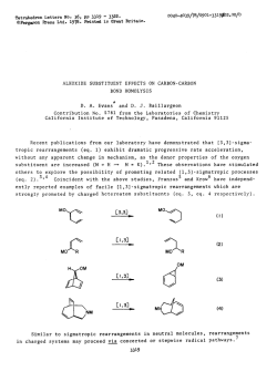
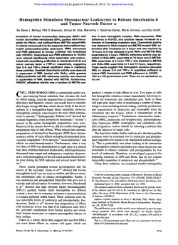
From www.bloodjournal.org by guest on February 6, 2015. For personal use only.
Adrenergic Modulation of Human Polymorphonuclear Leukocyte Activation.
Potentiating Effect of Adenosine
By Gianfranco Bazzoni, Elisabetta Dejana, and Aldo Del Maschio
The activation of polymorphonuclear leukocytes (PMN) is an
important step in the development of tissue damage associated with inflammatory and ischemic conditions. Catecholamines have been reported t o inhibit PMN functions, but the
high concentrations required cast doubt on their actual
relevance as a defense mechanism. We report here that
adenosine, which is actively released in ischemic conditions,
potentiates the effect of epinephrine and reduces the minimal active concentration required t o inhibit PMN activation
by at least two orders of magnitude. Epinephrine caused a
dose-related reduction of chemiluminescence, superoxide
anion generation, enzyme release (lysozyme and p-glucuronidase), and adhesion t o endothelial cell (EC)monolayers in
human PMN activated by N-formyl-methionyl-leucyl-phenylto
alanine (fMLP). This effect was only apparent at
mol/L. As expected, adenosine caused dose-dependent reductions of superoxide anion production and PMN adhesion t o
EC. Adenosine and epinephrine combined had an additive
effect on PMN superoxide production and adhesion t o EC.
The minimal effective concentration of epinephrine in combination with I O - * mol/L adenosine was in the range of lo-’’ t o
IO-’ mol/L. In contrast, adenosine inhibited only slightly
enzyme release and did not significantly enhance the inhibition by epinephrine on this parameter. Studies with adenosine analogs suggested that the potentiating effect of adenosine was mediated by A, receptors. The mechanism of
potentiation was not related t o additive effect on intracellular cyclic adenosine monophosphate levels. Epinephrine‘s
ability to modulate PMN activation and the potentiating
effect of adenosine may constitute a form of physiologic
protection against tissue injury in inflammatory and ischemic
processes.
o 1991 b y The American Society of Hematology.
R
sion to endothelial cells (EC).l3.l4Therefore, it was of
interest to investigate whether adenosine potentiated the
inhibitory effect of epinephrine on PMN responsiveness.
The results reported here show that at high concentrations epinephrine inhibited the PMN respiratory burst,
enzyme release, and adhesion to EC. Adenosine, at concentrations equal to or lower than those in blood, markedly
potentiated the inhibitory effect of epinephrine on PMN
activation bringing the active concentration of epinephrine
down to within the physiologic range.
These data support the hypothesis that distinct endogenous mediators released during tissue ischemia may cooperate to constitute a more effective defense mechanism
against harmful PMN activation.
ELEASE OF TOXIC oxygen free radicals and proteolytic enzymes by activated polymorphonuclear leukocytes (PMN) accounts for much of the tissue damage
described in inflammatory reactions’ and in ischemic conditions like myocardial infarction and “ischemia-reperfusion
Biochemical modifications that can interfere with
PMN functions might consequently condition the pathologic course of these diseases. The heightened sympathetic
activity that parallels the early phases of acute myocardial
infarction4 and results in elevated catecholamine levels in
blood’ might limit the effects of excessive PMN activation in
infarcted areas. Indeed, PMN have &-adrenergic receptors6whose stimulation by isoprenaline results in an adenyl
cyclase-mediated inhibitory effect on PMN activation.’.’”
However, very high doses of a selective synthetic agonist,
such as isoprenaline, needed to produce appreciable in
vitro inhibition raise some doubt on the physiologic relevance of the catecholamines’ effect on PMN.”
Little is known about whether non-adrenergic agents,
endogenously released in conditions of acute vascular
occlusion, influence the adrenergic control of leukocyte
activation. Among the biochemical responses to tissue
ischemia, the release of adenosine is one of the most
important.’* Adenosine can act as a physiologic defense
mechanism by inhibiting the PMN oxygen burst and adhe-
From the Laboratory of Vascular Biology, Istituto di Ricerche
Farmacologiche, “Mario Negri; ’’Milano, Italy.
Submitted June 26,1990; accepted January 4, 1991.
Supported by the Italian National Research Council (Project CNR
No. 89.01277.04).
Address reprint requests to Gianfranco Bazzoni, MD, Laboratory of
VascularBiology, Istituto di Ricerche Farmacologiche, “Mario Negn”’
E a Eritrea 62, 20157Milan0, Italy.
The publication costs of this article were defrayed in part by page
charge payment. This article must therefore be hereby marked
“advertisement” in accordance with 18 U.S.C. section 1734 solely to
indicate this fact.
0 1991 by The American Society of Hematology.
0006-4971191J7709-0023$3.OOlO
2042
MATERIALS AND METHODS
Chemicals and stimuli. The suppliers for chemicals were Sigma
Chemical Co (St Louis, MO) for N-formyl-methionyl-leucylphenylalanine (NLP), zymosan A (from Saccharomyces cerevisiae), epinephrine, isoprenaline, adenosine, NECA, CPA, phenylephrine, cytochalasin B, phenolphthalein standard solution,
phenolphthalein glucuronic acid, xanthine, xanthine oxidase, horseradish peroxidase, cytochrome c type 111, 4-(2-hydroxyethyl)-lpiperazine-ethane sulfonic acid (HEPES), and 5-amino-2,3-dihydro1,4-phthalazinedione (Luminol); Imperial Chemical Industries
(Macclesfield, England) for propranolol; Nycomed AS (Oslo,
Norway) for Lymphoprep; Boehringer Mannheim (Mannheim,
Germany) for Micrococcus luteus, lysozyme, and superoxide dismutase (SOD); Pharmacia Fine Chemical (Uppsala, Sweden) for
Dextran T 500; Kabi Vitrum (Stockholm, Sweden) for human
fibrinogen; Amersham (Buckinghamshire, England) for Na;’CrO,
and the cyclic adenosine monophosphate (CAMP)radioimmunoassay kit. Ro 20-1724 was a generous gift of Prodotti Roche (Milano,
Italy). All reagents for EC culture were purchased from GIBCOEurope (Paisley, Scotland). Tissue culture plates and flasks were
obtained from Cell Cult, Flow Laboratories (Milano, Italy). All
other materials were pure reagent grade.
fMLP was dissolved in dimethyl sulfoxide (DMSO) to a final
concentration of 50 mmol/L, stored at -20°C and diluted in
isotonic saline just before use; zymosan and opsonized zymosan
particles were prepared as previously described.15Epinephrine and
isoprenaline (the L -isomers of the bitartrate salt), propranolol (the
Blood, Vol77, No 9 (May 1). 1991: pp 2042-2048
From www.bloodjournal.org by guest on February 6, 2015. For personal use only.
2043
EPINEPHRINE,ADENOSINE, AND LEUKOCYTE FUNCTIONS
D,L-isomer of the hydrochloride salt), and adenosine were dissolved in isotonic saline as stock solutions (10 mmol/L) just before
use. Luminol was dissolved as stock solution (90 mmol/L) in
DMSO, further diluted in isotonic saline and stored at 4°C
protected from light. The final DMSO concentration never exceeded 0.001%.
PMNisolation. Venous blood from healthy donors who had not
received any medication for at least 2 weeks was anticoagulated
with trisodium citrate (3.8%, 1:9 vol/vol). PMN were isolated by
Dextran sedimentation followed by Lymphoprep gradient and
hypotonic lysis of erythrocytes.16PMN were washed and resuspended (5 x lo6 cells/mL) in ice-cold HEPES-Tyrode buffer (pH
7.4) containing (mmol/L): 129 NaC1, 9.9 NaHCO,, 2.8 KCI, 0.8
KH,PO,, 0.8 MgCI, . 6H,O, 5.6 dextrose, 1 CaCl,, and 10 HEPES.
Cell suspensions contained more than 97% viable PMN, as
evaluated by the Trypan blue exclusion test. PMN were used within
2 hours after their isolation.
Chemiluminescenceassay. PMN suspensions (5 x lo6cells/mL,
in a final volume of 1 mL), exposed to 90 nmol/L luminol and 2.5
pg/mL cytochalasin B, were preincubated at 37°C for the time
indicated under constant stirring, in the Chrono-Log lumiaggregometer (Havertown, PA), in the presence or absence of the
adrenergic agents. Changes in chemiluminescence (CL) were
recorded for 7 minutes after fMLP or opsonized zymosan stimulation.17 CL values reported in the present work are expressed in
millivolts and correspond to the maximal amplitude of the signal
recorded. Peak amplitude was measured in millimeters and transformed into millivolts, the maximal tracing excursion (160 mm)
corresponding to 1,600 mV. CL increased linearly with the concentration of the stimulus and the number of cells (ED,,: 68 f 8,
460 f 30, and > 1,000 x
M fMLP, for 5, 2.5, and 1.25 x lo6
PMN/mL, respectively). Basal CL was always negligible.
Control experiments showed that CL generated in a cell-free
system by xanthine
mol/L) plus xanthine oxidase (1 U/mL), in
the presence of horseradish peroxidase (9 U/mL), was not reduced
by epinephrine (483 f 26 mV and 494 f 35 mV, respectively, in
the presence and absence of
mol/L epinephrine (n = 3) or by
any of the other compounds used, thus excluding that light
quenching accounted for the inhibitory effect described.
Superoxide union generation. Superoxide anion generation was
determined by measuring the reduction of cytochrome c, as
described.'* PMN suspensions (5 x lo6 cells/mL) were preincubated at 22°C for 3 minutes with cytochrome c
mol/L) in the
presence or absence of epinephrine, stimulated with fMLP
mol/L), and then incubated for different times (10 to 60 minutes).
Cell suspensions were centrifuged (12,OOOg for 1 minute) and
aliquots of the supernatants were transferred into a 96-well
microtiter plate. Absorbance was measured simultaneously at 550
and 540 nm with a Multiskan spectrophotometer (Titertek, Flow
Laboratories, Great Britain). In some experiments, PMN were
preincubated (10 minutes at 22°C) with adenosine (lo-'' to
mol/L) or with the adenosine analogs NECA or CPA (10-lo to
mol/L) before exposure to epinephrine. In the presence of every
compound tested in this study, SOD (50 U/mL) almost completely
abolished cytochrome c reduction in both resting and fMLPstimulated PMN (eg, 0.8 f 0.2 v 15.7 f 0.5 nmol reduced, respectively, in the presence or absence of SOD).
Adhesion of PMN to EC. EC isolated from bovine thoracic
aorta were cultured in minimal essential medium (MEM) with 15%
fetal bovine serum, 10 mmol/L HEPES, 2 mmol/L glutamine, 100
U/mL penicillin, 100 pg/mL streptomycin, and 2.5 &mL fungizone. Cells were maintained in a 37"C, 5% CO, humidified
atmosphere and used between 8 and 16 in vitro passages. For the
adhesion assays, EC detached by brief exposure to trypsin (0.25%)-
EDTA (0.022%) were plated and grown to confluence in 96-well
plates.
PMN, prepared as described above, were radiolabeled for 1hour
at room temperature with Na,s1Cr04 (1 pCi/106 cells), washed
twice, and resuspended at 3 x lo6 cells/mL in HEPES-Tyrode
buffer. As described," radiolabeled PMN suspensions (100 p,L)
were added to each well with 0.38 mg/mL fibrinogen and 2.5 p,g/mL
cytochalasin B in the presence or absence of epinephrine. Three
minutes after exposure to epinephrine, cells were stimulated with
mol/L fMLP and incubated for 15 minutes at 37°C. Wells were
washed three times to remove nonadherent cells, and the remaining bound cells were lysed with sodium dodecyl sulfate (0.1% in 25
mmol/L NaOH) and the individual lysates were counted in a
gamma 5500 counter (Beckman, Fullerton, CA). Average binding
of unstimulated PMN was 86 f 7 cpmiwell, compared with 544 f
27 cpm/well for fMLP-treated PMN. In some experiments, PMN
were preincubated (for 10 minutes at 22°C) with adenosine (lO-'to
lo-' mol/L) before exposure to epinephrine.
Enzymatic release. P-glucuronidase enzymatic activity was determined using phenolphthalein glucuronate as substrate, monitoring
its cleavage at 540 nm.20Lysozyme concentration was determined
by measuring the lysis of Micrococcus luteus and comparing the
results with those obtained with known amounts of lysozyme.''
CAMP assay. cAMP determination was performed as described?' Briefly, PMN suspensions (5 X lo6 cells/mL) were incubated for 10 minutes at 37°C in the presence or absence of Ro
20-1724 (2 x
mow). At the indicated times after the addition
of the different compounds, samples were centrifuged at 12,OOOg
for 20 seconds at 4°C and the supernatants were decanted.
Aliquots (150 p,L) of ice-cold buffer containing Tris (5 x lo-'
mol/L) and EDTA (4 x lo-' mol/L) (pH 7.5) were then added to
the pellet. The samples were then boiled for 5 minutes and
centrifuged. The supernatants were subsequently assayed for
cAMP using a competitive protein binding assay.
Statistical analysis. Results reported here are means f SEM of
N experiments from different donors. Unpaired Student's t-test
was used to establish the significance of differences between the
means. Dose-response curves were processed by computer-assisted
analysis" to establish the stimulus concentration giving 50%
maximal response (ED,,) and the drug concentration giving 50%
maximal inhibition (ICso).
RESULTS
Effect of epinephrine on PMN respiratory burst and adhesion to EC. The effect of epinephrine on the fMLPinduced respiratory burst was first tested by luminoldependent CL. Stimulation of PMN by lo-' mol/L fMLP
gave a luminescent signal of 1,220 80 mV (at maximal
amplitude, 2 minutes after stimulation).
Incubation with epinephrine
to
mol/L) for 2
minutes before cell stimulation reduced fMLP-induced CL
in a dose-dependent manner (IC5,,,2.8 k 0.7 x lo-' mol/L).
CL triggered by a particulate stimulus such as opsonized
zymosan was evaluated in parallel. Unopsonized zymosan
had no effect, but stimulation of PMN with 1.5 x 10'
particles/mL of opsonized zymosan elicited a signal of 1,030
? 100 mV. Also this response was inhibited by epinephrine
(IC5', 3.9 ? 0.9 x
mol/L) in a concentration-dependent manner (Fig 1A).
The time course indicated that CL depended on the
preincubation time (0 t o 10 minutes) before exposure to
fMLP and that, on incubation of the cells with lo-' mol/L
*
From www.bloodjournal.org by guest on February 6, 2015. For personal use only.
2044
BAZZONI, DEJANA, AND DEL MASCHIO
c
-9 -8 -7 -6
log [ Epinephrine] ( M )
0
1
2
3
4
5
10
Time (min)
Fig 1. (A) Effect of epinephrine on CL in human PMN. PMN
suspensions (5 x 10' cells/mL) were preincubated (2 minutes, 37°C)
with epinephrine before stimulationwith lO-'mol/LfMLP (m) or 1.5 x
10' particles/mL of opsonized zymosan (0).Data (mean SEMI are
derived from five different experiments. *P < .05 and **P < .01,
significantly different from control (without epinephrine, c). (B) Time
dependence of epinephrine's inhibition of fMLP-induced CL. PMN
suspensions(5 x 10' cells/mL) were incubatedfor the times indicated
of lo-' mol/L epinephrine before
in the presence (m) or absence (0)
stimulationwith
mol/L fMLP. Each point represents the mean ?
SEM of three different experiments.
*
epinephrine, maximal reduction of the response was obtained between 2 and 4 minutes of incubation (Fig 1B).
Pharmacologic evidence was obtained that the inhibitory
effect was mediated by selective @-receptorstimulation: the
selective P-agonist isoprenaline was more effective than
epinephrine (IC5o,2 f 0.9 x
mol& n = 3, P < .05 v
epinephrine) and the a-agonist phenylephrine did not
affect the response (not shown). Moreover, the @-blocking
agent propranolol reversed the inhibition of epinephrine:
pretreatment of PMN with propranolol (lo-' to
mol/L)
before exposure to epinephrine (
mol/L) reversed the
latter's inhibitory effect on the fMLP-induced CL in a
concentration-dependent manner (70% ? 3% and
98% f 4% of control, respectively, in the presence of
and
mol/L propranolol; P < .01, n = 3).
Because CL results from the reaction of luminol with
multiple oxygen metabolites (eg, superoxide anion, hydrogen peroxide, and, principally, hypochlorous acid), superoxide dismutase-inhibitable cytochrome c reduction was used
in parallel to specifically assess the effect of epinephrine on
superoxide anion generation. As shown in Table 1, the
response increased with time plateauing 30 minutes after
stimulation with fMLP. Inhibition by epinephrine peaked
at 10 minutes of stimulation (IC5o,6.9 f 3 x lo-* mol/L)
and decreased at 30 minutes (IC5,,,4.6 & 1.5 x
mol/L),
while late-phase activation at 60 minutes was only slightly
affected (IC5o,1.0 f: 0.2 x
mol/L). Therefore, despite
differences in sensitivity and specificity for the oxygen
species and in the kinetics of the signals, an inhibitory effect
was similarly observed with both techniques.
Epinephrine's ability to modulate PMN adhesion to EC
was assessed by coincubating 5'Cr-labeled PMN with cultured EC from bovine aorta in the presence or absence of
epinephrine (lo-'' to lo-' mol/L). PMN adhesion in response to
mol/L fMLP was maximal at 15 minutes of
stimulation (62% & 6% of adherent cells). Epinephrine
inhibited PMN adhesion, at 15 minutes, in a dosedependent manner (ICso, 4.3 & 0.9 x
mol/L). PMN
adhesion to EC was never completely inhibited, even at the
highest concentration (
mol/L) of epinephrine used.
Finally, we found that at micromolar concentrations
epinephrine induced an appreciable inhibition of the enzymatic release in agreement with previous report^.',^
Potentiation of epinephrine's effect on PMN activation by
adenosine. To assess whether adenosine potentiated the
inhibitory effect of epinephrine on superoxide anion generation, PMN suspensions were preincubated with adenosine
(lo-'' to
mol/L), subsequently exposed to epinephrine
(lo-'' to
mol/L) for an additional 3 minutes of
incubation and finally stimulated with
mol/L fMLP. At
30 minutes of stimulation, cytochrome c reduction was
dose-dependently inhibited by both epinephrine and adenosine (Fig 2). The combination of both compounds resulted
in marked inhibition of the cellular response. Indeed, in the
mol/L), strong
presence of low doses of adenosine
inhibition was achieved even at physiologic concentrations
of epinephrine (IC5,,,0.48 5 0.1 x
mol/L).
Adenosine's ability to modulate epinephrine inhibition
of PMN adhesion to EC was assessed by preincubating
PMN suspensions with adenosine (W8to lod6mol/L)
before layering them onto EC monolayers. After 3 minutes
of incubation with epinephrine, PMN suspensions were
stimulated with lo-' mol/L fMLP. Cell adhesion was
dose-dependently reduced by both compounds (Fig 3).
Adenosine caused marked, dose-related enhancement of
epinephrine's inhibitory effect and PMN adhesion was
almost completely inhibited by micromolar concentrations
of both agents combined (78% f 8% of inhibition).
We next examined the effect of epinephrine and adenosine on the fMLP-induced release of lysozyme and @-glucuronidase. At 15 minutes of stimulation epinephrine caused
dose-dependent reduction of the release of lysozyme (Fig
4). At the highest dose of epinephrine
mol/L), the
release of lysozyme was reduced to 63% ? 4%. Adenosine
alone
to
mol/L) inhibited only slightly this
response and no significant enhancement of the epinephrine's effect was observed when PMN were preincubated
with adenosine before exposure to epinephrine (Fig 4).
Table 1. Effect of Epinephrineon fMLP-Induced Superoxide Anion
Generation
Time After Stimulation (min)
Epinephrine (mol/L)
-
10-10
10-9
10-8
10-7
10-6
10
7.4 2
6.6 2
5.9 2
4.9 2
2.6 ?
1.4 2
2.0
1.9
1.7
1.9
1.1*
0.3t
60
30
14.9
13.9
13.3
11.6
8.7
6.1
f 2.2
2 2.3
2 2.2
2 2.6
?
?
2.6*
1.8t
15.2 f 1.2
14.4 5 1.1
14.2 5 1.1
12.0 f 1.5*
9.3 5 0.3*
8.0 5 0.4t
PMN suspensions (5 x lO'cells/mL) were preincubated (3 minutes at
22°C) in the presence or absence of epinephrine, with 2.5 pg/mL
mol/L cytochrome c. Cells were then stimulated
cytochalasin B and
with lO-'mol/L fMLP. At the indicated timrs after stimulation, cells were
spun down at 12,OOOg for 1 minute, supernatants were collected, and
adsorption at 540 to 550 nm was determined. Data are expressed as
nanomoles of reduced cytochrome c and are means f SEM of four
separate experiments each performed in duplicate. *P < .05 and t P <
.01, significantly different from control values (in the absence of
epinephrine).
From www.bloodjournal.org by guest on February 6, 2015. For personal use only.
2045
EPINEPHRINE, ADENOSINE, AND LEUKOCYTE FUNCTIONS
100-
100-
75
-
80 -
50 -
25
60 -
-
'V
I
C
1
I
-10
I
-8
-9
0
I
-7
-6
c
log [ Epinephrine ] ( M )
100-
-
50 -
25
-
0'
I
I
c
-10
I
I
-9 -8
I
I
-9 -8
-7 -6 -5
log [ Epinephrine ] ( M )
Fig 2. Adenosine potentiates epinephrine's inhibition of fMLPinduced superoxide anion generation. PMN suspensions (5 x lo6
cells/mL) were preincubated (10 minutes) in the absence (m) or
presence of different doses of adenosine (0. lo-'" mol/L; A, lo-'
mol/L; and A, lo-' mol/L), subsequently exposed t o epinephrine for
another 3 minutes and finally stimulated with lo-' mol/L fMLP. Thirty
minutes after stimulation, supernatants were collected and absorption was measured at 540 t o 550 nm. Data (mean ? SEM, n = 3) are
expressed as percent of maximal response (in the absence of epinephrine and adenosine).
75
-10
I
I
-7 -6 -5
log [ Epinephrine ] ( M )
Fig 3. Adenosine potentiates epinephrine's inhibition of fMLPinduced adhesion of PMN t o EC. "Cr-labeled PMN suspensions
(3 x 10' cells/mL) were preincubated (10 minutes) in the absence (m)
or presence of different doses of adenosine (0. lo-' mol/L; A,
mol/L; and A,
mol/L), layered onto EC monolayers, exposed t o
epinephrine, and incubated for another 3 minutes before stimulation
mol/L fMLP. After incubation (15 minutes, 37°C. 5 % CO,),
with
nonadherent cells were removed and radioactivity associated with
adherent cells was determined. Each point represents the mean ?
SEM for three determinations in a single experiment. Two additional
experiments gave comparable results.
Fig 4. Adenosine does not potentiate epinephrine's inhibition of
fMLP-induced release of lysozyme. PMN suspensions (5 x 10' cells/
mL) were preincubated (10 minutes) in the absence ( W ) or presence of
different doses of adenosine (0. lo-' mol/L; and A, IO-' mol/L),
subsequently exposed t o epinephrine for another 3 minutes and
finally stimulated with lo-' mol/L fMLP. Fifteen minutes after stimulation, supernatants were collected and lysozyme release was determined as described. Data (mean f SEM, n = 3) are expressed as
percent of maximal response (in the absence of epinephrine and
adenosine).
Similarly, the release of P-glucuronidase was inhibited by
epinephrine in a dose-dependent way. Adenosine neither
inhibited per se the response nor potentiated epinephrine's
effect (data not shown).
Effect of adenosine receptor agonists on superoxide anion
generation. Adenosine modulates PMN functions through
two pharmacologically distinct surface receptors, which
have been identified with the A, and A, types. To define the
receptor through which adenosine potentiates the action of
epinephrine, we examined the effects of two highly selective
adenosine analogs, alone and in combination with epinephrine. NECA (lo-'' to
mol/L), the most potent agonist
for Az receptor^,'^ inhibited per se superoxide anion generation and potentiated the inhibitory effect of epinephrine in
a dose-dependent way (Fig 5A). In contrast, preincubation
of PMN with the highly selective A, receptor agonist CPA
(lo-'' to
mol/L) did not result in enhancement of the
inhibitory effect of epinephrine (Fig SB). These data
suggest that adenosine enhances the inhibitory effect of
epinephrine by occupancy of A, receptors.
Effect of epinephrine and adenosine on intracellular CAMP
levels. To evaluate whether amplification of the adenylate
cyclase response to epinephrine is the mechanism whereby
adenosine enhances the inhibitory effect of epinephrine,
intracellular CAMP levels were measured in resting and
fMLP-stimulated PMN (Table 2). In these experiments,
inhibition of CAMPphosphodiesterase with the nonmethylxanthine CAMP phosphodiesterase inhibitor Ro 20-1724
was necessary to magnify the CAMP response to the
From www.bloodjournal.org by guest on February 6, 2015. For personal use only.
BAZZONI, DEJANA, AND DEL MASCHIO
2046
1oc
7
I
;ip
v
c
._
6
A
A
75
100
75
4
T)
e,
50
50
25
25
E
P
.c
0
I
C
c
-10
-9
-8
-7
-6
C
log [ Epinephrine ] ( M )
-10
-9
-8
agonists. Indeed, in the absence of Ro 20-1724 no significant changes were evident after exposure to both agonists
(not shown).
Ro 20-1724 caused per se a substantial and persistent
increase of cAMP levels in resting PMN (1.1 f 0.2 v
7.9 f 0.5 pmol/107 PMN, respectively, in the absence and
presence of Ro 20-1724). In the presence of Ro 20-1724
both epinephrine and adenosine (
mol/L) increased
CAMP levels and their combination was only slightly more
effective than each agonist alone. Stimulation with fMLP
(lo-’ mol/L) caused twofold elevation in CAMPcontent and
amplified the accumulation of cAMP induced by epinephrine and adenosine. However, the combination of the two
agents did not further increase cAMP levels.
DISCUSSION
Among the multiple interactions of the sympathoadrenergic system with immune f~nctions,’~
the effect of
physiologic adrenergic agents on the functional activation
of PMN is important in view of a potential role in
modulating inflammatory reactions. We report here that
epinephrine, at high concentrations, reduced the fMLPinduced respiratory burst and adhesion to EC of human
Table 2. Effect of Epinephrine and Adenosine on cAMP Levels in
Human PMN
cAMP Content (pmolllO’ cells)
Additive(s)
Saline
Epinephrine (lo-’mol/L)
Adenosine
mol/L)
Epinephrine
mol/L)
mol/L)
-7
log [ Epinephrine ] ( M )
-fMLP
+fMLP
7.9 2 0.5 14.7 2 0.9
9.1 2 0.5 27.4 2 1.4’
10.1 2 0.3* 27.9 2 0.5*
+ adenosine (10.’
12.1 2 0.7* 28.3 2 0.5*
PMN suspensions (5 x 10‘ cells/mL) were preincubated for 10 minmol/L), exposed to
utes at 37°C in the presence of Ro 20-1724(2x
adenosine (or to an equivalent volume of saline) and further incubated
for another 2 minutes before addition of epinephrine and fMLP or
control buffer. One minute after stimulation with fMLP incubations were
terminated as described. Values represent the means t SEM of three
determinations, each performed in duplicate.
*P < .05 versus control (saline 2 fMLP).
-6
Fig 5. NECA but not CPA potentiates epinephrine‘s inhibition of fMLP-induced superoxide anion
generation. PMNsuspensions(5 x 10⁶cells/mL) were
preincubated (10 minutes) in the absence (M) or
presence of different doses of NECA (A) or CPA ( 8 )
(0,
lo-’’ mol/L; A, lo-* mol/L; and A, lo-* mol/L),
subsequently exposed to epinephrine for another 3
minutes and finally stimulated with
mol/L fMLP.
Thirty minutes after stimulation, supernatants were
collected and absorptionwas measured at 540 to 550
nm. Data (mean 2 SEM, n = 3) are expressed as
percent of maximal response (in the absence of
epinephrine and adenosine).
PMN. Addition of low concentrations of adenosine markedly potentiated the effect of epinephrine and reduced the
minimal active concentration of the catecholamine by at
least two orders of magnitude.
We first evaluated the effect of epinephrine alone on the
activation of PMN induced by the chemotactic peptide
fMLP. Short preincubation with epinephrine resulted in
dose-dependent inhibition of oxygen radical generation
evaluated by luminol-amplified CL and superoxide dismutase-inhibitable cytochrome c reduction.
Then we obtained evidence that epinephrine dosedependently reduced fMLP-stimulated adherence of PMN
to cultured endothelium. A general inhibitory effect of
epinephrine on leukocyte adhesiveness to EC is suggested
by the fact that epinephrine, as reported by Boxer et al,25
reduced also the basal adhesion of unstimulated PMN to
EC, which probably accounts for the detachment of PMN
from the marginating pool and the consequent neutrophilia
observed after administration of epinephrine in vivo.26
appreciable
However, in agreement with other
inhibition of PMN adhesion and release of oxygen radicals
and proteolytic enzymes was only seen at epinephrine
concentrations greater than lo-’ mol/L, a dose approximately two orders of magnitude higher than the physiologic
plasma levels of the ~atecholamine.~
Nothing is known about the effects of the interaction
between adrenergic mediators and adenine derivatives on
PMN activation. The role of adenosine is particularly
important because this nucleoside, produced mainly in
ischemic conditions,” prevents the PMN respiratory burst’’
and adhesion to EC.I4 One of the major findings of this
study was that preincubation of PMN with adenosine
dose-dependently potentiated the inhibitory effect of epinephrine on superoxide anion generation and on adhesion
to EC (Figs 2 and 3). Circulating levels of adenosine in
plasma are at least 0.3 pmol/LZ7and concentrations are
higher in hypoxic and ischemic tissues.” In the presence of
low doses of adenosine, even physiologic concentrations of
epinephrine (subnanomolar range) appreciably inhibited
both functional responses, indicating that the effects of
From www.bloodjournal.org by guest on February 6, 2015. For personal use only.
EPINEPHRINE, ADENOSINE, AND LEUKOCYTE FUNCTIONS
2047
catecholamines, which per se only slightly affect PMN
activation, may be amplified by endogenous nonadrenergic
agents.
The physiopathologic relevance of this finding is underlined by the fact that adenosine derives from damaged
cellsz and that oxygen-free radicals play a pivotal role in
cell injury.29Sublethal concentrations of hydrogen peroxide, an oxidant produced by PMN, induce the release of
cytoplasmic adenosine by EC.30Therefore, it can be postulated that the cooperative interaction of circulating epinephrine with IocalIy reIeased adenosine may dampen the
harmful effects of uncontrolled phagocytic activation.
The intracellular mechanism involved in the interaction
between epinephrine and adenosine remains to be elucidated. The effect of adenosine is probably mediated by
specific A, receptors, because the selective A, receptor
agonist NECA shared the same activity while the highly
selective A, receptor agonist CPA was ineffective?l
A2 receptors for adenosine were first described in brain
cells in vitro, where they mediated the accumulation of
CAMP?' Because p-adrenoceptor stimulation also increases CAMP levels in PMN; additive stimulation of
adenylate cyclase might be the mechanism underlying the
interaction between catecholamines and adenosine. However, this theory was not supported by experimental evidence. We report here that, although both epinephrine and
adenosine stimulated the accumulation of CAMP in resting
and fMLP-activated PMN, the combination of both agents
did not result in further, significant enhancement of CAMP
levels. Additional indirect evidence also supports this
finding. Adenosine apparently potentiates epinephrine
through A2 receptor occupancy, and A, receptor modulation of PMN activation has been demonstrated to be
CAMP-independent." In addition, adenosine increased the
epinephrine's inhibitory effect on PMN respiratory burst
and adhesion but not on degranulation, while agents that
stimulate adenylate cyclase, such as prostaglandins, catecholamines, and histamine, are potent inhibitors of enzyme
reIease.'~8~1'
In summary, we have found that epinephrine inhibits
PMN respiratory burst and adhesion to the endothelium in
response to a chemoattractant stimulus, and that these
effects were markedly potentiated by adenosine at concentrations normally found in plasma. Higher concentrations
of adenosine (that might be reached in cases of endothelial
damage or platelet a c t i ~ a t i o n ) ' ~and
* ~ ~elevated levels of
catecholamines (as ih pathological hyperadrenergic
almost completely inhibited PMN activation. We propose
that this interaction could easily be operative in many
pathophysiologic conditions and could constitute an important defense mechanism against harmful PMN activation.
ACKNOWLEDGMENT
The authors thank Judith Baggott and Vincenzo and Felice de
Ceglie for their help in the preparation of the manuscript.
REFERENCES
1. Weiss SJ: Tissue destruction by neutrophils. N Engl J Med
320:365,1989
2. Werns SW, Lucchesi BR: Inflammation and myocardial
infarction. Br Med Bull 43:460, 29&7
3. Ernst E, Hammerschmidt DE, Bagge U, Matrai A, Dormandy
JA: Leukocytesand the risk of ischemicdiseases.JAMA 2572318,1987
4. Cryer PE: Physiology and pathophysiology of the human
sympathoadrenal neuroendocrine system. N Engl J Med 303:436,
1980
5. Karlsberg RP, Cryer PE, Roberts R: Serial plasma catecholamine response early in the course of clinical acute myocardial
infarction: Relationship to infarct extent and mortality. Am Heart
J 102:24,1981
6. Galant SP, Underwood S, Duriseti L, Insel PA. Characterization of high-affinity @,-adrenergic receptor binding of ( -)-[3H]dihydroalprenolol to human polymorphonuclear cell particulates. J
Lab Clin Med 92:613,1978
7. Zurier RB, Weissmann G, Hoffstein S, Kammermann S, Tai
HH: Mechanisms of lysosomal enzyme release from human leukocytes. J Clin Invest 53:297,1974
8. Ignarro LJ, Colombo C Enzyme release from polymorphonuclear leukocyte lysosomes: Regulation by autonomic drugs and
cyclic nucleotides. Science 180:1181,1973
9. Schopf RE, LemmeI EM: Control of the reduction of oxygen
intermediates of human polymorphonuclear leukocytes and monocytes by p-adrenergic receptors. J Immunopharmacol5:203,1983
10. Nielson CP: @-Adrenergic modulation of polymorphonuclear leukocyte respiratory burst is dependent upon the mechanism
of cell activation, J Immunol 1392392,1987
11. Marone G, Thomas LL, Lichtenstein LM: The role of
agonists that activate adenylate cyclase in the control of CAMP
metabolism and enzyme release by human polymorphonuclear
leukocytes. J Immunol125:2277,1980
12. Headrick JP, Willis RJ: 5'-Nucleotidase activity and adenosine formation in stimulated, hypoxic and underperfused rat heart.
Biochem J 261:541,1989
13. Cronstein BN, Kramer SB, Weissmann GS, Hirschhorn R:
Adenosine: A physiological modulator of superoxide anion generation by human neutrophils. J Exp Med 158:1160,1983
14. Cronstein BN, Levin RI, Belanoff J, Weissmann G, Hirschhorn R: Adenosine: An endogenous inhibitor of neutrophilmediated injury to endothelial cells. J Clin Invest 78:760, 1986
15. Del Maschio A, Corvazier E, Maillet F, Kazatchkine MD,
Maclouf J: Platelet-dependent induction and amplification of
polymorphonuclear leucocyte lysosomal enzyme release. Br J
Haematol72329,1989
16. Bqiyum A: A one-stage procedure for isolation of granulocytes and lymphocytes from human blood. General sedimentation
properties of white blood cells in a 1 g gravity field. Scand J Clin
Lab Invesf 21:51,1968 (suppl97)
17. DeChatelet LR, Long GI), Shirley PS, Bass DA, Thomas
MJ, Henderson. FW, Cohen MS: Mechanism of the luminoldependent chemiluminescence of human neutrophils. J Immunol
129:1589,1982
18. Goldstein IM, Roos D, Kaplan HB, Weissmarm G: Complement and immunoglobulins stimulate superoxide production by
human leukocytes independently of phagocytosis. J Clin Invest
56:1155,1975
19. Breviario F, Bertocchi F, Dejana E, Bussolino F IL-1induced adhesion of polymorphonuclear leukocytes to cultured
human endothelial cells. Role of platelet-activating factor. J
Immunol141:3391,1988
From www.bloodjournal.org by guest on February 6, 2015. For personal use only.
2048
20. Stahl PD, Fishman WH: P-D-Glucuronidase, in Bergmeyer
HU (ed): Methods of Enzymatic Analysis. Weinheim, Germany,
Verlag Chemie, 1984, p 246
21. De Blasi A, Lipartiti M, Motulsky HJ, Insel PA, Fratelli M:
Agonist-induced redistribution of @-adrenergicreceptors on intact
human mononuclear leukocyte. Redistributed receptors are nonfunctional. J Clin Endocrinol Metab 61:1081, 1985
22. Sacchi-Landriani G, Guardabasso V, Rocchetti M: NL-FIT:
A microcomputer program for non-linear fitting. Comput Methods
Programs Biomed 16:35,1983
23. Cronstein BN, Rosenstein ED, Kramer SB, Weissmann G,
Hirschhom R: Adenosine: A physiologic modulator of superoxide
anion generation by human neutrophils. Adenosine acts via an Az
receptor on human neutrophils. J Immunol135:1366,1985
24. Felten DL, Felten SY, Bellinger DL, Carlson SL, Ackerman
KD, Madden KS, Olschowski JA, Livnat S: Noradrenergic sympathetic neural interactions with the immune system: Structure and
function. Immunol Rev 100:225, 1987
25. Boxer LA, Allen JM, Baehner RL: Diminished polymorphonuclear leukocyte adherence. Function dependent on release of
cyclic AMP by endothelial cells after stimulation of @-receptorsby
epinephrine. J Clin Invest 66:268, 1980
26. Steel CM, French EB, Aitchison WRC: Studies on adrenaline-induced leukocytosis in normal man. I. The role of the spleen
and the thoracic duct. Br J Haematol21:307,1975
BAZZONI, DEJANA, AND DEL MASCHIO
27. Hirshhorn R, Roegner-Maniscalco V, Kuritsky L, Rosen FS:
Bone marrow transplantation only partially restores purine metabolites to normal in adenosine-deaminase deficient patients. J Clin
Invest 68:1387,1981
28. Sacks T, Moldow CF, Craddock PR, Bowers TK, Jacob HS:
Oxygen radicals mediate endothelial cell damage by complementstimulated granulocytes. An in vitro model of immune vascular
damage. J Clin Invest 61:1161,1978
29. Babior BM: Oxidants from phagocytes: Agents of defense
and destruction. Blood 64:959, 1984
30. Ager A, Gordon J L Differential effects of hydrogen peroxide on indices of endothelial cell function. J Exp Med 159592,1984
31. Cronstein BN, Daguma L, Nichols D, Hutchison AJ, Williams
M: The adenosine/neutrophil paradox resolved: human neutrophils possess both A, and A, receptors that promote chemotaxis
and inhibit 0,-generation, respectively. J Clin Invest 85:1150, 1990
32. van Calker D, Mueller M, Hamprecht B: Adenosine regulates via two different types of receptors the accumulation of cyclic
AMP in cultured brain cells. J Neurochem 33:999,1979
33. Cronstein BN, Kramer SB, Rosenstein ED, Korchack HM,
Weissmann G, Hirshhorn R: Occupancy of adenosine receptors
raises cyclic AMP alone and in synergy with occupancy of chemoattractant receptors and inhibits membrane depolarization. Biochem
J 252:709,1988
From www.bloodjournal.org by guest on February 6, 2015. For personal use only.
1991 77: 2042-2048
Adrenergic modulation of human polymorphonuclear leukocyte
activation. Potentiating effect of adenosine
G Bazzoni, E Dejana and A Del Maschio
Updated information and services can be found at:
http://www.bloodjournal.org/content/77/9/2042.full.html
Articles on similar topics can be found in the following Blood collections
Information about reproducing this article in parts or in its entirety may be found online at:
http://www.bloodjournal.org/site/misc/rights.xhtml#repub_requests
Information about ordering reprints may be found online at:
http://www.bloodjournal.org/site/misc/rights.xhtml#reprints
Information about subscriptions and ASH membership may be found online at:
http://www.bloodjournal.org/site/subscriptions/index.xhtml
Blood (print ISSN 0006-4971, online ISSN 1528-0020), is published weekly by the American
Society of Hematology, 2021 L St, NW, Suite 900, Washington DC 20036.
Copyright 2011 by The American Society of Hematology; all rights reserved.


© Copyright 2026