
Strain-Dependent Variations in Stress Coping Behavior Are
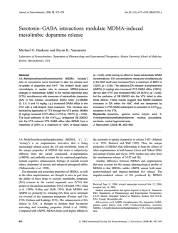
International Journal of Neuropsychopharmacology Advance Access published January 30, 2015
International Journal of Neuropsychopharmacology, 2015, 1–12
doi:10.1093/ijnp/pyu074
Research Article
research article
Strain-Dependent Variations in Stress Coping
Behavior Are Mediated by a 5-HT/GABA Interaction
within the Prefrontal Corticolimbic System
Diego Andolina, PhD; Dario Maran, MSc;Maria Teresa Viscomi, PhD;
Stefano Puglisi-Allegra, PhD
Santa Lucia Foundation, Rome, Italy (Drs Andolina, Viscomi, and Puglisi-Allegra); Dipartimento di Scienze Cliniche
Applicate e Biotecnologie, Universita` degli Studi dell’Aquila, Via Vetoio, L’Aquila, Italy (Dr Andolina); Dipartimento
di Psicologia and Centro ‘Daniel Bovet,’ Sapienza Università di Roma, Rome, Italy (Drs Maran and Puglisi-Allegra).
Correspondence: Diego Andolina, PhD, Dipartimento di Scienze Cliniche Applicate e Biotecnologie Universita` degli Studi dell’Aquila, Via Vetoio, L’Aquila
67010, Italy ([email protected]).
Abstract
Background: Serotonin and γ–aminobutyric acid (GABA) transmission is crucial in coping strategies.
Methods: Here, using mice from 2 inbred strains widely exploited in behavioral neurochemistry, we investigated whether
serotonin transmission in medial prefrontal cortex and GABA in basolateral amygdala determine strain-dependent liability
to stress response and differences in coping.
Results: C57BL/6J mice displayed greater immobility in the forced swimming test, higher serotonin outflow in medial
prefrontal cortex, higher GABA outflow in basolateral amygdala induced by stress, and higher serotonin 1A receptor levels
in medial prefrontal cortex accompanied by lower GABAb receptor levels in basolateral amygdala than DBA/2J mice. In
assessing whether serotonin in medial prefrontal cortex determines GABA functioning in response to stress and passive
coping behavior in C57BL/6J and DBA/2J mice, we observed that selective prefrontal serotonin depletion in C57BL/6J and
DBA/2J reduced stress-induced GABA outflow in basolateral amygdala and immobility in the forced swimming test.
Conclusions: These results show that strain-dependent prefrontal corticolimbic serotonin/GABA regulation determines the
strain differences in stress-coping behavior in the forced swimming test and point to a role of a specific neuronal system in
genetic susceptibility to stress that opens up new prospects for innovative therapies for stress disorders.
Keywords: serotonin, GABA, basolateral amygdala, medial prefrontal cortex, strain
Introduction
Comparative studies on neurotransmitter activity in different
brain regions of inbred strains of mice represent a major strategy
to investigate neurochemical mechanisms underlying behavioral
expression. The genetic stability of inbred strains over the years
and through laboratories has allowed much relevant information to be accumulated for several strains commonly used in the
field. Moreover, behavioral, pharmacological, physiological, and
biochemical comparisons between inbred strains represent a
preliminary stage of more thorough genetic research as quantitative trait loci, or recombinant inbred (available for C57BL/6J [C57]
and DBA/2J strains [DBA]) analyses to identify and map genes
in the mouse, a species characterized by broad gene homology
with humans. Various studies have shown that mice of the C57
and DBA strains differ in their behavior outcomes in the forced
Received: May 02, 2014; Revised: July 17, 2014; Accepted: July 31, 2014
© The Author 2015. Published by Oxford University Press on behalf of CINP.
This is an Open Access article distributed under the terms of the Creative Commons Attribution Non-Commercial License
(http://creativecommons.org/licenses/by-nc/4.0/), which permits non-commercial re-use, distribution, and reproduction in any
medium, provided the original work is properly cited. For commercial re-use, please contact [email protected]
1
2 | International Journal of Neuropsychopharmacology, 2015
swimming test (FST). C57 mice show high immobility and DBA
mice show low immobility (Cabib and Puglisi-Allegra, 1996; Alcaro
et al., 2002; Ventura et al., 2002), an index of passive coping and
depression-like phenotype. Pharmacological (Lucki et al., 2001) or
neurochemical (Ventura et al., 2002; Calcagno et al., 2007) studies have shown differences in brain neurotransmitters that have
been linked to strain differences in passive coping behavior. As
recently shown in C57 mice, a background commonly used in
molecular approaches, amygdalar γ–aminobutyric acid (GABA)
regulation by prefrontal serotonin (5-HT) is crucial in processing stressful experiences and determining passive coping outcomes measured by immobility in the FST (Andolina et al., 2013).
Indeed, sustained, stress-induced 5-HT outflow in the medial
prefrontal cortex (mpFC) and GABA outflow in the basolateral
amygdala (BLA) lead to sustained immobility, whereas prefrontal 5-HT to GABA BLA disconnection leads to low immobility in
the FST (Andolina et al., 2013). Moreover, we showed that 5-HT
depletion in mpFC determined an increase in c-fos expression
in the BLA during the FST, thus pointing to a control of prefrontal 5-HT transmission on neuronal activation and GABAergic
transmission in amygdala during stress (Andolina et al., 2013).
Genetic variation in cortico-amygdala system is strongly implicated in susceptibility to stress-related disorders such as anxiety
and depression as shown by a body of evidence (Drevets et al.,
1992; Holmes, 2009; Wellman et al., 2009). DBA mice are characterized by prefrontal 5-HT functioning that differs from that in the
C57 strain (Calcagno et al., 2007). In particular, DBA mice present
lower 5-HT transporter binding and lower immobility in the FST
than C57 (Sugimoto et al., 2008; Popova et al., 2009). Moreover, DBA
mice are homozygous for the 1473G allele of mouse TPH-2, linked
to low 5-HT synthesis rate, while C57BL/6 mice are homozygous
for the 1473C allele (Zhang et al., 2004; Cervo et al., 2005). This
allelic variant in DBA causes lower brain 5-HT synthesis than
in C57BL/6 mice carrying the “C” allele (Zhang et al., 2004; Cervo
et al., 2005). Moreover, differences between C57 and DBA mice
have been reported for amygdala functioning that have been
linked to strain-dependent difference in stress responsiveness
(Dubois et al., 2006; Yang et al., 2008; Mozhui et al., 2010). Thus,
these strains are a model of choice for investigating individual
differences in the prefrontal-amygdalar system and related serotonin-GABA functioning in stress-induced coping behavior.
Here, we investigated whether prefrontal/amygdala connectivity mediated by 5-HT and GABA transmission is a critical
neural substrate determining strain-dependent differences in
stress response and passive coping behavior. First, we phenotyped the 2 strains for 5-HT and GABA receptor subtype distribution in mpFC and BLA. Indeed, 5-HT1A and GABAb receptors
are known to be involved in stress response and stress-related
behavior (Heisler et al., 1998; Cousins and Seiden, 2000; Cryan and
Kaupmann, 2005; Car and Wisniewska, 2006; Frankowska et al.,
2007; Shishkina et al., 2012). Then, we assessed the response of
prefrontal 5-HT and amygdalar GABA to stress. To this aim, we
used intracerebral microdialysis to compare the response with an
acute stressor (restraint) of 5-HT in mpFC and GABA in BLA in the
2 strains. Restraint was chosen to evaluate the time-dependent
changes induced by stress in mpFC 5-HT and BLA GABA outflow
of C57 and DBA mice. Because we found that C57 and DBA mice
differ in 5-HT outflow in mpFC and GABA in the BLA in response
to stress, we hypothesized that these differences are responsible
for passive coping behaviors characterizing the 2 strains. Stressinduced GABA release in the BLA and behavior in the FST were
thus compared in C57 and DBA bearing a selective 5-HT depletion
in mpFC. To rule out nonspecific effects of prefrontal 5-HT depletion, locomotor activity and anxiety were also assessed.
Methods
Animals
Male mice of the inbred C57BL/6JIco (C57) and DBA/2JIco (DBA)
strains (Charles River, Italy), 8 to 9 weeks old at the time of
experiments, were housed as previously described (Ventura
et al., 2002). All experiments were conducted in accordance with
Italian national law (Decreto Legislativo no. 116, 1992) governing
the use of animals for research.
Drugs
Chloral hydrate, 5,7-dihydroxytryptamine (5,7-DHT), and
desipramine hydrochloride (DMI) were purchased from SigmaAldrich (St. Louis, MO). Chloral hydrate (350–450 mg/kg) and DMI
were dissolved in saline (0.9% NaCl) and injected intraperitoneally (i.p.) in a volume of 10 mL/kg. The 5,7-DHT was dissolved
in saline containing ascorbic acid (0.1%).
Stress Protocols
The restraint apparatus was formed by an adjustable neckblocking support mounted on a Plexiglas base and movable
U-shaped metal piece that could be fixed to the base at the
level of the animal’s hips, thus preventing the mouse from turning on its back (Cabib and Puglisi-Allegra, 1991). The FST was
as previously described (Alcaro et al., 2002; Cabib et al., 2002).
Briefly, mice were individually placed in a glass cylinder (diameter 18 cm, height 40 cm) containing 20 cm of fresh tap water
maintained at 28 ± 2°C during a single session lasting 10 minutes and tested only once to assess coping behavior in a new
stressful condition. The behavior of the animals was monitored
by a video system and scored by a trained observer blind to the
animals’ treatment.
Elevated Plus Maze
Emotional reactivity was measured by the behavioral responses
to the Elevated Plus Maze as previously described (Cabib et al.,
2003). Briefly, mice were individually tested in a single 5-minute
session. At the beginning of each test, the mouse was placed on
the center facing an open arm to initiate the test session. The
number of entries and the time (seconds) spent inside each type
of arm were recorded. Two measures were considered: the percentage of entries in the open arms (open entries/open closed
× 100) and the percentage of time spent in the open arms (time
in open/open closed × 100).
Locomotor Activity
Locomotor activity was measured by a single session of open
field test. The apparatus was a circular open field, 60 cm in
diameter and 20 cm in height. Each mouse was introduced individually for 5 minutes and distance travelled (centimeters) was
recorded. The apparatus was cleaned between subjects.
Microdialysis
Animals were anaesthetized with chloral hydrate, mounted in a
stereotaxic frame (David Kopf Instruments, Tujunga, CA) equipped
with a mouse adapter, and implanted unilaterally with a guide
cannula (stainless steel, shaft outer diameter 0.38 mm, Metalant
AB, Stockholm, Sweden) in mpFC or BLA. Guide cannula implantation in mpFC and BLA was counterbalanced for left and right
Andolina et al. | 3
hemisphere. The length of the guide cannula was 1 mm (mpFC)
and 4.5 mm (BLA) for C57 and 1 mm (mpFC) and 4.0 mm (BLA) for
DBA. The guide cannula was fixed with epoxy glue, and dental
cement was added for further stabilization. Since the brains of
C57 and DBA mice are different in size and weight, different coordinates for probe implantation were used for the 2 backgrounds.
The coordinates from bregma (measured according to the atlas of
Franklin and Paxinos, 1997 and Mouse Brain Atlases, The Mouse
Brain Library, www.nervenet.org/mbl/) were: mpFC: C57 = 2.5 AP,
0.6 L; DBA = 2.0 AP, 0.6 L. BLA: C57 = -1.16 AP, -3.55 L; DBA = -0.9 AP,
-3.05 L. The probe (dialysis membrane length: 2 mm for mpFC and
1 mm for BLA; outer diatmer 0.24 mm, MAB 4 cuprophane microdialysis probe, Metalant AB) was introduced 24 hours after implantation of the guide cannula. The animals were lightly anaesthetized
to facilitate manual insertion of the microdialysis probe into the
guide cannula. The day before use, the membranes were tested to
verify in vitro 5-HT and GABA recovery. The microdialysis probe
was connected to a CMA/100 pump (Carnegie Medicine, Stockholm,
Sweden) through PE-20 tubing and an ultra-low torque dual channel liquid swivel (Model 375/D/22QM, Instech Laboratories, Inc.,
Plymouth Meeting, PA) to allow free movement. Artificial cerebrospinal fluid was pumped through the dialysis probe at a constant flow rate of 2 μL/min. Experiments were carried out 22 to
24 hours after probe placement as previously described (Pascucci
et al., 2009). The mean concentration of the 3 samples collected
immediately before treatment (<10% variation) was taken as basal
concentration. Twenty microliters of the dialysate samples was
analyzed by high-performance liquid chromatography (HPLC). The
HPLC analysis of 5-HT concentration in the dialysates was as previously described (Pascucci et al., 2009). GABA concentrations in the
dialysates were determined as described by Rea et al. (2005). The
detection limit of the assay was 4.2 and 0.1 pg/20 L (signal-to-noise
ratio 2) for GABA and 5-HT, respectively.
Selective 5-HT Depletion in the mpFC
Animals were i.p. injected with DMI (35 mg/kg) 30 minutes before
5,7-DHT microinjection to protect noradrenergic neurons. Bilateral
injection of 5,7-DHT (2.5 μg/0.2 μL/4 min for each side) or vehicle was
made into the mpFC [coordinates: C57: 2.52 AP; ±0.6 L; 2.0 V; DBA: 2.0
AP; ±0.6 L; 2.0 V with respect to bregma (Franklin and Paxinos, 1997;
Mouse Brain Atlases, The Mouse Brain Library, www.nervenet.org/
mbl/)] through a stainless-steel cannula (0.15 mm outer diameter,
UNIMED, Lausanne, Switzerland) connected to a 1-μL syringe by a
polyethylene tube and driven by a CMA/100 pump (Figure 1). The
cannula was left in place for an additional 2 minutes after the end
of the infusion. Sham animals (sham) were subjected to the same
treatment but received intracerebral vehicle. Note that preliminary
experiments showed that naive mice were not significantly different from sham in our testing conditions. Animals were implanted
with a guide cannula in the mpFC and the BLA and used for microdialysis experiment 7 days after surgery, as previously described.
5-HT, norepinephrine (NE), and dopamine (DA) tissue levels
in the mpFC (prelimbic [PL] or infralimbic, Franklin and Paxinos,
1997; Mouse Brain Atlases, The Mouse Brain Library, www.nervenet.org/mbl/) were assessed to evaluate the amount and
extent of 5,7-DHT–induced depletion. Punches were obtained
from brain slices (coronal sections) not thicker than 300 μm
(Puglisi-Allegra et al., 2000). Stainless-steel tubes of 0.5 or
1.0 mm inside diameter were used. The coordinates were measured according to the atlas of Franklin and Paxinos (1997) and
Mouse Brain Atlases (The Mouse Brain Library, www.nervenet.
org/mbl/). Monoamine tissue level analysis was as previously
described (Ventura et al, 2007; Andolina et al., 2013).
Figure 1. Representative positions of approximate location of 5,7-dihydroxytryptamine (5,7-DHT) point microinjection (white dot) in medial prefrontal cortex (mpFC) in C57BL/6J (C57) and DBA/2J (DBA) (A). The arrow indicates the point
of microinjection. Representative positions of the probe in the mpFC (B) and
basolateral amygdala (BLA) (C) in DBA mice and C57 mice. The segment of the
membrane probe is also shown.
Probe Placement
At the end of the experiment, mice were killed by decapitation. Brains were postfixed in 4% paraformaldehyde, and correct probe placements were checked by visual inspection of the
probe tracks on Nissl-stained coronal sections (40 μm). Only
mice with correct probe placement in BLA and mpFC were considered in the results (Figure 1).
Immunohistochemistry
Animals were transcardially perfused with 50 mL of saline followed by 40 mL of 4% paraformaldehyde under anesthesia that
was induced by i.p. injections of chloral hydrate. Each brain was
immediately removed, postfixed in the same paraformaldehyde
solution for 2 hours, and after 3 washes in phosphate-buffered
saline (PBS) was transferred to a 30% sucrose solution at 4°C
4 | International Journal of Neuropsychopharmacology, 2015
until it sank. Each brainstem was cut into 4 series of 30-μm–
thick transverse sections using a freezing microtome, and slices
were collected in PBS. The following primary antibodies were
used: rabbit polyclonal anti-5-HT1A receptor (Immunological
Sciences; 1:200), mouse monoclonal anti GABAbR1 receptor
(Immunological Sciences; 1:100), and rabbit polyclonal anti-parvalbumin (Immunological Sciences; 1:200). One series of sections
(3 sections, distance approximately C57: 2.71, 2.22, and 1.98 mm;
DBA: 2.80, 2.40, and 1.70 from bregma for PL area; C57: -1.28,
-1.64, and -2.12 mm, DBA: -1.06, -1.46, and -1.70 from bregma
for BLA, according to the above-mentioned atlas (Franklin and
Paxinos, 1997; Mouse Brain Atlases, The Mouse Brain Library,
www.nervenet.org/mbl/) was incubated overnight with 5-HT1A
primary antibody or a cocktail of GABAbR1 and parvalbumin.
All primary antibody solutions were prepared in PBS and 0.3%
Triton X-100 and incubated overnight at 4°C. After 3 washes in
PBS, sections were incubated 2 hours at room temperature with
a cocktail of secondary antibodies, including Cy2-conjugated
donkey anti-mouse IgG (1:200; Jackson Immunoresearch, West
Grove, PA) and Cy3-conjugated donkey anti-rabbit IgG (1:200;
Jackson Immunoresearch, West Grove, PA) and counterstained
with NeuroTrace 640/660 fluorescent Nissl (Invitrogen). Sections
were examined under a confocal laser scanning microscope
(Zeiss, LSM700, Germany) equipped with 4 laser lines: violet
diode emitting at 405 nm (for DAPI), argon emitting at 488 nm,
and helium/neon emitting at 543 and 633 nm. Quantification
of the 5-HT1A receptor in PL area and GABAb receptor in BLA
was performed by densitometric analyses. After background
subtraction, 5-HT1A receptor- and GABAb receptor-associated
signals were quantified by manually outlining individual cells
and measuring cell-associated fluorescence intensity with
the ImageJ software (http://rsb.info.nih.gov/ij/). The ratio F/A
defines mean fluorescence of individual cells (F) normalized to
total cellular surface (A). Quantification was done on 3 sections
for PL (C57: 2.71, 2.22, and 1.98 mm; DBA: 2.80, 2.40, and 1.70
from bregma) and on 3 sections for BLA (C57: -1.28, -1.64, and
-2.12 mm, DBA: -1.06, -1.46, and -1.70 from bregma) per animal
(n = 6/group).
Statistics
Immunohistochemistry
C57 vs DBA differences in 5-HT1A in mpFC and GABAb in BLA
expression were evaluated by Student’s t test for each brain
structure (PL and BLA).
Microdialysis
Data on the effect of restraint stress on 5-HT release in
mpFC and GABA release in BLA were statistically analyzed
by repeated-measures analysis of variance (ANOVA) with 2
between factors (strain, 2 levels: C57 and DBA; treatment, 2
levels: sham and 5-HT depleted) and one within factor (minutes, 7 levels: 0, 20, 40, 60, 80, 100, and 120 minutes of restraint).
Statistical analyses were performed on raw data (concentration of pg/20 μL). Simple effects were assessed by 1-way ANOVA
for each time point. The effects of strain on basal extracellular
5-HT levels in mpFC and GABA levels in BLA and the effect of
selective 5-HT depletion in mpFC on basal extracellular 5-HT
levels in mpFC and basal extracellular GABA levels were analyzed in each group (C57 sham, C57 5-HT depleted, DBA sham,
DBA 5-HT depleted). Duncan’s test was used post hoc in this
case.
FST
Statistical analyses for the FST were run on the duration (second) of immobility, swimming, and struggling behavior during
a 10-minute test. Data were analyzed by 2-way ANOVA (factors:
strain, 2 levels: C57 and DBA; treatment, 2 levels: sham and 5-HT
depleted).
5-HT Depletion in mpFC
The effects of prefrontal 5-HT depletion on tissue levels of 5-HT,
DA, and NE in mpFC (PL or IF) in C57 and DBA mice were analyzed by 2-way ANOVA. The factors were: treatment (2 levels:
C57 sham and C57 5-HT depleted) for C57, and 2 levels (DBA
sham and DBA 5-HT depleted) for DBA and experiment (2 levels: FST experiment and microdialysis experiments). Statistical
analyses were carried out on data from the FST and microdialysis experiments.
Elevated Plus Maze
Statistical analyses for Elevated Plus Maze were run on the
percentage of entries in the open arms (open entries/open
closed × 100) and the percentage of time spent in the open
arms (time in open/open closed × 100) during a 5-minute
test. Data were analyzed by 2-way ANOVA (factors: strain,
2 levels: C57 and DBA; treatment, 2 levels: sham and 5-HT
depleted).
Locomotor Activity
Statistical analyses for the open field test were run as distance
travelled (cm) during a 5-minute test. Data were analyzed by
2-way ANOVA (factors: strain, 2 levels: C57 and DBA; treatment,
2 levels: sham and 5-HT depleted).
For experiments (microdialysis, FST, 5-HT depletion, Plus
Maze, open field), individual between-group comparisons were
carried out when appropriate by a posthoc test (Duncan’s multiple-range test).
Results
Effect of Strain on 5-HT1A Receptor Expression in
mpFC and GABAb Receptor Expression in BLA
Densitometric analysis in the PL area revealed that C57 mice
showed significantly higher levels of 5-HT1A receptor expression than DBA mice (t = 2.53, df = 10; P < .05) (Figure 2), whereas
it showed in the BLA GABAb receptor that C57 mice had a significantly lower level of GABAb receptors than DBA mice (t = 3.06,
df = 10; P < .05) (Figure 3).
Effects of 5-HT Depletion in mpFC on Stress-Induced
5-HT Outflow in mpFC and GABA Outflow in BLA in
C57 and DBA Mice
First, a significant difference was found in basal extracellular
5-HT levels in mpFC and GABA levels in the BLA of C57 and DBA
mice, whereas selective 5-HT depletion of mpFC in C57 and DBA
mice did not change basal extracellular 5-HT levels in mpFC and
GABA levels in BLA compared with C57 sham and DBA sham,
respectively [5-HT: F3.27 = 30.08; P < .01; C57 sham (n = 8), 0.92 ± 0.073
pg/20 μL; DBA sham (n = 8), 0.36 ± 0.07 pg/20 μL; C57 5-HT-depl (n = 8),
1.08 ± 0.079 pg/20 μL; DBA 5-HT depl (n = 7), 0.33 ± 0.48 pg/20 μL;
Andolina et al. | 5
Figure 2. Serotonin (5-HT1A) immunoreactivity in the medial prefrontal cortex (mpFC) prelimbic area (PL) of C57BL/6J (C57) sham and DBA/2J (DBA) sham doublelabeled confocal images of NeuroTrace counterstaining (A: blue) and 5-HT1A labeling (B: red) plus merged (C). Panels D and E are higher magnification pictures from
PL of C57 sham (D) and DBA sham (E) reacted with 5-HT1A antibody. Histogram of densitometric values (F) of 5-HT1A immunofluorescence expressed as mean fluorescence of individual cells normalized to total cellular surface (F/A). Data are reported as means ± SD (n = 6 animal/group). *P < .05; scale bars: A-C = 200 μm; D-E = 50 μm.
GABA: F3.28 = 8.23; P < .01; C57 sham (n = 8), 57.9 ± 6.4 pg/20 μL; DBA
sham (n = 8), 27.16 ± 4.4 pg/20 μL; C57 5-HT depl (n = 8), 63.87 ± 9.1
pg/20 μL; DBA 5-HT depl (n = 8), 29.44 ± 5.8 pg/20 μL]. The effects
of restraint on 5-HT release in mpFC and GABA release in BLA
are shown in Figure 4. Statistical analyses revealed a significant
interaction of strain × treatment × time for both the 5-HT outflow
in mpFC and GABA outflow in BLA (5-HT: F6,19 = 2.01; P < .01; GABA:
F6,19 = 1.73; P < .05). Restraint produced a time-dependent increase
in 5-HT outflow in mpFC and in GABA outflow in the BLA in both
strains, but C57 sham mice displayed significantly higher 5-HT levels in mpFC and higher GABA levels in BLA than DBA sham mice.
Selective 5-HT depletion in mpFC of C57 and DBA mice dramatically reduced the increase in 5-HT outflow in mpFC and of GABA
in BLA. Note that no significant differences were evident between
the left and right sides of the mpFC or BLA probe implanted on
5-HT and GABA outflow, respectively. Prefrontal 5,7-DHT in C57
and DBA mice produced a significant decrease in 5-HT tissue levels in PL and infralimbic, whereas it spared NE and DA (Table 1).
The fact that selective 5-HT depletion of mpFC does not
change basal extracellular 5-HT levels suggests that spared serotonergic afferents develop a compensatory response that leads
to an extracellular 5-HT outflow similar to that of sham animals, in agreement with previous studies (Kirby et al., 1995; Hall
et al., 1999). Whether this compensatory response depends on
increased neurotransmitter synthesis or on other mechanisms
remains to be ascertained. However, 5-HT depletion abolished
the serotonergic response to stress in the mpFC, possibly indicating that compensatory response does not allow additional
increase in 5-HT outflow after stress challenge.
Effects of Prefrontal Cortical 5-HT Depletion on
Stress-Coping Behavior in the FST in C57 and
DBA Mice
As for immobility, although 2-way ANOVA did not reveal significant interaction strain × treatment (F1,28 = 0.83, P = .36), analysis
6 | International Journal of Neuropsychopharmacology, 2015
Figure 3. GABAb immunoreactivity in the basolateral amygdala (BLA) of C57BL/6J (C57) sham and DBA/2J (DBA) sham. Triple-labeled confocal images of NeuroTrace
counterstaining (A: blue), and parvalbumin labeling (B: red), GABAb (C: green) plus merged (D) of BLA. Panels E and F are higher magnification pictures from BLA of C57
(E) and DBA (F) reacted with GABAb antibody. Histogram of densitometric values (G) of GABAb immunofluorescence expressed as mean fluorescence of individual cells
normalized to total cellular surface (F/A). Data are reported as means ± SD (n = 6 animals/group). *P < .05. Scale bars: A-D = 200 μm; E-F = 50 μm.
showed significant effects of strain (F1,28 = 31.85, P < .001) and
treatment (F1,28 = 14.32, P < .001). Duncan’s test showed in C57
mice significantly higher levels of immobility than in DBA mice,
while in mpFC 5-HT–depleted C57 and DBA mice, significantly
lower immobility was evident compared with C57 sham and
DBA mice, respectively [C57 sham (n = 8), 395.57 ± 17.48 seconds; DBA sham (n = 8), 251.40 ± 11.45 seconds; C57 5-HT depl
(n = 8), 292.27 ± 37.40 seconds; DBA 5-HT depl (n = 8), 188.30 ± 9.86]
(Figure 5). It should be noted that the decrease in immobility behavior in C57 and DBA mice receiving the selective 5-HT
depletion in mpFC is accompanied by a significant increase in
swimming behavior (Table 2).
Effects of Prefrontal Cortical 5-HT Depletion on
Elevated Plus Maze in C57 and DBA Mice
As for the elevated plus maze, 2-way ANOVA revealed significant
effects of strain (percent time open/tot: F1,28 = 32.78, P < .001; percent time closed/tot: F1,28 = 25.04, P < .001; percent entries open/tot:
F1,28 = 43.92, P < .001; entries closed/tot: F1,28 = 43.92, P < .001) and no
Andolina et al. | 7
Figure 4. Effects of 120-minute restraint stress on serotonin (5-HT) outflow in the medial prefrontal cortex (mpFC) (A) and GABA outflow in the basolateral amygdala (BLA)
(B) of sham C57 and DBA mice and of C57 and DBA mice bearing a selective prefrontal cortical 5-HT depletion in the mpFC (C57 5-HT Depl, DBA 5-HT Depl). Arrows indicate
the beginning of the restraint. §P < .05 from the basal values. *P < .05, C57 sham in comparison with the corresponding time point of DBA sham group. #P < .05 C57 sham
in comparison with the corresponding time point of C57 5-HT Depl group. +P < .05 DBA sham in comparison with the corresponding time point of DBA 5-HT Depl group.
Table 1. Effects of 5,7-DHT Infusion in mpFC on 5-HT, NE, and DA
Tissue Levels (ng/g Wet Weight) in mpFC Areas (Prelimbic Cortex and
Infralimbic Cortex) of the C57 Sham, C57 5-HT–Depleted, DBA Sham,
and DBA 5-HT–Depleted Groups
Sham
Prelimbic cortex (C57)
5-HT
NE
DA
Infralimbic cortex (C57)
5-HT
NE
DA
Prelimbic cortex (DBA)
5-HT
NE
DA
Infralimbic cortex (DBA)
5-HT
NE
DA
5-HT depleted
815 ± 122
655 ± 16
119 ± 61
147 ± 42**
644 ± 54
108 ± 38
432 ± 90
398 ± 29
105 ± 26
161 ± 37*
405 ± 32
128 ± 22
808 ± 41
945 ± 48
95 ± 3
97 ± 19**
1056 ± 50
91 ± 4
600 ± 39
875 ± 46
156 ± 21
91 ± 13**
1028 ± 70
177 ± 21
Abbreviations: DA, dopamine; DBA, DBA/2J; 5,7-DHT, 5,7-dihydroxytryptamine;
5-HT, serotonin; mpFC, medial prefrontal cortex; NE, noradrenaline.
*P < .01; **P < .001.
significant effects of treatment (percent time open/tot: F1,28 = 1.51,
P = .22; percent time closed/tot: F1,28 = 3.34, P = .08; percent entries
open/tot: F1,28 = 0.01, P = .90; entries closed/tot: F1,28 = 0.01, P = .90) and
strain × treatment interaction (percent time open/tot: F1,28 = 1.78,
P = .19; percent time closed/tot: F1,28 = 0.05, P = .81; percent entries
open/tot: F1,28 = 0.61, P = .43; entries closed/tot: F1,28 = 0.61, P = .43).
Duncan’s test showed a significant effect of strain (C57 sham,
DBA sham) for the percent of time spent in open or closed arms
and for the percent of entries in open or closed arms. Selective
5-HT depletion in C57 and DBA did not produce any significant
effect for the percent of time spent in open or closed arms, and
for total entries in open or closed arms compared with C57 sham
and DBA sham, respectively. The data are reported in Figure 6.
Effects of Prefrontal Cortical 5-HT Depletion on
Locomotor Activity in C57 and DBA Mice
As for the locomotor activity analysis on open field, 2-way
ANOVA revealed no significant effects of strain (F1,28 = 3.11, P = .08)
and treatment (F1,28 = 0.30, P = .58) and interaction (F1,28 = 0.7, P = .93)
on distance travelled (cm) (data are reported in Figure 7).
Discussion
Evidence points to a clear-cut strain-dependent role of prefrontal 5-HT and amygdalar GABA transmission in stress response
and coping behavior. C57 mice are characterized by higher
5-HT1A receptor levels in mpFC accompanied by lower GABAb
receptor levels in BLA than in DBA mice. Moreover, C57 mice
present higher 5-HT steady levels in mpFC and higher GABA
steady levels in BLA than DBA. We have shown that, in mice, a
stressful experience such as restraint induces a time-dependent
increase of 5-HT output in the mpFC and of GABA in the BLA, in
agreement with previous reports (Reznikov et al., 2009; Pascucci
et al., 2009; Andolina et al., 2013). These results are the first evidence from an in vivo study showing that acute restraint stress
produces different effects on mpFC 5-HT and BLA GABA release
in C57 and DBA mice, thus affirming that genetic background
causes differences in the response of the prefrontal/amygdala
5-HT/GABA system to stress. Interestingly, the 5-HT and GABA
release in mpFC and BLA, respectively, are clearly related to sustained immobility in the FST. In fact, selective prefrontal 5-HT
depletion in C57 and DBA mice reduces the amine outflow in
the mpFC and GABA release in the BLA in response to restraint
stress and leads to a dramatic decrease of immobility in both
C57 and DBA mice. These results, in line with previous data
(Andolina et al., 2013), support the fact that low serotonergic
and GABAergic tone in mpFC and BLA is negatively related to
passive coping in a stressful condition such as the FST. A causal
relationship between serotonergic and GABAergic tone in mpFC
and BLA and immobility in the FST has been previously demonstrated. In fact, we showed that the FST induced a clear-cut
time-related increase of GABA output in BLA of mice, whereas
selective 5-HT depletion in mpFC caused a dramatic decrease of
GABA output throughout (Andolina et al., 2013). Note that the
results obtained by the open field test showed that the observed
differences in immobility behavior in the FST cannot be ascribed
to differences in spontaneous locomotor activity. Moreover, they
point to a prefrontal/amygdala system in which 5-HT and GABA
are strain-dependently orchestrated to control stress-induced
adaptive behavioral outcomes in accordance with other evidence showing a major influence of genetic variation in cortico-amygdala serotonin function and individual differences
in stress response and risk for stress-related disease (Holmes,
2009; Homberg, 2012).
In line with previous studies, we report that sham DBA mice
displayed higher basal anxiety than sham C57 mice in the elevated plus maze (Võikar et al., 2005; Mozhui et al., 2010), whereas
selective 5-HT depletion in mpFC did not affect anxiety in either
8 | International Journal of Neuropsychopharmacology, 2015
Figure 5. Effects of strain (C57 sham, DBA sham) and of selective prefrontal cortical serotonin (5-HT) depletion in the medial prefrontal cortex (mpFC) of C57
and DBA mice (C57 5-HT Depl; DBA 5-HT Depl) on immobility in the forced swimming test (FST). Results are expressed as mean ± SE duration (second) of immobility. *P < .05. C57 sham in comparison with DBA sham group, C57 5-HT-Depl
group and DBA sham group in comparison with DBA 5-HT-Depl group.
Table 2. Level of Swimming and Struggling Behavior in the FST in
C57 Sham, C57 5-HT Depleted in mpFC, DBA Sham, and DBA 5-HT
depleted in mpFC Groups
C57 sham
C57 5-HT depleted
DBA sham
DBA 5-HT depleted
Swimming
Struggling
139.91 ± 23.34a,b
221.28 ± 28.96
257.91 ± 16.66c
345.8 ± 15.89
64.51 ± 10.46
86.34 ±15.38
90.69 ± 16.96
64.79 ± 6.77
Abbreviations: C57, C57BL/6J; DA, dopamine; DBA, DBA/2J; FST, forced swimming test; 5-HT, serotonin; mpFC, medial prefrontal cortex.
C57 sham in comparison with C57 5-HT–depleted group.
C57 sham in comparison with DBA sham group.
c
DBA sham in comparison with DBA 5-HT–depleted group.
a
b
C57 or DBA mice. These results suggest that a decrease of immobility behavior in the FST in DBA and C57 mice bearing a selective prefrontal 5-HT depletion in the mpFC cannot be ascribed
to an alteration of emotional reactivity. Although in this study
we evaluated the role of genotype and of the 5-HT prefrontal
GABArgic amygdala system in neural and behavioral responses
to the first exposure to an aversive experience, the reported
differences between C57 and DBA mice could be the basis of
differences in fear memory reported in these strains (Waddell
et al., 2004). In fact, alteration of fear memory, for example, an
impaired fear extinction, is a core symptom of anxiety disorders,
such as posttraumatic stress disorder; several studies reported
that mpFC 5-HT and BLA GABAergic transmission are involved
in this behavioral phenotype (Akirav and Maroun, 2007; Shin
and Liberzon, 2010).
Differences in the 5-HT system in C57 and DBA mice
have been extensively studied and indicate that these differences underlie strain differences in immobility. For instance,
Sugimoto et al. (2008) showed that the amount of 5-HT transporter and 5-HT1A binding in prefrontal cortex was linked to
baseline immobility time in C57 and DBA mice (Sugimoto et al.,
2008; Popova et al., 2009). In line with these data, our results suggest that differences in prefrontal cortical 5-HT release, which
controls BLA GABA release, contribute to strain differences in
immobility in these 2 strains. Indeed, selective prefrontal 5-HT
depletion in C57 and DBA mice reduces immobility in the FST.
Consistent with these data, C57 mice showed a higher level of
5-HT1A receptors in PL cortex compared with DBA mice. Several
subtypes of the 5-HT receptor family have been associated with
stress response and depressive disorders. However, within this
family, the 5-HT1A receptor has attracted increasing interest in
the auxiliary therapy of depression. It has been reported that
the 5-HT1A receptor is expressed on pre- and postsynaptic sites.
Presynaptic high-density 5-HT1A autoreceptors are located
somatodendritically in the raphe nuclei (Albert et al., 1990; Blier
and de Montigny, 1990; Ago et al., 2003). Postsynaptic 5-HT1A
receptors are widely expressed as heteroreceptors on glutamatergic pyramidal cells and on GABAergic interneurons in the
hippocampus, cortical regions, septum, amygdala, and hypothalamus (Pompeiano et al., 1992; Chessell et al., 1993; Wedzony
et al., 2007), where their activation leads to decreased firing
rates (Sprouse and Aghajanian, 1988; Blier and de Montigny,
1990; Tanaka and North, 1993). Thus, the majority of pyramidal neurons are functionally inhibited by 5-HT in a 5-HT1A–
dependent manner (Beique et al., 2004; Zhang and Arsenault,
2005; Goodfellow et al., 2009; Zhong and Yan, 2011), suggesting that 5-HT1A receptors play a dominant role in regulating
pyramidal neuron activity. Depressive symptoms and passive
coping behavior are associated with prefrontal cortex hypoactivity and changes in 5-HT receptor levels (Hariri et al., 2002;
Amat et al., 2005; Canli et al., 2005; Heinz et al., 2005; Pezawas
et al., 2005; Firk and Markus, 2007; Maier and Watkins, 2010).
Because we found that DBA mice display both lower expression
of 5-HT1A in the PL area and lower levels of 5-HT in mpFC mice
compared with C57 mice, it is likely that the main consequence
of increased extracellular 5-HT in the mpFC of C57 mice is an
overinhibition of mpFC neuronal activity through 5-HT1A, leading to an increase in passive-coping behavior in the FST. Thus,
our data are consistent with the hypothesis that high 5-HT1A
receptor levels in mpFC (PL area) sustain immobility in the FST.
Indeed, we found that C57 mice showed both higher levels of
5-HT1A receptor in mpFC and a higher level of immobility in
the FST than DBA mice. It is therefore likely that selective 5-HT
depletion in mpFC consistently reduces immobility in C57 and
DBA mice through consequent reduced stimulation of 5-HT1A
receptors. It should be noted that DBA mice (homozygous for
the Tph2 allele 1473G) show low immobility in comparison with
C57BL/6 mice, which are homozygous for the Tph2 1473C allele
(Zhang et al., 2004; Cervo et al., 2005). Moreover, introduction
of the 1473G/G single-nucleotide polymorphisms (SNPs) into
a C57BL/6N genetic background has been reported to cause
a desensitization of 5-HT1A (Berger et al., 2012). In line with
this evidence, we found that DBA mice show lower levels of
5-HT1A receptors than C57 mice. The mpFC plays a crucial role
in the regulation of stimulus-driven amygdala response, partly
through glutamatergic projections to populations of GABAergic
neurons within the amygdala (Quirk et al., 2003; Likhtik et al.,
2005; Shin et al., 2005). Variability in the structure and function
of this corticolimbic circuitry has been associated with individual differences in personality measures, reflecting sensitivity to
environmental threat and related risk of psychopathology (Etkin
et al., 2004; Pezawas et al., 2005; Shin et al., 2005; Fakra et al.,
2009; Holmes, 2009; Homberg, 2012). In particular, human studies have shown that individual differences in prefrontal cortical
5-HT are related to amygdala activity (Fisher et al., 2009, 2011).
The GABAergic system is the principal modulator of amygdala
and substantial clinical and preclinical evidence implicates a
dysfunction of the GABA system in depression (Krystal et al.,
2002; Brambilla et al., 2003). Rodent studies have shown that
GABA in the amygdala is involved in depression-like behavior
in the FST (Ebner et al., 2005; Andolina et al., 2013). For instance,
evidence suggests that GABAb receptor agonist baclofen inhibit
central neural circuits and peripheral sympathetic nervous system that are involved in the stress response (Bolser et al., 1995).
Andolina et al. | 9
Figure 6. Effects of strain (C57 sham, DBA sham) and of selective prefrontal cortical serotonin (5-HT) depletion in the medial prefrontal cortex (mpFC) of C57 and DBA
mice (C57 5-HT Depl; DBA 5-HT Depl) on anxiety behavior in the elevated plus maze. Data are expressed as mean percent time spent or mean percent entries made in
the open arm and as mean percent time spent or mean percent entries made in closed arms ± SE. *P < .05.
Figure 7. Effects of strain (C57 sham, DBA sham) and of selective prefrontal cortical serotonin (5-HT) depletion in the medial prefrontal cortex (mpFC) of C57
and DBA mice (C57 5-HT Depl; DBA 5-HT Depl) on locomotor activity in the open
field. Results are expressed as mean ± SE distance moved (cm).
Moreover, high levels of GABAb receptor expression in the BLA
have been shown to play an important role in regulating emotional behavior and depression (McDonald et al., 2004; Cryan
and Kaupmann, 2005), and there is evidence of baclofen having antidepressant effects in the FST (Cryan and Kaupmann,
2005; Car and Wiśniewska, 2006; Frankowska et al., 2007). In line
with this evidence we found that C57 mice show higher levels
of immobility behavior in the FST and lower GABAb expression
in BLA than in DBA mice. Interestingly, an antidepressant effect
of baclofen could be mediated by a decrease of GABA release. It
has been reported that baclofen produces a decrease of GABA
release (Rea et al., 2005). In support of this hypothesis, we
found that DBA show lower levels of immobility behavior in the
FST and lower GABA levels in BLA accompanied by increased
expression of GABAb than C57 mice.
Many studies suggest that prefrontal cortex-amygdala system is highly implicated in stress response and stress-related
disturbances (Siegle et al., 2002; Phillips et al., 2003; Phelps et al.,
2004; Akirav and Maroun, 2007; Qi et al., 2008). Moreover, dysfunctions of this neural circuit have been associated with individual differences in risk for psychopathology (Drevets et al.,
1992; Pezawas et al., 2005, Holmes, 2009). mpFC is considered to
play a critical role in regulation of amygdala-mediated arousal
in response to emotionally salient stimuli (Quirk et al., 2003;
Likhtik et al., 2005) through 5-HT (LeDoux, 2000; Martín-Ruiz
et al., 2001; Fisher et al., 2009, 2011). A growing body of evidence,
in particular rising from genetically modified or inbred mice, has
provided significant insight into the way genetic variation in the
5-HT system can affect the development and functioning of
mpFC-amygdala circuitry (Holmes, 2009). For instance, Wellman
et al. (2007) showed that the loss of 5-HTT gene function, leading
to a marked increase in extracellular levels of 5-HT in different
brain region including frontal cortex (Mathews et al., 2004), compromises the capacity to cope with environmental stress and
causes morphological abnormalities in both BLA and mpFC. Our
results demonstrate that mpFC controls amygdala to moderate
stress response and its intensity through 5-HT transmission
in mpFC modulating amygdalar GABA depending on genetic
background.
The prefrontal-amygdala system envisaged here involves
a parallel stress-induced increase in 5-HT and GABA transmission in the 2 brain regions, the former controlling the latter through neural pathways (circuitry) that can be validly
hypothesized (Amat et al., 1998; Rainnie, 1999). Evidence suggests that stress-induced increase of prefrontal cortical 5-HT
release overinhibits mpFC glutamatergic neuronal activity
through 5-HT1A (for review, see Puig and Gulledge, 2011), thus
inhibiting GABA release within the DRN, leading to permissive
effects on 5-HT neurons in DRN and producing increased 5-HT
10 | International Journal of Neuropsychopharmacology, 2015
release in BLA. Since 5-HT is a modulator of GABA release in
BLA (Rainnie, 1999), increased 5-HT is likely to increase GABA
activity in this area and increase passive coping behavior. This
hypothesis is supported by data showing that passive coping
is related to pronounced 5-HT immunostaining in the BLA
(Lehner et al., 2006). It should be noted that various studies
suggest a link between the GABAergic and monoaminergic
hypotheses of depression (Pilc and Lloyd, 1984; Lloyd et al.,
1985; Slattery et al., 2005). Thus, taken together, the data presented herein point to a role of 5-HT and GABA neurotransmission in the prefrontal cortex-amygdala system in stress
response and coping outcomes. Moreover, they point to a role
of 5-HT and GABAergic neurotransmission in the prefrontal
cortex/amygdala system in strain-dependent susceptibility to
stress response and stress coping and suggest a way of developing therapeutic approaches for the treatment of depression
by combining actions on different neurotransmitter systems,
especially 5-HT and GABA.
Acknowledgments
We thank Dr. Sergio Papalia for his skillful assistance. This
research was supported by Ricerca Corrente, Italian Ministry of
Health and Ateneo 2012, Sapienza University of Rome, and by
Ministero della Ricerca Scientifica e Tecnologica (FIRB 2010).
Statement of Interest
None.
References
Ago Y, Koyama Y, Baba A, Matsuda T (2003) Regulation by 5-HT1A
receptors of the in vivo release of 5-HT and DA in mouse frontal cortex. Neuropharmacology 45:1050–1056.
Akirav I, Maroun M (2007) The role of the medial prefrontal
cortex-amygdala circuit in stress effects on the extinction of
fear. Neural Plast 2007:30873.
Albert PR, Zhou QY, Van Tol HH, Bunzow JR, Civelli O (1990) Cloning, functional expression, and mRNA tissue distribution of
the rat 5-hydroxytryptamine1A receptor gene. J Biol Chem
265:5825–5832.
Alcaro A, Cabib S, Ventura R, Puglisi-Allegra S (2002) Genotype
and experience dependent susceptibility to depressive-like
responses in the forced-swimming test. Psychopharmacology (Berl) 164:138–143.
Amat J, Matus-Amat P, Watkins LR, Maier SF (1998) Escapable
and inescapable stress differentially alter extracellular levels of 5-HT in the basolateral amygdala of the rat. Brain Res
812:113–120.
Amat J, Baratta MV, Paul E, Bland ST, Watkins LR Maier SF (2005)
Medial prefrontal cortex determines how stressor controllability affects behavior and dorsal raphe nucleus. Nat Neurosci 8:365–371.
Andolina D, Maran D, Valzania A, Conversi D Puglisi-Allegra S
(2013) Prefrontal/amygdalar system determines stress coping
behavior through 5-HT/GABA connection. Neuropsychopharmacology 38:2057–2067.
Beique JC, Campbell B, Perring P, Walker P, Mladenovic L Andrade
R (2004) Serotonergic regulation of membrane potential in
developing rat prefrontal cortex: coordinated expression of
5-hydroxytryptamine (5-HT)1A, 5-HT2A, and 5- HT7 receptors. J Neurosci 24:4807–4817.
Berger SM, Weber T, Perreau-Lenz S, Vogt MA, Gartside SE, MaserGluth C, Lanfumey L, Gass P, Spanagel R, Bartsch D (2012). A
functional Tph2 C1473G polymorphism causes an anxiety
phenotype via compensatory changes in the serotonergic
system. Neuropsychopharmacology 37:1986–1998.
Blier P, de Montigny C (1990) Electrophysiological investigation
of the adaptiveresponse of the 5-HT system to the administration of 5-HT1A receptor agonists. J Cardiovasc Pharmacol
15:S42–S48.
Bolser DC, Blythin DJ, Chapman RW, Egan RW, Hey JA, Rizzo C,
Kuo SC, Kreutner W (1995) The pharmacology of SCH 50911: a
novel, orally active GABA-B receptor antagonist. J Pharmacol
Exp Ther. 274:1393–1398.
Brambilla P, Harenski K, Nicoletti M, Sassi RB, Mallinger AG,
Frank E, Kupfer DJ, Keshavan MS, Soares JC (2003) MRI investigation of temporal lobe structures in bipolar patients. J Psychiatr Res 37:287–295.
Cabib S, Ventura R, Puglisi-Allegra S (2002) Opposite imbalances between mesocortical and mesoaccumbens dopamine
responses to stress by the same genotype depending on living conditions. Behav Brain Res 129:179–185.
Cabib S, Pascucci T, Ventura R, Romano V, Puglisi-Allegra S
(2003) The behavioral profile of severe mental retardation
in a genetic mouse model of phenylketonuria. Behav Genet
33:301–310.
Cabib S, Puglisi-Allegra S (1991) Genotype-dependent effects of
chronic stress on apomorphine-induced alterations of striatal
and mesolimbic dopamine metabolism. Brain Res 542:91–96.
Cabib S, Puglisi-Allegra S (1996) Stress, depression and the mesolimbic dopamine system. Psychopharmacology (Berl) 128:331–342.
Calcagno E, Canetta A, Guzzetti S, Cervo L Invernizzi RW (2007)
Strain differences in basal and post-citalopram extracellular
5-HT in the mouse medial prefrontal cortex and dorsal hippocampus: relation with tryptophan hydroxylase-2 activity. J
Neurochem 103:1111–1120.
Canli T, Omura K, Haas BW, Fallgatter A, Constable RT, Lesch KP
(2005) Beyond affect: a role for genetic variation of the serotonin transporter in neural activation during a cognitive attention task. Proc Natl Acad Sci USA 102:12224–12229.
Car H, Wisniewska RJ (2006) Antidepressant-like effects of
baclofen and LY367385 in the forced swim test in rats. Pharmacol Rep 58:758–764.
Cervo L, Canetta A, Calcagno E, Burbassi S, Sacchetti G, Caccia S, Fracasso C, Albani D, Forloni G, Invernizzi RW (2005)
Genotype-dependent activity of tryptophan hydroxylase-2
determines the response to citalopram in a mouse model of
depression. J Neurosci 25:8165–8172.
Chessell IP, Francis PT, Pangalos MN, Pearson RC, Bowen DM
(1993) Localisation of muscarinic (m1) and other neurotransmitter receptors on corticofugal-projecting pyramidal neurones. Brain Res 632:86–94.
Cousins MS, Seiden LS (2000) The serotonin-1A receptor antagonist WAY-100635 modifies fluoxetine’s antidepressant-like
profile on the differential reinforcement of low rates 72-s
schedule in rats. Psychopharmacology (Berl) 148:438–442.
Cryan JF, Kaupmann K (2005) Don’t worry ‘B’ happy!: a role for
GABA(B) receptors in anxiety and depression. Trends Pharmacol Sci. 26:36–43.
Drevets WC, Videen TO, Price JL, Preskorn SH, Carmichael ST,
Raichle ME (1992) A functional anatomical study of unipolar
depression. J Neurosci. 12:3628–3641.
DuBois DW, Perlegas A, Floyd DW, Weiner JL, McCool BA (2006)
Distinct functional characteristics of the lateral/basolateral
Andolina et al. | 11
amygdala GABAergic system in C57BL/6J and DBA/2J mice. J
Pharmacol Exp Ther 318:629–640.
Ebner K, Bosch JO, Kromer SA, Singewald N, Neumann ID (2005)
Release of oxytocin in the rat central amygdala modulates
stress coping behavior and the release of excitator amino
acids. Neuropsychopharmacology 30:223–230.
Etkin A, Klemenhagen KC, Dudman JT, Rogan MT, Hen R, Kandel ER, Hirsch J (2004) Individual differences in trait anxiety
predict the response of the basolateral amygdala to unconsciously processed fearful faces. Neuron 44:1043–1055.
Fakra E, Hyde LW, Gorka A, Fisher PM, Munoz KE, Kimak M, Halder I, Ferrell RE, Manuck SB, Hariri AR (2009) Effects of HTR1A
C(-1019)G on amygdala reactivity and trait anxiety. Arch Gen
Psychiatry 66:33–40.
Firk C, Markus CR (2007) Review: Serotonin by stress interaction:
a susceptibility factor for the development of depression? J
Psychopharmacol 21:538–544.
Fisher PM, Meltzer CC, Price JC, Coleman RL, Ziolko SK, Becker C,
Moses-Kolko EL, Berga SL Hariri AR (2009) Medial prefrontal
cortex 5-HT(2A) density is correlated with amygdala reactivity, response habituation, and functional coupling. Cereb Cortex 19:2499–2507.
Fisher PM, Price JC, Meltzer CC, Moses-Kolko EL, Becker C, Berga
SL, Hariri AR (2011) Medial prefrontal cortex serotonin 1A and
2A receptor binding interacts to predict threatrelated amygdala reactivity. Biol Mood Anxiety Disord 1:2.
Franklin KBJ, Paxinos G (1997) The Mouse Brain: In Stereotaxic
Coordinates (Academic, San Diego).
Frankowska M, Filip M, Przegaliński E (2007) Effects of GABAB
receptor ligands in animal tests of depression and anxiety.
Pharmacol Rep 59:645–655.
Goodfellow NM, Benekareddy M, Vaidya VA, Lambe EK (2009)
Layer II/III of the prefrontal cortex: inhibition by the serotonin 5-HT1A receptor in development and stress. J Neurosci
29:10094–10103.
Hall FS, Devries AC, Fong GW, Huang S, Pert A (1999) Effects of
5,7-dihydroxytryptamine depletion of tissue serotonin levels
on extracellular serotonin in the striatum assessed with in
vivo microdialysis: relationship to behavior. Synapse 33:16–25.
Hariri AR, Mattay VS, Tessitore A, Kolachana B, Fera F, Goldman D, Egan MF, Weinberger DR (2002) Serotonin transporter
genetic variation and the response of the human amygdala.
Science 297:400–403.
Heinz A, Braus DF, Smolka MN, Wrase J, Puls I, Hermann D, Klein
S, Grusser SM, Flor H, Schumann G, Mann K, Buchel C (2005)
Amygdala-prefrontal coupling depends on a genetic variation of the serotonin transporter. Nat Neurosci 8:20–21.
Heisler LK, Chu HM, Brennan TJ, Danao JA, Bajwa P, Parsons LH,
Tecott LH (1998) Elevated anxiety and antidepressant-like
responses in serotonin 5-HT1A receptor mutant mice. Proc
Natl Acad Sci USA 95:15049–15054.
Holmes A (2009) Genetic variation in cortico-amygdala serotonin function and risk for stressrelated disease. Neurosci
Biobehav Rev 32:1293–1314.
Homberg JR (2012) The stress-coping (mis)match hypothesis for
nature x nurture interactions. Brain Res 1432:114–121.
Kirby LG, Kreiss DS, Singh A, Lucki I (1995) Effect of destruction of
serotonin neurons on basal and fenfluramine-induced serotonin release in striatum. Synapse 20:99–105.
Krystal JH, Sanacora G, Blumberg H, Anand A, Charney DS, Marek
G, Epperson CN, Goddard A, Mason GF (2002) Glutamate and
GABA systems as targets for novel antidepressant and mood
stabilizing treatments. Mol Psychiatry 1:S71–80.
LeDoux JE (2000) Emotion circuits in the brain. Annu Rev Neurosci 23:155–184.
Lehner M, Taracha E, Skorzewska A, Maciejak P, WislowskaStanek A, Zienowicz M, Szyndler J, Bidziński A, Płaźnik A
(2006) Behavioral, immunocytochemical and biochemical
studies in rats differing in their sensitivity to pain. Behav
Brain Res 171:189–198.
Likhtik E, Pelletier JG, Paz R, Pare D (2005) Prefrontal control of
the amygdala. J Neurosci 25:7429–7437.
Lloyd KG, Thuret F, Pilc A (1985) Upregulation of gamma-aminobutyric acid (GABA) B binding sites in rat frontal cortex:
a common action of repeated administration of different
classes of antidepressants and electroshock. J Pharmacol Exp
Ther 235:191–199.
Lucki I, Dalvi A, Mayorga AJ (2001) Sensitivity to the effects of
pharmacologically selective antidepressants in different
strains of mice. Psychopharmacology (Berl) 155:315–322.
Maier SF, Watkins LR (2010) Role of the medial prefrontal cortex
in coping and resilience. Brain Res 1355:52–60.
Martín-Ruiz R, Puig MV, Celada P, Shapiro DA, Roth BL, Mengod
G, Artigas F (2001) Control of serotonergic function in medial
prefrontal cortex by serotonin-2A receptors through a glutamatedependent mechanism. J Neurosci 21:9856–9866.
Mathews TA, Fedele DE, Coppelli FM, Avila AM, Murphy DL,
Andrews AM (2004) Gene dose-dependent alterations in
extraneuronal serotonin but not dopamine in mice with
reduced serotonin transporter expression. J Neurosci Methods 140:169–181.
McDonald AJ, Mascagni F, Muller JF (2004) Immunocytochemical
localization of GABABR1 receptor subunits in the basolateral
amygdala. Brain Res 1018:147–158.
Mozhui K, Karlsson RM, Kash TL, Ihne J, Norcross M, Patel S, Farrell MR, Hill EE, Graybeal C, Martin KP, Camp M, Fitzgerald
PJ, Ciobanu DC, Sprengel R, Mishina M, Wellman CL, Winder
DG, Williams RW, Holmes A (2010) Strain differences in stress
responsivity are associated with divergent amygdala gene
expression and glutamate-mediated neuronal excitability. J
Neurosci 30:5357–5367.
Pascucci T, Andolina D, La Mela I, Conversi D, Latagliata C,
Ventura R, Puglisi-Allegra S, Cabib S (2009) 5-Hydroxytryptophan rescues serotonin response to stress in prefrontal
cortex of hyperphenylalaninaemic mice. Int J Neuropsychopharmacol 14:479–489.
Pezawas L, Meyer-Lindenberg A, Drabant EM, Verchinski BA,
Munoz KE, Kolachana BS, Egan MF, Mattay VS, Hariri AR,
Weinberger DR (2005) 5-HTTLPR polymorphism impacts
human cingulate-amygdala interactions: a genetic susceptibility mechanism for depression. Nat Neurosci 8:828–834.
Phelps EA, Delgado MR, Nearing KI, LeDoux JE (2004) Extinction
learning in humans: role of the amygdala and vmPFC. Neuron 43:897–905.
Phillips ML, Drevets WC, Rauch SL, Lane R (2003) Neurobiology
of emotion perception II: implications for major psychiatric
disorders. Biol Psychiatry 54:515–528.
Pilc A, Lloyd KG (1984) Chronic antidepressants and GABA “B”
receptors: a GABA hypothesis of antidepressant drug action.
Life Sci 35:2149–2154.
Pompeiano M, Palacios JM, Mengod G (1992) Distribution and
cellular localization of mRNA coding for 5-HT1A receptor in
the rat brain: correlation with receptor binding. J Neurosci
12:440–453.
Popova NK, Naumenko VS, Tibeikina MA, Kulikov AV (2009) Serotonin transporter, 5-HT1A receptor, and behavior in DBA/2J
12 | International Journal of Neuropsychopharmacology, 2015
mice incomparison with four inbred mouse strains. J Neurosci Res 87:3649–3657.
Puig MV, Gulledge AT (2011) Serotonin and prefrontal cortex
function: neurons, networks, and circuits. Mol Neurobiol
44:449–464.
Puglisi-Allegra S, Cabib S, Pascucci T, Ventura R, Cali F, Romano V
(2000) Dramatic brain aminergic deficits in a genetic mouse
model of phenyketonuria. Neuroreport 11:1361–1364.
Qi X, Lin W, Li J, Li H, Wang W, Wang D, Sun M (2008) Fluoxetine
increases the activity of the ERK-CREB signal system and alleviates the depressive-like behavior in rats exposed to chronic
forced swim stress. Neurobiol Dis 31:278–285.
Quirk GJ, Likhtik E, Pelletier JG, Pare D (2003) Stimulation of
medial prefrontal cortex decreases the responsiveness of
central amygdala output neurons. J Neurosci 23:8800–8807.
Rainnie DG (1999) Serotonergic modulation of neurotransmission in the rat basolateral amygdala. J Neurophysiol 82:69–85.
Rea K, Cremers TI, Westerink BH (2005) HPLC conditions are critical for the detection of GABA by microdialysis. J Neurochem
94:672–679.
Reznikov LR, Reagan LP, Fadel JR (2009) Effects of acute and
repeated restraint stress on GABA efflux in the rat basolateral
and central amygdala. Brain Res 1256:61–68.
Shin LM, Liberzon I (2010) The neurocircuitry of fear, stress, and
anxiety disorders. Neuropsychopharmacology. 35:169–191.
Shin LM, Wright CI, Cannistraro PA, Wedig MM, McMullin K, Martis B, Macklin ML, Lasko NB, Cavanagh SR, Krangel TS, Orr SP,
Pitman RK, Whalen PJ, Rauch SL (2005) A functional magnetic
resonance imaging study of amygdala and medial prefrontal
cortex responses to overtly presented fearful faces in posttraumatic stress disorder. Arch Gen Psychiatry 62:273–281.
Shishkina GT, Kalinina TS, Dygalo NN (2012) Effects of swim
stress and fluoxetine on 5-HT1A receptor gene expression
and monoamine metabolism in the rat brain regions. Cell Mol
Neurobiol 32:787–794.
Siegle GJ, Steinhauer SR, Thase ME, Stenger VA, Carter CS (2002)
Can’t shake that feeling: event-related fMRI assessment of
sustained amygdala activity in response to emotional information in depressed individuals. Biol Psychiatry 51:693–707.
Slattery DA, Desrayaud S, and Cryan JF (2005) GABAB receptor
antagonist-mediated antidepressant-like behavior is serotonin-dependent. J Pharmacol Exp Ther 312:290–296.
Sprouse JS, Aghajanian GK (1988) Responses of hippocampal
pyramidal cells to putative serotonin 5-HT1A and 5-HT1B
agonists: a comparative study with dorsal raphe neurons.
Neuropharmacology 27:707–715.
Sugimoto Y, Kajiwara Y, Hirano K, Yamada S, Tagawa N, Kobayashi Y, Hotta Y, Yamada J (2008) Mouse strain differences
in immobility and sensitivity to fluvoxamine and desipramine in the forced swimming test: analysis of serotonin
and noradrenaline transporter binding. Eur J Pharmacol 592:
116–122.
Tanaka E, North RA (1993) Actions of 5-hydroxytryptamine on neurons of the rat cingulate cortex. J Neurophysiol 69:1749–1757.
Ventura R, Cabib S, Puglisi-Allegra S (2002) Genetic susceptibility of mesocortical dopamine to stress determines liability
to inhibition of mesoaccumbens dopamine and to behavioral ‘despair’ in a mouse model of depression. Neuroscience
115:999–1007.
Ventura R, Morrone C, Puglisi-Allegra S (2007) Prefrontal/accumbal catecholamine system determines motivational salience
attribution to both reward- and aversion-related stimuli. Proc
Natl Acad Sci USA 104:5181–5186.
Võikar V, Polus A, Vasar E, Rauvala H (2005) Long-term individual
housing in C57BL/6J and DBA/2 mice: assessment of behavioral consequences. Genes Brain Behav 4:240–252.
Yang RJ, Mozhui K, Karlsson RM, Cameron HA, Williams RW, Holmes A (2008) Variation in mouse basolateral amygdala volume is associated with differences in stress reactivity and
fear learning. Neuropsychopharmacology 33:2595–2604.
Waddell J, Dunnett C, Falls WA (2004) C57BL/6J and DBA/2J mice
differ in extinction and renewal of extinguished conditioned
fear. Behav Brain Res 154:567–576.
Wedzony K, Chocyk A, Kolasiewicz W, Mackowiak M (2007) Glutamatergic neurons of rat medial prefrontal cortex innervating
the ventral tegmental area are positive for serotonin 5- HT1A
receptor protein. J Physiol Pharmacol 58:611–624.
Wellman CL, Izquierdo A, Garrett JE, Martin KP, Carroll J, Millstein
R, Lesch KP, Murphy DL, Holmes A (2007). Impaired stresscoping and fear extinction and abnormal corticolimbic morphology in serotonin transporter knock-out mice. J Neurosci.
27:684–691.
Zhang X, Beaulieu JM, Sotnikova TD, Gainetdinov RR, Caron MG
(2004) Tryptophan hydroxylase-2 controls brain serotonin
synthesis. Science 305:217.
Zhang ZW, Arsenault D (2005) Gain modulation by serotonin
in pyramidal neurones of the rat prefrontal cortex. J Physiol
566:379–394.
Zhong P, Yan Z (2011) Differential regulation of the excitability of prefrontal cortical fastspiking interneurons and
pyramidal neurons by serotonin and fluoxetine. PLoS One
6:e16970.

© Copyright 2026