
PDF (1.2 MB) - Nuclear Medicine and Biology
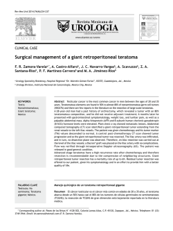
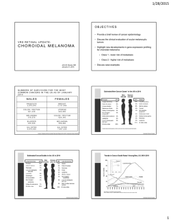
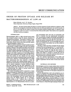
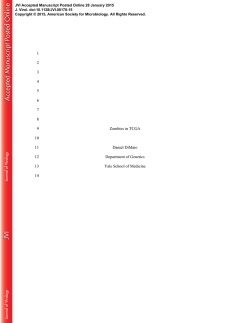
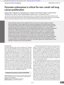
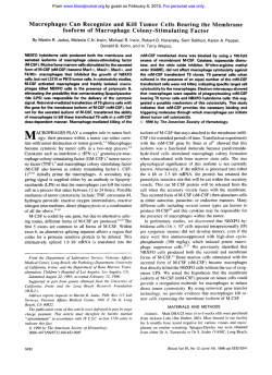
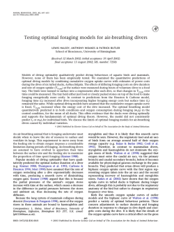
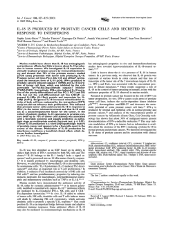
Nuclear Medicine and Biology 40 (2013) 795–800
Contents lists available at SciVerse ScienceDirect
Nuclear Medicine and Biology
journal homepage: www.elsevier.com/locate/nucmedbio
Uptake of O-(2-[ 18F]fluoroethyl)-L-tyrosine in reactive astrocytosis in the vicinity of
cerebral gliomas
Marc D. Piroth a, f, 1, Jeyakamalini Prasath b, f, 1, Antje Willuweit b, f, Gabriele Stoffels b, f,
Bernd Sellhaus c, f, Ansel van Osterhout d, Stefanie Geisler b, f, Nadim J. Shah b, f, Michael J. Eble a, f,
Heinz H. Coenen b, f, Karl-Josef Langen b, e, f,⁎
a
Department of Radiation Oncology, RWTH Aachen University Hospital, Aachen, Germany
Institute of Neuroscience and Medicine, Forschungszentrum Jülich, Germany
c
Institute of Neuropathology, RWTH Aachen University Hospital, Aachen, Germany
d
Department of Neurosurgery, RWTH Aachen University Hospital, Aachen, Germany
e
Department of Nuclear Medicine, RWTH Aachen University Hospital, Aachen, Germany
f
Jülich-Aachen Research Alliance (JARA) – Section JARA-Brain
b
a r t i c l e
i n f o
Article history:
Received 16 March 2013
Received in revised form 5 May 2013
Accepted 9 May 2013
Keywords:
Cerebral glioma
PET
Astrogliosis
O-(2-[18F]fluoroethyl)-L-tyrosine
Autoradiography
a b s t r a c t
PET using O-(2-[ 18F]fluoroethyl)-L-tyrosine (18F-FET) allows improved imaging of tumor extent of cerebral
gliomas in comparison to MRI. In experimental brain infarction and hematoma, an unspecific accumulation of
18
F-FET has been detected in the area of reactive astrogliosis which is a common cellular reaction in the
vicinity of cerebral gliomas. The aim of this study was to investigate possible 18F-FET uptake in the area of
reactive gliosis in the vicinity of untreated and irradiated rat gliomas.
Methods: F98-glioma cells were implanted into the caudate nucleus of 33 Fisher CDF rats. Sixteen animals
remained untreated and in 17 animals the tumor was irradiated by Gamma Knife 5–8 days after implantation
(2/50 Gy, 3/75 Gy, 6/100 Gy, 6/150 Gy). After 8–17 days of tumor growth the animals were sacrificed following
injection of 18F-FET. Brains were removed, cut in coronal sections and autoradiograms of 18F-FET distribution
were produced and compared with histology (toluidine blue) and reactive astrogliosis (GFAP staining). 18F-FET
uptake in the tumors and in areas of reactive astrocytosis was evaluated by lesion to brain ratios (L/B).
Results: Large F98-gliomas were present in all animals showing increased 18F-FET-uptake which was similar
in irradiated and non-irradiated tumors (L/B: 3.9 ± 0.8 vs. 4.0 ± 1.3). A pronounced reactive astrogliosis
was noted in the vicinity of all tumors that showed significantly lower 18F-FET-uptake than the tumors (L/B:
1.5 ± 0.4 vs. 3.9 ± 1.1). The area of 18F-FET-uptake in the tumor was congruent with histological tumor extent
in 31/33 animals. In 2 rats irradiated with 150 Gy, however, high 18F-FET uptake was noted in the area of
astrogliosis which led to an overestimation of the tumor size.
Conclusions: Reactive astrogliosis in the vicinity of gliomas generally leads to only a slight 18F-FET-enrichment
that appears not to affect the correct definition of tumor extent for treatment planning.
© 2013 Elsevier Inc. All rights reserved.
1. Introduction
PET using radiolabelled amino acids is a valuable method to
improve the diagnostic accuracy of cerebral glioma in combination
with MRI. This concerns in particular an improved delineation of
the solid tumor tissue for biopsy guidance and treatment planning
[1–7] as well as treatment monitoring, and diagnosis of brain
tumor recurrence [8–11]. Amino acid PET is especially helpful to
⁎ Corresponding author. Institute of Neuroscience and Medicine 4, Forschungszentrum Jülich, and Department of Nuclear Medicine, RWTH Aachen University Hospital,
D-52425 Jülich, Germany. Tel.: + 49 2461 61 5900; fax: + 49 2461 61 8261.
E-mail address: [email protected] (K.-J. Langen).
1
Both authors contributed equally to this work.
0969-8051/$ – see front matter © 2013 Elsevier Inc. All rights reserved.
http://dx.doi.org/10.1016/j.nucmedbio.2013.05.001
delineate tumor parts with intact blood–brain barrier that exhibit
no enhancement in MRI after application of paramagnetic contrast
media and is under clinical evaluation for radiation treatment
planning [12–17].
O-(2-[ 18F]fluoroethyl)-L-tyrosine ( 18F-FET) has become a wellestablished 18F-labelled amino acid for PET (half-life, 110 min) that
shows logistic advantages for clinical practice compared with the
short lived L-Methyl-[ 11C]-methionine ( 11C-MET) (half-life 20 min)
[18–21]. Clinical results in brain tumors with PET using 11C-MET and
18
F-FET have been reported to be very similar [22–24].
Although the uptake of radioactive amino acids is considered
relatively specific for neoplastic masses, the possibility of non-specific
enhancement must be borne in mind. There have been reports of focal
18
F-FET and 11C-MET uptake around hematomas and in cerebral
ischemia, as well as tracer accumulation in ring enhancing lesions like
796
M.D. Piroth et al. / Nuclear Medicine and Biology 40 (2013) 795–800
brain abscesses and acute inflammatory demyelination, in sarcoidosis
and also in radiation necrosis [25–27].
The cellular components that may cause “false positive” uptake of
amino acids in non-neoplastic lesions have been explored in experimental brain injuries in rats such as brain abscesses, cortical infarctions and cerebral hematoma [28–30] and a temporary
enrichment of both 18F-FET and 11C-MET was observed in areas
with reactive astrocytosis.
Reactive astrocytosis is a well-known phenomenon in brain
injuries that can also be found in the surroundings of brain tumors
[31–33]. Furthermore, radiation therapy is known to evoke cellular
responses resulting in astrogliosis [34,35]. Increased 18F-FET-uptake
in such areas could lead to an overestimation of tumor size which may
be of crucial importance for the use of this method for surgical
resection and radiation treatment planning in both primary and
recurrent gliomas. The aim of this study was to explore possible “false
positive” uptake of 18F-FET in the area of reactive gliosis in the vicinity
of untreated and irradiated rat gliomas.
2. Material and methods
2.1. Animals
A total of thirty-three male Fischer 344 CDF rats (age 7–11 weeks,
weight 200–380 g, Charles River Wiga, Sulzfeld Germany) were used
in this study. The experiments were approved by the district govern-
ment according to the German Law on the Protection of Animals
(Recklinghausen/Germany No. 8-87-50.10.35.08.228). The animals
were kept under standard housing conditions with free access to food
and water.
Brain tumors were produced in all animals as described below.
Sixteen tumor bearing rats remained untreated and 17 animals received a single shot radiotherapy. Further details on the timing of
the experiments and radiation doses are given in Table 1.
2.2. Cell line and tumor implantation
The F98 rat glioma cell line was grown as a permanent cell
culture as described previously [23]. F98 tumors have been
described histologically as anaplastic or undifferentiated glioma
and exhibit an invasive growth pattern and a weak immunogenic
response. The biological properties of this cell line closely
resemble those of human glioblastoma [36]. The cells were
cultured as a monolayer in Dulbecco's modified Eagle's medium
high glucose (DMEM) supplemented with 5% (v/v) heat-inactivated fetal calf serum (FCS), 100 units/mL penicillin, 0.1 mg/mL
streptomycin and 2 mM glutamate (cell growth medium) in an
atmosphere of humidified 5% CO2 at 37 °C. For cell passaging F98
cells were washed with pre-warmed Dulbecco's phosphate
buffered saline (DPBS) and trypsinized by using 1-fold trypsin/
EDTA for ~ 5 min at 37 °C. Trypsin was inactivated by adding prewarmed cell growth medium.
Table 1
Data on animals with F98 Gliomas.
Rat No.
RTx-dosea
[Gy]
Timing of RTxb
[d]
Autoradiographyc
[d]
Tumor size
[mm]
FET uptake
Tumor
[L/B]d
FET uptake
Astrogliosis
[L/B]
1
2
3
4
5
6
7
8
9
10
11
12
13
14
15
16
17
18
19
20
21
22
23
24
25
26
27
28
29
30
31
32
33
Mean
SD
50
50
75
75
75
100
100
100
100
100
100
150
150
150
150
150
150
6
7
7
6
6
5
5
7
7
7
7
6
7
8
8
6
6
6.5
0.9
8
9
9
14
12
11
13
13
14
14
12
12
13
13
13
13
17
12
12
13
13
13
13
12
12
11
11
14
14
13
13
13
13
12.5
1.7
5.4
6.0
4.9
8.2
9.0
6.6
3.6
2.1
9.8
9.4
8.4
6.9
2.5
8.5
3.7
5.3
6.1
5.2
7.6
7.3
8.3
8.2
9.0
2.7
8.1
6.2
5.9
2.7
1.9
9.7
8.3
3.7
5.3
6.3
2.4
5.7
3.9
5.2
3.4
4.1
3.5
2.7
3.0
3.0
3.1
4.0
3.6
4.3
3.5
3.7
4.1
5.1
3.5
5.1
5.0
3.7
7.2
6.2
2.2
3.4
4.3
4.5
2.5
3.6
3.4
4.0
2.5
2.7
3.9
1.1
1.4
1.4
1.6
0.9
2.0
1.2
1.7
1.7
1.2
1.7
1.4
1.8
1.3
1.6
1.4
1.3
1.4
2.1
1.3
1.4
1.5
1.2
1.2
1.2
1.4
1.0
1.2
1.5
1.4
1.0
2.2
2.4
2.6
1.5
0.4
a
b
c
d
RTx = radiation dose to the target volume delivered by Gamma Knife.
Timing of RTx = time period between tumor implantation and radiotherapy.
Autoradiography = time period between tumor implantation and autoradiography.
L/B = radioactivity concentration in the lesion divided by normal brain tissue.
M.D. Piroth et al. / Nuclear Medicine and Biology 40 (2013) 795–800
797
Fig. 1. Coronal brain slices of a non-irradiated F98-rat glioma after 9 days of tumor growth (rat no. 2). Nuclear staining with DAPI on the left depicts the tumor which shows identical
extent in the autoradiography of 18F-FET uptake (middle). GFAP staining marking reactive astrogliosis around the tumor is shown on the right. Arrows indicate areas with strong
astrocytosis in the vicinity of the tumor. 18F-FET uptake in that area is not increased.
For stereotaxic tumor implantation, animals were sedated in a
2–5% atmosphere of isoflurane and anesthetized subsequently with
an intraperitoneal injection of a mixture of ketamine (100 mg/kg
bodyweight) and xylazine (10 mg/kg bodyweight). The head was
fixed in a stereotactic frame and the skin of the skull was incised to
expose the cranial bone. A hole was drilled into the skull with a
diameter of 0.9 mm using a micro-drill 2 mm lateral right from the
bregma. The F98 rat glioma cells (30000–100000 cells in 5–10 μL)
were injected into the right basal ganglia at a depth of 5 mm from the
skull surface using a 10-μL syringe.
2.3. Gamma Knife irradiation
Seventeen animals were irradiated by a Leksell Gamma Knife®
Model B (Elekta AB, Stockholm, Sweden). Six to eight days after
tumor implantation the rats were sedated in an isoflurane
atmosphere (2%–5%) and were fixed in a dedicated rat positioning
system [37]. The device was adapted to the stereotactical frame and
was set on the positioning system of the Gamma Knife within an
8 mm collimator helmet. Radiotherapy was performed using a
standard treatment plan based on an MRI scan. The images were
transferred to the Elekta workstation and co-registrated. The
planning process was performed using the Leksell GammaPlan®
software (version 5.32). The single isocenter was focused to the left
striatum. The radiation doses (50–150 Gy, irradiation time: 47–
86 min) were prescribed to the 90% isodose involving the left
hemisphere as target volume.
2.4. Radiotracer
The amino acid derivative O-(2-[ 18F]fluoroethyl)-L-tyrosine was
produced via aminopolyether supported nucleophilic 18F-fluorination
of N-trityl-O-(2-tosyl-oxyethyl)-L-tyrosine tert-butylester and subsequent deprotection with a specific radioactivity of N200 GBq/μmol
[38]. The uncorrected yield of tracer was about 35% and radiochemical
purity N 98%. The tracer was administered as isotonic neutral solution.
2.5. Autoradiography
Eight to seventeen days after tumor implantation the rats were reanesthesized for tracer injection. The majority of animals (n = 30)
were sacrificed approximately two weeks after tumor implantation
with some variation (11–17 days) due to logistic reasons (availability
of 18F-FET synthesis, timing of gamma knife irradiation)(see Table 1).
Three animals without radiation therapy (animal no. 1–3) were
already investigated at 8 to 9 days after tumor implantation to explore possible differences of 18F-FET uptake in the early phase of
tumor growth. The animals received an intravenous injection of
40 MBq 18F-FET via the tail vein. One hour after tracer injection, rats
were sacrificed and the brains were removed immediately and frozen
in isopentane (2-methylbutane) at − 50 °C. Brains were cut in
coronal sections (40 μm thickness) with a cryostat microtome (CM
3050; Leica Mikrosysteme Vertrieb GmbH, Bensheim, Germany).
The tissue sections were placed on phosphor imaging plates (BASSR 2025, Raytest-Fuji, Straubenhardt, Germany) within two hours
after tracer injection along with in-house made calibrated 18F liver
paste standards. Upon exposure, the imaging plates were scanned
with a high performance imaging plate reader (BAS 5000 BioImage
Analyzer, Raytest-Fuji, Straubenhardt, Germany). Quantitative autoradiograms were generated (Bq/mg wet weight of the tissue) using
the software provided by the manufacturer and the known
radioactivity concentrations of the standards. The autoradiograms
were evaluated by circular regions of interest (ROIs) placed on areas
with maximal tracer uptake in the vicinity of the tumors (size:
1 mm 2) and the contralateral normal brain tissue (size: 3 mm 2).
Standardized uptake values (SUV) were calculated by normalization
of the average uptake in the ROIs of maximal tracer uptake in the
lesion (SUVmax) resp. average uptake in the contralateral gray
matter (SUVbr) to injected dose and body weight. Lesion to brain
Fig. 2. Coronal brain slices of F98-rat glioma after 12 days of tumor growth and 5 days after gamma knife irradiation with 75 Gy (rat no. 19). Tumor extent is identical as depicted by
histological toluidine blue staining (left) and autoradiography of 18F-FET uptake (middle). GFAP staining marks reactive astrogliosis around the tumor (right). Areas with strong
astrocytosis in the vicinity of the tumor are indicated by arrows, whereas 18F-FET uptake in that area is not increased.
798
M.D. Piroth et al. / Nuclear Medicine and Biology 40 (2013) 795–800
Fig. 3. Coronal brain slices of a 13 days old F98-rat glioma, 6 days after gamma knife irradiation with 150 Gy (rat no. 32). Nuclear staining with DAPI on the left depicts the tumor.
Delineation of the tumor is projected onto the autoradiography of 18F-FET uptake (middle, dotted line). Tumor size is overestimated by 18F-FET due to increased uptake in the area of
reactive astrogliosis as shown in the GFAP staining (right).
ratios (L/B) were calculated by dividing the SUVmax in the lesion
by SUVbr.
2.6. Double immunofluorescence labeling and histologic staining
Double immunofluorescence labeling was performed to identify
specific subtypes of cells involved in the process of tracer uptake.
Reactive astrocytes in brain slices were detected by staining for glial
fibrillary acidic protein (GFAP) using rabbit anti-rat GFAP polyclonal
antibody (1:1000; abcam, Cambridge, UK). As secondary antibodies,
goat anti-rabbit Alexa Fluor 568 or goat anti-mouse Alexa Fluor 488
(1:300; Invitrogen, Karlsruhe, Germany) was used. In all slices, cell
nuclei were counterstained with 4′6′-diamidino-2-phenylindole
hydrochloride (2 μg/mL DAPI; Sigma-Aldrich Chemie GmbH, Munich,
Germany). In addition, tissue slices were stained histologically by
toluidine blue in serial slices using a standard procedure.
2.7. Anatomical correlations
The autoradiograms and tissue stainings were visually compared
for tumor extent and tracer uptake, and the maximal diameter of the
tumors was measured. To identify discrepancies in tumor extent
between the 18F-FET autoradiograms and histologically stained
sections a circumference ROI was drawn along the borders of the
brain slices and the tumors based on histological stainings (DAPI and
toluidine blue). After adapting the size of the corresponding autoradiogram to the circumference region, the tumor ROI was reprojected on the autoradiogram, which allowed the identification of
tracer uptake outside the tumor (Fig. 3).
to the tumors (L/B: 1.5 ± 0.4 vs. 3.9 ± 1.1) (Fig. 4). ROC analysis
yielded an optimal cut-off of the L/B ratio of 2.19 to differentiate
between tumor and astrogliosis (area under curve: 0.96; 95% CI:
0.98–1.00; p b 0.0001; sensitivity 98%; specificity 96%). The area of
increased 18F-FET-uptake was congruent with histological tumor
extent in 31/33 animals. However, in 2 rats irradiated with 150 Gy,
prominent 18F-FET uptake similar to that in the tumor was noted in
an area of reactive astrogliosis. This “false positive” 18F-FET accumulation led to an overestimation of tumor size in these animals
(animal no 32 and 33) (Fig. 3, Table 1). No significant correlation
was observed between the radiation dose on the one hand and the
size of the tumor, 18F-FET uptake in the tumor or 18F-FET uptake in
reactive astrogliosis on the other hand. Nevertheless, tumors with
high dose irradiation (150 Gy) showed prominent histological abnormalities indicating the efficiency of irradiation (Fig. 5). The L/B
ratio of 18F-FET-uptake in the area of reactive astrogliosis in the
vicinity of gliomas was significantly lower than that previously observed in experimental infarction and hematoma (1.5 ± 0.4 vs.
2.2 ± 0.3 and 2.4 ± 1.0, p b 0.01) (Fig. 4).
4. Discussion
The results of this study indicate that astrogliosis in the vicinity of
cerebral gliomas in rats does not lead to increased 18F-FET uptake in
general. Therefore, the 18F-FET-based delineation of tumor volumes in
2.8. Statistical analysis
Values are expressed as mean ± standard deviation. Statistical
methods used were t-test or Mann–Whitney Rank Sum Test for group
comparisons. Correlation analyses were performed with Pearson
correlation coefficient. Probability values less than 0.05 were
considered significant. The diagnostic accuracy of the L/B of 18F-FET
uptake for differentiation of tumor and astrogliosis was evaluated by
receiver-operating-characteristic (ROC) curve analyses (SigmaPlot
Version 11.0, Systat Software Inc., San Jose, CA). Decision cut-off was
considered optimal when the product of paired values for sensitivity
and specificity reached its maximum.
3. Results
Large F98-gliomas (size 6.3 ± 2.4 mm) were present in all
animals. All tumors showed increased 18F-FET-uptake which was
similar in irradiated and non-irradiated tumors (L/B: 4.1 ± 1.4
versus 3.8 ± 0.8, n.s.) (Figs. 1–3). As expected a pronounced reactive astrogliosis was noted in the vicinity of all tumors, detected by
immunofluorescence staining against GFAP. 18F-FET-uptake was
significantly lower in areas of reactive astrogliosis in comparison
Fig. 4. Comparison of lesion to normal brain ratios (L/B) of 18F-FET uptake in areas of
reactive astrocytosis in the vicinity of F98 gliomas (this study) to those in the vicinity of
hematomas [28] or cortical infarctions [30] and in the tumor tissue of F98 gliomas (this
study). 18F-FET uptake in the astrogliosis surrounding F98 gliomas is significantly lower
than uptake in the tumor area or than uptake in astrogliosis around other lesions. The
dotted line indicates the optimal cut-off value of 2.19 (ROC analysis) that separates best
between tumor and astrogliosis.
M.D. Piroth et al. / Nuclear Medicine and Biology 40 (2013) 795–800
799
Fig. 5. Left: F98 glioma, still displaying high cell density after 50 Gy irradiation (rat no. 17). The nuclei of tumor cells have intact nuclear membranes and normally distributed
chromatin. The nuclear/cytoplasmic ratio is shifted in favour of the cell nuclei. The cytoplasmic processes of the cells appear intact and are readily visible. Right: F98 glioma after
150 Gy irradiation (rat no. 28). The nuclei of tumor cells appear rather weakly stained and do not show normally distributed chromatin. In many cells the nuclei also appear
somewhat necrotic as a result of irradiation.
human gliomas is probably not influenced by unspecific 18F-FET
uptake in the area of reactive astrogliosis. These results are in line
with a biopsy controlled study in humans which demonstrated a
slightly increased 18F-FET uptake in peritumoral tissue with a L/B of
1.2 ± 0.4 which was significantly lower than that of human gliomas
(2.6 ± 0.9). In that study, the tumor could be separated reliably from
peritumoral tissue using a cut-off value of 1.6 [2]. In our study, the
cut-off value as determined by ROC analysis was slightly higher (2.2).
This may be explained by higher 18F-FET uptake in the F98 gliomas
(3.9 ± 1.1) in comparison to the human study. 18F-FET uptake in the
area of astrogliosis in the vicinity of experimental gliomas was only
slightly higher than that observed in peritumoral tissue in humans
(1.5 ± 0.4 vs. 1.2 ± 0.4). Therefore, the cut-off used for delineating
human gliomas in the treatment planning process should rely on the
data obtained in humans.
It is an interesting finding that 18F-FET uptake in reactive astrogliosis in this rapidly growing brain tumor during the first two weeks
is significantly lower than that observed in other experimental brain
lesions such as ischemia or hematoma. After focal cortical ischemia
we observed 18F-FET uptake in reactive astrogliosis with an L/B ratio
N2.0 up to 7 days after ischemia [30]. This abnormal 18F-FET uptake
in reactive astrogliosis decreased to L/B ratios of less than 1.6 approximately two weeks after infarction. Similarly, we observed abnormal
18
F-FET uptake in areas of reactive astrogliosis in the vicinity of
cerebral hematoma with maximal tracer uptake 5 to 9 days after
bleeding [28]. 18F-FET accumulation decreased to normal values 2 to
3 weeks after bleeding although GFAP staining remained positive
[28]. It appears that increased uptake of 18F-FET occurs only in certain
phases of reactive astrogliosis in the area of brain lesions. Reactive
astrogliosis is not a simple all-or-none phenomenon but is a finely
gradated continuum of changes that occur in context-dependent
manners regulated by specific signalling events [31]. Further studies
are needed to explore the specific conditions that induce 18F-FET
uptake in reactive astrocytes. Our findings suggest that especially
during the phase of acute tumor growth 18F-FET uptake in reactive
astrogliosis is generally low and does not influence the definition of
tumor volume for therapy planning. On the other hand, in two
animals irradiated with 150 Gy we observed high 18F-FET uptake in
the area of reactive astrocytosis which led to an overestimation of
tumor size based on the 18F-FET autoradiogram. Although this
radiation dose is far above the doses applied in humans it demonstrates that under certain circumstances false positive 18F-FET uptake
caused by astrogliosis may occur. In a case report, prominent 18F-FET
uptake has been reported in a brain lesion in the temporal lobe
15 years after local radiotherapy of an adjacent head and neck tumor
[27]. The lesion showed strong astrocytosis and the patient was
diagnosed with radiation-induced leukoencephalopathy. This finding
indicates the possibility of false positive 18F-FET uptake in late tissue
reactions after irradiation. The majority of studies, however, indicate
that 18F-FET PET is rather reliable in differentiating recurrent tumor
versus posttherapeutic reactive changes. A previous study in rats
demonstrated that radiation necrosis induced by proton irradiation
(150 or 250 Gy) in healthy rats led to only a slightly increased 18F-FET
uptake [39] that did not hamper the differentiation of radiation
necrosis from tumor recurrence. Additionally, retrospective studies in
humans indicated a high specificity of 18F-FET PET to differentiate
recurrent tumor from radionecrosis [10,40]. Furthermore, follow-up
studies showed that 18F-FET PET is able to assess treatment response
at an early stage of disease and predicts outcome more reliably than
conventional MRI [9,11]. Thus, the current clinical studies do not
suggest that “false positive” 18F-FET uptake in post-therapeutic
changes represents a major problem in clinical practice.
5. Conclusion
Reactive astrogliosis in the vicinity of gliomas generally leads to
only a slight 18F-FET-enrichment that appears not to affect the correct
definition of tumor extent for treatment planning. After high-dose
irradiation unspecific 18F-FET uptake in the vicinity of tumors could be
observed in single animals, which appears to be of minor relevance in
clinical practice.
Acknowledgments
The authors wish to thank Mr. Michael Schoeneck for technical
assistance, Mr. Norbert Hartwigsen, Tanja Juraschek and Larissa
Damm for animal husbandry, Mrs. Erika Wabbals, Mrs. Silke
Grafmüller and Mr. Sascha Rehbein for technical assistance in
radiosynthesis of 18F-FET.
References
[1] Kracht LW, Miletic H, Busch S, Jacobs AH, Voges J, Hoevels M, et al. Delineation of
brain tumor extent with [11C]L-methionine positron emission tomography: local
comparison with stereotactic histopathology. Clin Cancer Res 2004;10:7163–70.
[2] Pauleit D, Floeth F, Hamacher K, Riemenschneider MJ, Reifenberger G, Muller HW,
et al. O-(2-[18F]fluoroethyl)-L-tyrosine PET combined with MRI improves the
diagnostic assessment of cerebral gliomas. Brain: a journal of neurology 2005;128:
678–87.
800
M.D. Piroth et al. / Nuclear Medicine and Biology 40 (2013) 795–800
[3] Pauleit D, Stoffels G, Bachofner A, Floeth FW, Sabel M, Herzog H, et al. Comparison
of 18F-FET and 18F-FDG PET in brain tumors. Nucl Med Biol 2009;36:779–87.
[4] Pirotte B, Goldman S, Massager N, David P, Wikler D, Lipszyc M, et al. Combined
use of 18F-fluorodeoxyglucose and 11C-methionine in 45 positron emission
tomography-guided stereotactic brain biopsies. J Neurosurg 2004;101:476–83.
[5] Piroth MD, Holy R, Pinkawa M, Stoffels G, Kaiser HJ, Galldiks N, et al. Prognostic
impact of postoperative, pre-irradiation (18)F-fluoroethyl-l-tyrosine uptake in
glioblastoma patients treated with radiochemotherapy. Radiother Oncol 2011;99:
218–24.
[6] Floeth FW, Sabel M, Ewelt C, Stummer W, Felsberg J, Reifenberger G, et al.
Comparison of 18F-FET PET and 5-ALA fluorescence in cerebral gliomas. Eur J Nucl
Med Mol Imaging 2011;38:731–41.
[7] Rapp M, Heinzel A, Galldiks N, Stoffels G, Felsberg J, Ewelt C, et al. Diagnostic
Performance of 18F-FET PET in Newly Diagnosed Cerebral Lesions Suggestive of
Glioma. J Nucl Med 2013;54:229–35.
[8] Galldiks N, Kracht LW, Burghaus L, Thomas A, Jacobs AH, Heiss WD, et al. Use of
11
C-methionine PET to monitor the effects of temozolomide chemotherapy in
malignant gliomas. Eur J Nucl Med Mol Imaging 2006;33:516–24.
[9] Piroth MD, Pinkawa M, Holy R, Klotz J, Nussen S, Stoffels G, et al. Prognostic value of
early [18F]fluoroethyltyrosine positron emission tomography after radiochemotherapy in glioblastoma multiforme. Int J Radiat Oncol Biol Phys 2011;80:176–84.
[10] Popperl G, Gotz C, Rachinger W, Gildehaus FJ, Tonn JC, Tatsch K. Value of O-(2-[18F]
fluoroethyl)-L-tyrosine PET for the diagnosis of recurrent glioma. Eur J Nucl Med
Mol Imaging 2004;31:1464–70.
[11] Galldiks N, Langen KJ, Holy R, Pinkawa M, Stoffels G, Nolte KW, et al. Assessment
of treatment response in patients with glioblastoma using O-(2-18F-fluoroethyl)L-tyrosine PET in comparison to MRI. J Nucl Med 2012;53:1048–57.
[12] Grosu AL, Weber WA. PET for radiation treatment planning of brain tumours.
Radiother Oncol 2010;96:325–7.
[13] Grosu AL, Weber WA, Astner ST, Adam M, Krause BJ, Schwaiger M, et al. 11Cmethionine PET improves the target volume delineation of meningiomas treated
with stereotactic fractionated radiotherapy. Int J Radiat Oncol Biol Phys 2006;66:
339–44.
[14] Piroth MD, Pinkawa M, Holy R, Stoffels G, Demirel C, Attieh C, et al. Integratedboost IMRT or 3-D-CRT using FET-PET based auto-contoured target volume
delineation for glioblastoma multiforme–a dosimetric comparison. Radiat Oncol
2009;4:57.
[15] Rickhey M, Moravek Z, Eilles C, Koelbl O, Bogner L. 18F-FET-PET-based dose
painting by numbers with protons. Strahlenther Onkol 2010;186:320–6.
[16] Weber DC, Zilli T, Buchegger F, Casanova N, Haller G, Rouzaud M, et al. [18F]
Fluoroethyltyrosine- positron emission tomography-guided radiotherapy for
high-grade glioma. Radiat Oncol 2008;3:44.
[17] Piroth MD, Pinkawa M, Holy R, Klotz J, Schaar S, Stoffels G, et al. Integrated boost
IMRT with FET-PET-adapted local dose escalation in glioblastomas. Results of a
prospective phase II study. Strahlenther Onkol 2012;188:334–9.
[18] Langen KJ, Hamacher K, Weckesser M, Floeth F, Stoffels G, Bauer D, et al. O-(2-[18F]
fluoroethyl)-L-tyrosine: uptake mechanisms and clinical applications. Nucl Med
Biol 2006;33:287–94.
[19] Wester HJ, Herz M, Weber W, Heiss P, Senekowitsch-Schmidtke R, Schwaiger M,
et al. Synthesis and radiopharmacology of O-(2-[18F]fluoroethyl)-L-tyrosine for
tumor imaging. J Nucl Med 1999;40:205–12.
[20] Galldiks N, Langen KJ. Use of amino acid PET in the Diagnostic and Treatment
Management of cerebral gliomas. Fortschr Neurol Psychiatr 2012;80:17–23.
[21] Langen KJ, Bartenstein P, Boecker H, Brust P, Coenen HH, Drzezga A, et al. German
guidelines for brain tumour imaging by PET and SPECT using labelled amino acids.
Nuklearmedizin 2011;50:167–73.
[22] Grosu AL, Astner ST, Riedel E, Nieder C, Wiedenmann N, Heinemann F, et al. An
Interindividual Comparison of O-(2-[18F]Fluoroethyl)-L-Tyrosine (FET)- and
L-[Methyl-11C]Methionine (MET)-PET in Patients With Brain Gliomas and
Metastases. Int J Radiat Oncol Biol Phys 2011;81:1049–58.
[23] Langen KJ, Jarosch M, Mühlensiepen H, Hamacher K, Broer S, Jansen P, et al.
Comparison of fluorotyrosines and methionine uptake in F98 rat gliomas. Nucl
Med Biol 2003;30:501–8.
[24] Weber WA, Wester HJ, Grosu AL, Herz M, Dzewas B, Feldmann HJ, et al. O-(2-[18F]
fluoroethyl)-L-tyrosine and L-[methyl-11C]methionine uptake in brain tumours:
initial results of a comparative study. Eur J Nucl Med 2000;27:542–9.
[25] Floeth FW, Pauleit D, Sabel M, Reifenberger G, Stoffels G, Stummer W, et al. 18FFET PET differentiation of ring-enhancing brain lesions. J Nucl Med 2006;47:
776–82.
[26] Singhal T, Narayanan TK, Jain V, Mukherjee J, Mantil J. 11C-L-methionine positron
emission tomography in the clinical management of cerebral gliomas. Mol
Imaging Biol 2008;10:1–18.
[27] Pichler R, Wurm G, Nussbaumer K, Kalev O, Silye R, Weis S. Sarcoidois and
radiation-induced astrogliosis causes pitfalls in neuro-oncologic positron emission tomography imaging by O-(2-[18F]fluoroethyl)-L-tyrosine. J Clin Oncol
2010;28:e753–5.
[28] Salber D, Stoffels G, Oros-Peusquens AM, Shah NJ, Reifenberger G, Hamacher K,
et al. Comparison of O-(2-18F-fluoroethyl)-L-tyrosine and L-3H-methionine
uptake in cerebral hematomas. J Nucl Med 2010;51:790–7.
[29] Salber D, Stoffels G, Pauleit D, Oros-Peusquens AM, Shah NJ, Klauth P, et al.
Differential uptake of O-(2-18F-fluoroethyl)-L-tyrosine, L-3H-methionine, and 3Hdeoxyglucose in brain abscesses. J Nucl Med 2007;48:2056–62.
[30] Salber D, Stoffels G, Pauleit D, Reifenberger G, Sabel M, Shah NJ, et al. Differential
uptake of [18F]FET and L-[3H]-methionine in focal cortical ischemia. Nucl Med Biol
2006;33:1029–35.
[31] Sofroniew MV, Vinters HV. Astrocytes: biology and pathology. Acta Neuropathol
2010;119:7–35.
[32] Lee J, Borboa AK, Baird A, Eliceiri BP. Non-invasive quantification of brain tumorinduced astrogliosis. BMC Neurosci 2011;12:9.
[33] Chekhonin VP, Baklaushev VP, Yusubalieva GM, Pavlov KA, Ukhova OV, Gurina OI.
Modeling and immunohistochemical analysis of C6 glioma in vivo. Bull Exp Biol
Med 2007;143:501–9.
[34] Hwang SY, Jung JS, Kim TH, Lim SJ, Oh ES, Kim JY, et al. Ionizing radiation induces
astrocyte gliosis through microglia activation. Neurobiol Dis 2006;21:457–67.
[35] Yang T, Wu SL, Liang JC, Rao ZR, Ju G. Time-dependent astroglial changes after
gamma knife radiosurgery in the rat forebrain. Neurosurgery 2000;47:407–15.
[36] Barth RF, Kaur B. Rat brain tumor models in experimental neuro-oncology: the
C6, 9L, T9, RG2, F98, BT4C, RT-2 and CNS-1 gliomas. J Neurooncol 2009;94:
299–312.
[37] Im YS, Nam DH, Kim JS, Ju SG, Lim DH, Lee JI. Stereotactic device for Gamma Knife
radiosurgery in experimental animals: technical note. Stereotact Funct Neurosurg
2006;84:97–102.
[38] Hamacher K, Coenen HH. Efficient routine production of the 18F-labelled amino
acid O-2-18F fluoroethyl-L-tyrosine. Appl Radiat Isot 2002;57:853–6.
[39] Spaeth N, Wyss MT, Weber B, Scheidegger S, Lutz A, Verwey J, et al. Uptake of 18Ffluorocholine, 18F-fluoroethyl-L-tyrosine, and 18F-FDG in acute cerebral radiation
injury in the rat: implications for separation of radiation necrosis from tumor
recurrence. J Nucl Med 2004;45:1931–8.
[40] Rachinger W, Goetz C, Popperl G, Gildehaus FJ, Kreth FW, Holtmannspotter M,
et al. Positron emission tomography with O-(2-[18F]fluoroethyl)-l-tyrosine versus
magnetic resonance imaging in the diagnosis of recurrent gliomas. Neurosurgery
2005;57:505–11.
© Copyright 2026