
Ethanol-induced in vitro invasion of breast cancer
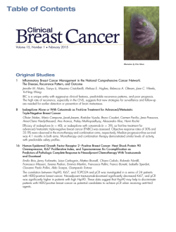
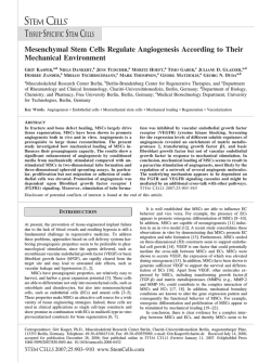
Int. J. Cancer: 112, 738 –746 (2004) © 2004 Wiley-Liss, Inc. Publication of the International Union Against Cancer FAST TRACK ETHANOL-INDUCED IN VITRO INVASION OF BREAST CANCER CELLS: THE CONTRIBUTION OF MMP-2 BY FIBROBLASTS Moe Moe AYE1, Cuiling MA2, Hong LIN2, Kimberly A. BOWER2, Richard C. WIGGINS1 and Jia LUO2,3* 1 Department of Neurobiology and Anatomy, West Virginia University School of Medicine, Robert C. Byrd Health Sciences Center, Morgantown, WV, USA 2 Department of Microbiology, Immunology and Cell Biology, West Virginia University School of Medicine, Robert C. Byrd Health Sciences Center, Morgantown, WV, USA 3 Institute for Nutritional Sciences, SIBS, Chinese Academy of Sciences, Shanghai, People’s Republic of China Ethanol is a tumor promoter and may promote metastasis of breast cancer. However, the underlying cellular/molecular mechanisms remain unknown. Overexpression and high activity of matrix metalloproteinase-2 (MMP-2) are frequently associated with metastatic breast cancers and serve as a prognostic indicator of clinical outcome. MMP-2 is predominantly expressed in stromal fibroblasts and plays a pivotal role in regulating the invasive behavior of breast tumor cells. We hypothesized that ethanol may enhance the invasion of breast tumor cells by modulating the activity of fibroblastic MMP-2. With in vitro models (HS68 and CCD1056SK human fibroblasts), we showed that ethanol at physiologically relevant concentrations (50 –200 mg/dl) activated MMP-2; conversely, at a higher concentration (400 mg/dl), it inhibited the MMP-2 activity. Consistently, conditioned medium collected from ethanol (50 –200 mg/dl)-exposed fibroblasts markedly enhanced the invasive potential of breast cancer cells and mammary epithelial cells overexpressing ErbB2/HER2 (BT474, SKBR-3 and HB2ErbB2 cells) but had little effect on cells with low ErbB2 levels (BT20 and HB2 cells). In contrast, conditioned medium obtained from ethanol (400 mg/dl)treated fibroblasts inhibited cell invasion. Selective inhibitors of MMP-2 (SB-3CT and OA-Hy) eliminated ethanol-stimulated invasion, indicating that the effect of ethanol was mediated by MMP-2. Ethanol activated conventional PKCs and JNKs in fibroblasts; inhibitors of PKC (Go6850 and Go6976) and JNKs (SP600125) significantly inhibited ethanol-mediated MMP-2 activation as well as cell invasion, indicating that PKCs and JNKs play a role in ethanol-induced MMP-2 activation and cell invasion in vitro. Thus, ethanol-promoted breast cancer cell invasion may be mediated by the modulation of fibroblastic MMP-2. © 2004 Wiley-Liss, Inc. Key words: alcohol; ErbB; metastasis; proteinases; signal transduction Breast cancer is a leading cause of morbidity and mortality in women.1 The endogenous and environmental factors that contribute to its etiology remain elusive. Despite being responsive to hormonal manipulation and chemotherapy, relapse after treatment is common, particularly in patients presenting with metastatic disease.2 The metastatic process involves the degradation of different macromolecular components of the extracellular matrix (ECM) and basement membranes and is regulated by intrinsic properties of the tumor cells as well as microenvironmental factors. Alcohol is a tumor promoter; there is a positive correlation between alcohol intake and the risk of several human cancers, including mouth/oropharyngeal cancer, oesophageal cancer, liver cancer and breast cancer.3–11 Epidemiologic studies indicate that alcohol consumption is associated with advanced and invasive breast tumors,12–14 suggesting that alcohol may enhance tumor development and metastasis. These epidemiologic results are supported by experimental studies using animal models and cell culture systems, which show that ethanol promotes mammary tumorigenesis and stimulates proliferation as well as invasion of breast cancer cells.15–21 The molecular mechanisms underlying ethanol action, however, remain to be determined. Abnormal communication between the mammary epithelium and stromal cells promotes tumorigenesis and development of breast carcinomas.22 Cancer-stroma interaction is mediated at least in part through the matrix metalloproteinases (MMPs). MMPs are a family of zinc-dependent endopeptidases that collectively are capable of degrading all components of the ECM. MMPs have been implicated in normal matrix remodeling events such as development of the mammary gland23 and in pathologic conditions, including tumor invasion and metastasis.24 Coupled with their function in metastasis, the MMPs also have a role in carcinogenesis.25,26 High levels of MMP-2 and MMP-9 have been found to correlate with enhanced metastasis and poor prognosis in patients with breast cancer.26 –31 Furthermore, MMP-2 activity is associated with the risk for a relapse in breast cancer patients.30 Interestingly, in most cases the two MMPs are not produced by malignant epithelium itself, but rather by surrounding tumor stroma.32–35 In particular, MMP-2 is mainly produced by stromal fibroblasts of breast tumors.35–37 It has been demonstrated that malignant breast tumor cells can enhance MMP-2 production in surrounding fibroblasts; cancer cells can subsequently use MMP-2 produced by the fibroblasts to facilitate their egress from tumor mass and entry into new sites.38 Breast tumor cells may not be the only target of ethanol action; ethanol may affect stromal cells and disrupt the tumor-stroma interaction. We hypothesize that ethanol may enhance the invasion of breast tumor cells by modulating the expression/activity of MMP-2 in fibroblasts. With in vitro models, we demonstrate that ethanol activates MMP-2 of fibroblasts in a concentration-dependent manner; conditioned medium collected from ethanol-exposed fibroblasts significantly alters invasive behavior of breast cancer cells and mammary epithelial cells. Our study suggests that Grant sponsor: National Institutes of Health; Grant numbers: AA12968 and CA90385; Grant sponsor: Alcohol Beverage Medical Research Foundation. Dr. Wiggins’s current address is: U.S. Environmental Protection Agency, Neurotoxicology Division, MD B105-03, Research Triangle Park, NC 27711. The first two authors contributed equally to this work. *Correspondence to: Department of Microbiology, Immunology and Cell Biology, West Virginia University School of Medicine, Morgantown, WV 26506. Fax: ϩ304-293-7823. E-mail: [email protected] Received 10 May 2004; Accepted after revision 23 June 2004 DOI 10.1002/ijc.20497 Published online 15 July 2004 in Wiley InterScience (www.interscience. wiley.com). 739 ETHANOL ACTIVATES MMP-2 IN FIBROBLASTS MMP-2 plays a critical role in the ethanol-stimulated invasion of breast cancer cells. MATERIAL AND METHODS Cell culture and treatment Human breast cancer cell lines (BT20, BT474 and SKBR-3), human fibrosarcoma cell line (HT1080) and human breast fibroblast cells (CCD1056SK) were purchased from American Type Culture Collection (ATCC, Rockville, MD). Human dermal fibroblast cells (HS68) was kindly provided by Dr. Daniel Flynn (Department of Microbiology, Immunology and Cell Biology, West Virginia University). BT20, BT474, SKBR-3, HT1080 and HS68 cells were grown in Dulbecco’s Modified Eagle’s Medium (DMEM) supplemented with 10% fetal bovine serum (FBS), penicillin (100 U/ml)/streptomycin (100 g/ml). Human breast epithelial cell line, HB2, is a clonal derivative of a nontumorigenic mammary epithelial cell line MTSV1-7 and expresses many of the markers typical of luminal mammary epithelial cells.39 HB2ErbB2 cells are HB2 cells overexpressing ErbB2.40 These cells were grown in the Eagle’s MEM containing 10% FBS, 2 mM Lglutamine, 25 g/ml gentamycin and 5 g/ml hydrocortisone. The cells are maintained at 37°C with 5% CO2. To block MMP-2 activity and other signaling components, the cells were pretreated with various inhibitors for 30 min. Inhibitors for MMP-2/MMP-9 (SB-3CT), MMP-2 (OA-Hy), JNK (SP600125), Protein kinase C (Go6850 and Go6976), MEK1 (PD98059) and p38 MAPK were purchased from Calbiochem (La Jolla, CA). Selective PI3K inhibitor (LY294002) was purchased from Promega (Madison, WI). Ethanol exposure method Cells were treated with ethanol at different concentrations (0, 25, 50, 100, 200 or 400 mg/dl). A method using a sealed container41 was used to maintain ethanol levels accurately in the culture medium. The containers were incubated in a humid atmosphere of 5% CO2 at 37°C. Preparation of conditioned media and cell lysates Fibroblast cells (3 ϫ 106 cells) were plated in 60 mm dishes and grown in the medium containing 10% FBS for 24 hr. Then the medium was replaced with a fresh medium without serum, and the cells were maintained in this medium for 24 – 48 hr. Fibroblastconditioned medium was collected and stored at Ϫ70°C. Cells were washed with PBS and lysed with RIPA buffer [150 mM, 50 mM Tris pH 8, 1% Nonidet P-40, 0.1% SDS, 0.5% deoxycholic acid sodium, 0.1 mg/ml phenylmethylsulfonyl fluoride (PMSF), 1 mM sodium orthovanadate and 15 g/ml aprotinin] for 10 min. Solubilized cells were centrifuged, proteinaceous supernatant was collected, and protein concentrations in the conditioned medium and cell lysates were determined.21 Gelatin zymography Fibroblast-conditioned medium and cell lysates were analyzed for gelatinase activity by the zymography. Proteins in cell lysates and medium samples were separated by electrophoresis under nonreducing conditions on 10% SDS- polyacrylamide gel containing 0.1% gelatin. The gels were rinsed with 2.5% Triton X-100 followed by washing with a buffer containing 50 mM Tris HCl, 0.1M NaCl, 2.5% Triton X-100, then treated with reaction buffer (50 mM Tris-HCl pH 8 and 10 mM CaCl2) overnight at 37°C. The gels were stained with a solution containing 0.25% Coomassie blue R-250, 10% methanol and 10% acetic acid and detained in the same solution without dye. Western blots Cellular proteins were separated by 10% SDS-polyacrylamide gel electrophoresis and transferred to nitrocellulose membranes. Membranes were blocked with 5% nonfat dry milk in 0.01 M PBS (pH 7.4) and 0.05% Tween-20 (TPBS) at room temperature for 1 hr, then incubated with a primary antibody directed against MMP-2 (diluted at 1:200 in TPBS) (Santa Cruz Biotech, Santa Cruz, CA), followed by 2 washes with TPBS. The membrane was incubated with a secondary antibody (diluted at 1:1,000 in TPBS) at room temperature for 1 hr. The immune complexes were detected by an enhanced chemiluminescence method (Amersham, Arlington Heights, IL). In vitro cell invasion A bioassay for in vitro cell invasion using Matrigel Invasion Chambers (Fisher Scientific, Pittsburgh, PA) was performed as described previously.16,21 Statistical analysis Differences among treatment groups were tested using analysis of variance (ANOVA). Differences in which p was less than 0.05 were considered statistically significant. In cases where significant differences were detected, specific post-hoc comparisons between treatment groups were examined with Student-Newman-Keuls tests. RESULTS Conditioned medium from ethanol-exposed fibroblasts stimulates invasion of breast cancer cells and mammary epithelial cells HB2ErbB2, BT474 and SKBR3 are cell lines overexpressing ErbB2, whereas BT20 and HB2 cells have low expression of ErbB2. In general, cells with upregulated ErbB2 expression exhibited higher invasive potential (Fig. 1a). Conditioned medium collected from HS68 fibroblast cells enhanced the invasion of breast cancer cells and mammary epithelial cells (Fig. 1b). Interestingly, it appeared that the increase in cell invasion occurred in cells expressing high levels of ErbB2. Furthermore, conditioned medium collected from ethanol-exposed HS68 cells significantly altered the invasive potential of breast cancer cells and mammary epithelial cells in a concentration-dependent manner (Fig. 1c). At concentrations of 100 and 200 mg/dl, ethanol treatment stimulated cell invasion; conversely, at 400 mg/dl, it either did not affect or inhibited cell invasion. In addition, treatment of ethanol stimulated the invasion of cells expressing higher levels of ErbB2, namely, HB2ErbB2, BT474 and SKBR3 cells. Ethanol-stimulated cell invasion is mediated by fibroblastic MMP-2 MMP-2 is produced as an inactive enzyme, pro-MMP2 (72 kDa). Its activation is mediated by cleavage of the propeptide domain.42 Previous studies indicate that MMP-2 activation is accompanied by the expression of active forms of 64/62 kDa and 59 kDa.43,44 Gelatin zymography revealed that HS68 and CCD1056SK cells expressed high levels of pro-MMP-2 (72 kDa) and a small amount of 2 active forms of MMP-2 (62 and 59 kDa) (Fig. 2a). The identity of MMP-2 was further confirmed by Western blot using a specific anti-MMP-2 antibody (Fig. 2a). HT1080 cells, which are known for the production of MMP-2 and MMP-9, were used as a reference for MMP expression. Zymography showed that HT1080 cells expressed MMP-2 (72 kDa and 62 kDa) and pro-MMP-9 (92 kDa). MMP-9 expression in HS68 and CCD1056SK cells was quite weak and barely detectable. Next, we examined the effect of ethanol on fibroblastic MMPs. HS68 cells were exposed to ethanol (100 – 400 mg/dl) for 24 – 48 hr. The conditioned medium and cell lysate were collected and subjected to gelatin zymography. The results of ethanol exposure for 24 and 48 hr were similar; only data collected from 48 hr of ethanol exposure are presented. As shown in Figure 2b, at concentrations of 100 and 200 mg/dl, ethanol significantly increased the amount of active forms of MMP-2 (62 and 59 kDa), indicating the activation of MMP-2. In contrast, ethanol at 400 mg/dl decreased the amount of active forms of MMP-2. In HS68 cells, ethanol at concentrations less than 100 mg/dl did not significantly affect MMP-2 activation (data no shown). Ethanol-induced alterations in MMP-2 activation did not result from changes in cell viability because ethanol did not significantly affect viability of these cells as determined by the MTT assay (data not shown). Therefore, the 740 AYE ET AL. FIGURE 1 – Effect of fibroblast-conditioned medium on the invasion of breast cancer and mammary epithelial cells. (a) Human breast cancer cells (BT20, BT474 and SKBR3) and mammary epithelial cells lines (HB2 and HB2ErbB2) were maintained in serum-free medium (SFM). The invasive potential of these cells during a period of 48 hr was determined by a Matrigel in vitro invasion assay as described in Material and Methods. The result was the mean of 3 replications. (b) HS68 human dermal fibroblast cells were cultured in serum-free medium for 48 hr and conditioned medium was collected. BT20, BT474 and SKBR3, HB2 and HB2ErbB2 cells were maintained in either SFM or HS68 cell-conditioned medium (CM). The invasive potential of these cells during a period of 48 hr was determined. The invasive potential of the cells maintained in the CM was quantified and expressed relative to that in the SFM. The result was the mean of 3 replications. Asterisk denotes significant difference from SFM groups, p Ͻ 0.05. (c) HS68 cells grown in serum-free medium were exposed to ethanol (0 – 400 mg/dl) for 48 hr, and conditioned medium was collected. The effect of these conditioned media on the invasion of BT20, BT474, SKBR3, HB2 and HB2ErbB2 cells was determined as described above. Ethanol-induced alteration in the cell invasion was expressed relative to untreated groups. The result was the mean of 3 replications. Asterisk denotes significant difference from untreated groups, p Ͻ 0.05. ETHANOL ACTIVATES MMP-2 IN FIBROBLASTS pattern of ethanol modulation of MMP-2 activation was consistent with its effect on cell invasion. MMP-9 expression in HS68 cells was weak and was not affected by ethanol exposure (data not shown). Similarly, ethanol modulated MMP-2 activation in CCD1056SK breast fibroblast cells in a concentration-dependent manner (Fig. 2c). CCD1056SK cells were more sensitive to ethanol; the effect of ethanol was evident at 50 –100 mg/dl and diminished at 200 mg/dl. At 400 mg/dl, ethanol also inhibited MMP-2 activation in CCD1056SK cells (data not shown). To verify the involvement of MMP-2 in ethanol-stimulated cell invasion, we employed 2 selective MMP inhibitors to block MMP-2 activity. SB-3CT inhibits MMP-2 activity by directly targeting the catalytic zinc ion of MMP-2.45 OA-Hy, another specific inhibitor for MMP-2, has been previously used to block MMP-2-mediated cell invasion.46,47 As shown in Figure 2d and e, both inhibitors significantly blocked ethanol-stimulated invasion of HB2ErbB2 cells; it appeared that SB-3CT was more effective. SB-3CT also modestly and significantly inhibited basal cell invasion (the invasion not stimulated by ethanol). Ethanol-induced MMP-2 activation is mediated by PKC We further investigated the effect of ethanol on intracellular signaling in fibroblasts. There are multiple PKC isoforms including: conventional PKCs (␣,  and ␥), which are regulated by calcium, diacylglycerol (DAG), phorbol esters (TPA or PMA) and phosphatidylserine (PS); novel PKCs (␦, ⑀, , and ), which are calcium-independent but are regulated by DAG, TPA and PS; and atypical PKCs ( and ), which are calcium-independent and do not require DAG, TPA and PS. Anti-phospho-PKC(pan) antibody detects phosphorylated PKC␣/, ␦, ⑀ and isoforms. Anti-phospho-PKC␣/II antibody recognizes phosphorylated PKC␣/II at Thr638/641. Using both antibodies, we observed the ethanolstimulated PKC phosphorylation. As shown in Figure 3, ethanolenhanced PKC phosphorylation was detected as early as 15 min after ethanol exposure; the stimulation lasted for at least 2 hr. Ethanol had little effect on the phosphorylation of PKC ␦, and /. Ethanol induced a transient phosphorylation of JNKs without affecting ERK, p38 MAPK and Akt (Fig. 3). Using selective inhibitors, we sought to determine the potential signaling components that are responsible for ethanol-induced MMP-2 activation. Go6976 is a selective inhibitor of PKC␣ and PKC;48 Go6850 (Bisindolylmaleimide I; Bis-I) is a broad inhibitor of PKC.49 Consistent with the results showing that ethanol activated PKC␣/ II, PKC inhibitors (Go6850 and Go6976) blocked ethanol-induced MMP-2 activation (Fig. 4b). It was noted that Go6976 also FIGURE 2 – MMP-2 activation and ethanol-stimulated invasion. (a) Cell extracts were analyzed for the expression of gelatinases by Western blots (lane 1) and gelatin zymography (lanes 2– 4). Lanes 1 and 2, HS68 cells; lane 3, CCD1056SK cells; lane 4, HT1080 cells. (b) HS68 cells grown in the serum-free medium were exposed to ethanol (0 – 400 mg/dl) for 48 hr. Conditioned medium and cell extract were collected, and protein concentration was determined. The expression and activation of MMP-2 was analyzed by gelatin zymography (top panel). The relative amount of MMP-2 was measured microdensitometrically using SigmaGel software (SPSS, Chicago, IL) and expressed relative to the control (bottom panel). The experiment was replicated 3 times. (c) CCD1056SK cells were grown in serum-free medium and exposed to ethanol (0 –200 mg/dl) for 24 hr. Conditioned media were collected and subject to gelatin zymography (top panel). The relative amount of MMP-2 was quantified and expressed relative to the control (bottom panel). (d) HS68 cells grown in serum-free medium were exposed to ethanol (100 or 200 mg/dl) with/without MMP-2 inhibitors (OA-Hy, 20 M; SB-3CT, 4 M) for 48 hr. Conditioned media were collected; the effect of these conditioned media on the invasion of HB2ErbB2 cells was determined as described in Figure 1. The result was the mean of 3 replications. Asterisk denotes significant difference from untreated groups (Ct), p Ͻ 0.05; double asterisks denote significant difference from paired ethanol-treated groups, p Ͻ 0.05. (e) CCD1056SK cells grown in serum-free medium were exposed to ethanol (100 mg/dl) with/without MMP-2 inhibitors (OA-Hy, 20 M; SB-3CT, 4 M) for 24 hr. Notations are as in panel c. 741 742 AYE ET AL. FIGURE 2 – CONTINUED. sought to determine whether blocking PKCs and JNKs was sufficient to inhibit ethanol-mediated cells invasion. As shown in Figure 5, Go6976 inhibited ethanol-stimulated invasion; SP600125 also decreased ethanol-induced cell invasion but to a lesser extent. DISCUSSION FIGURE 3 – Effect of ethanol on intracellular signaling. HS68 cells were grown in serum-free medium and exposed to ethanol (200 mg/dl) for 5–120 min. The phosphorylation of various protein kinases was assessed by Western blots using phospho-specific antibodies. The blot was stripped and reprobed with an anti-actin antibody. The experiment was replicated 3 times. blocked basal activation of MMP-2 (activation not stimulated by ethanol) (Fig. 4a). Blocking JNK activity by SP600125 also inhibited MMP-2 activation. On the other hand, inhibition of ERK, p38 MAPK and Akt had little effect on ethanol-mediated MMP-2 activation. Since MMP-2 activation, which was essential for ethanol-stimulated invasion, was regulated by PKCs and JNKs, we Reciprocal cellular interaction between epithelial and stromal cells plays an important role in tumor growth, progression and metastasis.22,50,51 Several lines of evidence indicate that stromal cells may regulate tumor invasion and dissemination via ECM remodeling and degradation.38,51–54 MMPs, particularly MMP-2, are strongly implicated in this process.55,56 Fibroblasts, the most abundant stromal cell type in desmoplastic tumors, are the major source of MMP-2. It has been shown that breast cancer cellconditioned medium enhances MMP-2 but not MMP-9 production by fibroblasts.35 Similarly, Saad et al.38 report that co-culture of breast cancer cells and bone marrow fibroblasts (BMFs) induces the release of MMP-2 from BMFs. MMP-2 is expressed in the very early stage of breast cancer and its levels are consistent with increasing tumor grade.29,57 Malignant cells could use MMP-2 produced by adjacent fibroblasts to facilitate their invasion of normal tissues.38 Our results indicate that HS68 and CCD1056SK fibroblasts have a high expression of MMP-2 but weak levels of MMP-9, consistent with the literature showing that fibroblasts are an important source of MMP-2. The fibroblast-conditioned medium enhances the invasion of some breast cancer cells. In addition, conditioned media collected from ethanol-exposed fibroblasts significantly alter invasive behavior of breast cancer cells and mammary epithelial cells in a concentration-dependent manner. At physiologically relevant concentrations (50 –200 mg/dl), ethanol markedly enhances the invasive potential of breast cancer cells or mammary epithelial cells; however, at a relatively high concentration of 400 mg/dl, it inhibits the invasion of some breast cancer cells. This biphasic effect of ethanol is also observed on MMP-2 activation; ethanol at modest concentrations activates MMP-2, whereas it inhibits MMP-2 activation at a high concentration. It appears that CCD1056SK cells are more sensitive to ethanol than HS68 cells; ethanol at 50 mg/dl activates MMP-2 in CCD1056SK, whereas 100 mg/dl is necessary to activate MMP-2 in HS68 cells. The mechanism underlying this differential sensitivity to ethanol is currently unknown; it may be due to the difference in the genetic background and signaling components that these cells inherited. For example, it is shown that the status of growth factor receptor and intracellular signaling components determine cellular sensitivity to ethanol.21,41 Despite the differential sensitivity, these cells respond to ethanol in a similar pattern. Furthermore, selective inhibitors of MMP-2 (SB-3CT and OA-Hy) eliminate ethanolstimulated invasion. Taken together, these results indicated fibro- ETHANOL ACTIVATES MMP-2 IN FIBROBLASTS 743 FIGURE 4 – Effect of protein kinase inhibitors on MMP-2 activation. HS68 cells were grown in serum-free medium and exposed to ethanol (200 mg/dl; 48 hr) with/without specific protein kinase inhibitors. The expression and activation of MMP-2 was analyzed by gelatin zymography. The experiment was replicated 3 times. The inhibitors are: Go6976 (1 M), PKC␣/ inhibitor; Go6850 (Bis-I, 1 M), pan-PKC inhibitor; LY294002 (LY, 10 M), PI3K inhibitors; PD98059 (PD, 50 M), MEK1 inhibitor; SB202190 (SB, 10 M), p38 MAPK inhibitor; SP600125 (SP, 25 M), JNK inhibitor. FIGURE 5 – Effect of PKC and JNK inhibitors on ethanol-stimulated cell invasion. (a) HS68 cells grown in serum-free medium were exposed to ethanol (200 mg/dl) with/without Go6970 (Go) and SP600125 (SP) for 48 hr. The conditioned media were collected; the effect of these conditioned media on the invasion of HB2ErbB2 cells was determined as described in Figure 1. The experiment was replicated 3 times. Asterisk denotes significant difference from untreated group (Ct), p Ͻ 0.05; double asterisks denote significant difference from paired ethanol-treated group (Et), p Ͻ 0.05. (b) CCD1056SK cells maintained in serum-free medium and exposed to ethanol (200 mg/dl) with/without Go6970 (Go) and SP600125 (SP) for 24 hr. The conditioned media were collected; the effect of these conditioned media on the invasion of HB2ErbB2 cells was assayed. Other notations are as in panel a. blastic MMP-2 plays a critical role in regulating invasiveness of breast cancer and mammary epithelial cells in vitro. Two in vivo studies examine the effect of ethanol on MMPs. Lois et al.58 demonstrated that ethanol exposure increases MMP-2 and MMP-9 activity but not their production in rat lungs. Similarly, ethanol consumption upregulates the enzymatic activity of MMP-2 in rat aortas.59 Although these studies do not identify the source of MMP-2, they support our finding that MMP-2 is a target of ethanol. Therefore, it is conceivable that ethanol may stimulate stromal fibroblasts surrounding breast carcinomas, which are a rich source of MMP-2, and render MMP-2 activation. We demonstrate that the fibroblast-conditioned medium enhances the invasion of breast cancer cells and mammary epithelial cells overexpressing ErbB2. Interestingly, ethanolstimulated invasion only occurs in the cells of ErbB2 overexpression. The epidermal growth factor receptor (EGFR) family contains EGFR (ErbB1/HER1), ErbB2 (HER2/Neu), ErbB3 (HER3) and ErbB4 (HER4), which play an important role in regulation of cell proliferation, differentiation, survival, adhesion, migration and invasion.60,61 ErbB2 is a potent oncoprotein, despite the fact that no known ligand binds to it with high affinity. Although a normal level of ErbB2 is required for the regulation of normal breast growth and development,62 amplification and overexpression of ErbB2 is the most common mechanism leading to the disruption of normal cellular control and the formation of aggressive tumor cells in breast tissue.60,61 ErbB2 has been shown to facilitate the invasion of breast cancer cells in vitro and in vivo.21,63– 65 The status of ErbB2 expression in a given cell is critical in determining the cellular response to various exogenous stimuli.21,66 – 68 Overexpression of ErbB2 could result in autophosphorylation, which initiates intracellular signaling; it is suggested that ErbB2 controls stroma-toepithelium crosstalk.69 Although underlying mechanisms are currently unknown, our results suggest that the status of ErbB2 744 AYE ET AL. expression may modulate the interaction between breast tumor cells and fibroblasts. Ethanol has been shown to modulate PKC activity in vitro and in vivo.70 –73 Our study shows that ethanol promotes phosphorylation of conventional PKCs in fibroblasts. Anti-phosphoPKC(pan)(II Ser660) antibody detects PKC␣/, ␦, ⑀ and isoforms phosphorylated at a C-terminal residue homologous to Ser660 of PKC II. Anti-phospho-PKC␣/II antibody recognizes phosphorylated PKC␣/II at Thr638/641. Using these antibodies, we can conclude that ethanol stimulates ␣ and/or  isoforms. It is not clear whether ethanol also affects other conventional PKCs. Using selective inhibitors of PKCs, our study verifies that PKCs, probably the PKC␣ and/or  isoform, are involved in ethanolinduced MMP-2 activation because a specific inhibitor of PKC␣/ (Go6976) is sufficient to block ethanol-induced MMP-2 activation. Furthermore, Go6976 significantly inhibits ethanol-mediated cell invasion, validating that the PKC-MMP-2 pathway plays an important role in ethanol-stimulated cell invasion. Calcium is a key regulator of conventional PKCs. Ethanol is shown to disrupt intracellular calcium homeostasis.74,75 Therefore, it is possible that ethanol activates conventional PKCs through its modulation of intracellular calcium concentration. PKCs have been implicated in MMP-2 expression/activation; however, its involvement in the regulation of MMP-2 appears to be cell type-dependent. For example, it has been shown that phorbol 12-myristate 13-acetate (PMA), a PKC activator, induces MMP-2 activation and promotes invasiveness of glioblastoma cells in vitro; blocking PKC activity by selective PKC inhibitors eliminates PMA-induced MMP-2 activation and cell invasion.76,77 On the other hand, Chang et al.78 demonstrate that PKC inhibitors (H7 and Go6976) inhibit MMP-2 production in cultured human pulp cells, suggesting PKCs are involved in the regulation of MMP-2 expression. In contrast, Yeung and Hurta79 report that PMA downregulates MMP-2 mRNA in the H-ras-transformed mouse fibroblast cell line. Ethanol also induces a transient activation of both JNK1 and JNK2, which lasts for about 30 min. JNKs are members of mitogenactivated protein kinases and are important mediators of cellular response to stress stimuli. Blockage of JNK activity significantly inhibits ethanol-mediated MMP-2 activation and cell invasion, suggesting that JNKs are also involved in these processes. Several potential mechanisms may contribute to the involvement of MMP-2 in ethanol-stimulated cell invasion. First, activated MMP-2 can degrade Matrigel and facilitate the invasion of breast cancer cells. Alternatively, MMP-2 may interact with cell surface integrins and enhance cell mobility and invasion.80 In addition, activated MMP-2 is also shown to regulate the activity of other cell surface proteinases and increase cell invasiveness. For example, MMP-2 can activate MMP-9, which is produced by breast cancer cells.81 In conclusion, our study indicates that fibroblastic MMP-2 is a target of ethanol. MMP-2 is an important mediator for the invasion and metastasis of breast tumor cells. Since stromal fibroblasts are the major source of MMP-2 in mammary tissues, our results suggest that ethanol may promote tumor metastasis by activating stromal MMP-2. REFERENCES 1. 2. 3. 4. 5. 6. 7. 8. 9. 10. 11. 12. 13. 14. 15. 16. 17. Kelsey JL, Horn-Ross PL. Breast cancer: magnitude of the problem and descriptive epidemiology Epidemiol Rev 1993;15:7–16. Honig S. Treatment of metastatic disease: hormonal therapy and chemotheraphy In: Harris J, Lippman M, Morrow M, Hellman S, eds. Disease of the breast. Philadelphia: Lippinoctt-Raven, 1996. 669 – 734. Hiatt RA. Alcohol consumption and breast cancer. Med Oncol Tumor Pharmacother 1990;7:143–51. Plant ML. Alcohol and breast cancer: a review. Int J Addict 1992;27: 107–28. Mufti SI. International Society for Biomedical Research on Alcoholism: relationship of cell necrosis and proliferation, free radicals and other agents to alcohol-related cancers. J Cancer Res Clin Oncol 1993;19:304 –5. Mufti SI. Alcohol-stimulated promotion of tumors in the gastrointestinal tract. Cancer Detect Prev 1998;22:195–203. Rosenberg L, Metzger LS, Palmer JR. Alcohol consumption and risk of breast cancer: a review of the epidemiologic evidence. Epidemiol Rev 1993;15:133– 44. Longnecker MP, Paganini-Hill A, Ross RK. Lifetime alcohol consumption and breast cancer risk among postmenopausal women in Los Angeles. Cancer Epidemiol Biomarkers Prev 1995;4:721–5 Longnecker MP, Newcomb PA, Mittendorf R, Greenberg ER, Clapp RW, Bogdan GF, Baron J, MacMahon B, Willett WC. Risk of breast cancer in relation to lifetime alcohol consumption. J Natl Cancer Inst 1995;87:923–9. Singletary KW, Gapstur SM. Alcohol and breast cancer: review of epidemiologic and experimental evidence and potential mechanisms. JAMA 2001;2862143–51. Rehm J, Room R, Graham K, Monteiro M, Gmel G, Sempos CT. The relationship of average volume of alcohol consumption and patterns of drinking to burden of disease: an overview. Addiction 2003;98: 1209 –28. Weiss HA, Brinton LA, Brogan D, Coates RJ, Gammon MD, Malone KE, Schoenberg JB, Swanson CA. Epidemiology of in situ and invasive breast cancer in women aged under 45. Br J Cancer 1996; 73:1298 –305. Vaeth PA, Satariano WA. Alcohol consumption and breast cancer stage at diagnosis. Alcohol Clin Exp Res 1998;22:928 –34. Stoll BA. Alcohol intake and late-stage promotion of breast cancer. Eur J Cancer 1999;35:1653– 8. Singletary K. Ethanol and experimental breast cancer: a review. Alcohol Clin Exp Res 1997;21:334 –9. Luo J, Miller MW. Ethanol enhances erbB-mediated migration of human breast cancer cells in culture. Breast Cancer Res Treat 2000; 63:61–9. Meng Q, Gao B, Goldberg ID, Rosen EM, Fan S. Stimulation of cell 18. 19. 20. 21. 22. 23. 24. 25. 26. 27. 28. 29. 30. invasion and migration by alcohol in breast cancer cells. Biochem Biophys Res Commun 2000;273:448 –53. Watabiki T, Okii Y, Tokiyasu T, Yoshimura S, Yoshida M, Akane A, Shikata N, Tsubura A. Long-term ethanol consumption in ICR mice causes mammary tumor in females and liver fibrosis in males. Alcohol Clin Exp Res 2000;24:117S–22S. Singletary KW, Frey RS, Yan W. Effect of ethanol on proliferation and estrogen receptor-alpha expression in human breast cancer cells. Cancer Lett 2001;165:131–7. Izevbigie EB, Ekunwe SI, Jordan J, Howard CB. Ethanol modulates the growth of human breast cancer cells in vitro. Exp Biol Med (Maywood) 20002;227:260 –5. Ma C, Lin H, Leonard SS, Shi X, Ye J, Luo J. Overexpression of ErbB2 enhances ethanol-stimulated intracellular signaling and invasion of human mammary epithelial and breast cancer cells in vitro. Oncogene 2003;22:5281–90. Wiseman BS, Werb Z. Stromal effects on mammary gland development and breast cancer Science 2002;296:1046 –9. Werb Z, Ashkenas J, MacAuley A, Wiesen JF. Extracellular matrix remodeling as a regulator of stromal-epithelial interactions during mammary gland development, involution and carcinogenesis. Braz J Med Biol Res 1996;29:1087–97. McCawley LJ, Matrisian LM. Matrix metalloproteinases: they’re not just for matrix anymore! Curr Opin Cell Biol 2001;13:534 – 40. Coussens LM, Tinkle CL, Hanahan D, Werb Z. MMP-9 supplied by bone marrow-derived cells contributes to skin carcinogenesis. Cell 2000;103:481–90. Duffy MJ, Maguire TM, Hill A, McDermott E, O’Higgins N. Metalloproteinases: role in breast carcinogenesis, invasion and metastasis. Breast Cancer Res 2000;2:252–7. Kossakowska AE, Huchcroft SA, Urbanski SJ, Edwards DR. Comparative analysis of the expression patterns of metalloproteinases and their inhibitors in breast neoplasia, sporadic colorectal neoplasia, pulmonary carcinomas and malignant non-Hodgkin’s lymphomas in humans. Br J Cancer 1996;73:1401– 8. Hanemaaijer R, Verheijen JH, Maguire TM, Visser H, Toet K, McDermott E, O’Higgins N, Duffy MJ. Increased gelatinase-A and gelatinase-B activities in malignant vs. benign breast tumors. Int J Cancer 2000;86:204 –7. Talvensaari-Mattila A, Paakko P, Hoyhtya M, Blanco-Sequeiros G, Turpeenniemi-Hujanen T. Matrix metalloproteinase-2 immunoreactive protein: a marker of aggressiveness in breast carcinoma. Cancer 1998;83:1153– 62. Talvensaari-Mattila A, Paakko P, Blanco-Sequeiros G, TurpeenniemiHujanen T. Matrix metalloproteinase-2 (MMP-2) is associated with the risk for a relapse in postmenopausal patients with node-positive ETHANOL ACTIVATES MMP-2 IN FIBROBLASTS 31. 32. 33. 34. 35. 36. 37. 38. 39. 40. 41. 42. 43. 44. 45. 46. 47. 48. 49. 50. 51. breast carcinoma treated with antiestrogen adjuvant therapy. Breast Cancer Res Treat 2001;65:55– 61. Talvensaari-Mattila A, Paakko P, Turpeenniemi-Hujanen T. Matrix metalloproteinase-2 (MMP-2) is associated with survival in breast carcinoma. Br J Cancer 2003;89:1270 –5. Basset P, Bellocq JP, Wolf C, Stoll I, Hutin P, Limacher JM, Podhajcer OL, Chenard MP, Rio MC, Chambon P. A novel metalloproteinase gene specifically expressed in stromal cells of breast carcinomas. Nature 1990;348:699 –704. Pyke C, Ralfkiaer E, Huhtala P, Hurskainen T, Dano K, Tryggvason K. Localization of messenger RNA for Mr 72,000 and 92,000 type IV collagenases in human skin cancers by in situ hybridization. Cancer Res 1992;52:1336 – 41. Crawford HC, Matrisian LM. Tumor and stromal expression of matrix metalloproteinases and their role in tumor progression. Invasion Metastasis 1995;14:234 – 45. Singer CF, Kronsteiner N, Marton E, Kubista M, Cullen KJ, Hirtenlehner K, Seifert M, Kubista E. MMP-2 and MMP-9 expression in breast cancer-derived human fibroblasts is differentially regulated by stromal-epithelial interactions. Breast Cancer Res Treat 2002;72:69 – 77. Noel AC, Polette M, Lewalle JM, Munaut C, Emonard HP, Birembaut P, Foidart JM. Coordinate enhancement of gelatinase A mRNA and activity levels in human fibroblasts in response to breast-adenocarcinoma cells. Int J Cancer 1994;56:331– 6. Heppner KJ, Matrisian LM, Jensen RA, Rodgers WH. Expression of most matrix metalloproteinase family members in breast cancer represents a tumor-induced host response. Am J Pathol 1996;149:273– 82. Saad S, Gottlieb DJ, Bradstock KF, Overall CM, Bendall LJ. Cancer cell-associated fibronectin induces release of matrix metalloproteinase-2 from normal fibroblasts. Cancer Res 2002;62:283–9. Alford D, Baeckstrom D, Geyp M, Pitha P, Taylor-Papadimitriou J. Integrin-matrix interactions affect the form of the structures developing from human mammary epithelial cells in collagen or fibrin gels. J Cell Sci 1998;111:521–32. Ye J, Xu RH, Taylor-Papadimitriou J, Pitha PM. Sp1 binding plays a critical role in Erb-B2- and v-ras-mediated downregulation of alpha2integrin expression in human mammary epithelial cells. Mol Cell Biol 1996;16:6178 – 89. Luo J, Miller MW. Differential sensitivity of human neuroblastoma cell lines to ethanol: correlations with their proliferative responses to mitogenic growth factors and expression of growth factor receptors. Alcohol Clin Exp Res 1997;21:1186 –94. Nagase H. Activation mechanisms of matrix metalloproteinases. Biol Chem 1997;378:151– 60. Gilles C, Bassuk JA, Pulyaeva H, Sage EH, Foidart JM, Thompson EW. SPARC/osteonectin induces matrix metalloproteinase 2 activation in human breast cancer cell lines. Cancer Res 1998;58: 5529 –36. Esparza J, Vilardell C, Calvo J, Juan M, Vives J, Urbano-Marquez A, Yague J, Cid MC. Fibronectin upregulates gelatinase B (MMP-9) and induces coordinated expression of gelatinase A (MMP-2) and its activator MT1-MMP (MMP-14) by human T lymphocyte cell lines. A process repressed through RAS/MAP kinase signaling pathways. Blood 1999;94:2754 – 66. Kleifeld O, Kotra LP, Gervasi DC, Brown S, Bernardo MM, Fridman R, Mobashery S, Sagi I. X-ray absorption studies of human matrix metalloproteinase-2 (MMP-2) bound to a highly selective mechanismbased inhibitor comparison with the latent and active forms of the enzyme. J Biol Chem 2001;276:17125–31. Emonard H, Marcq V, Mirand C, Hornebeck W. Inhibition of gelatinase A by oleic acid. Ann NY Acad Sci 1999;878:647–9 Liao X, Thrasher JB, Pelling J, Holzbeierlein J, Sang QX, Li B. Androgen stimulates matrix metalloproteinase-2 expression in human prostate cancer. Endocrinology 2003;144:1656 – 63. Martiny-Baron G, Kazanietz MG, Mischak H, Blumberg PM, Kochs G, Hug H, Marme D, Schachtele C. Selective inhibition of protein kinase C isozymes by the indolocarbazole Go 6976. J Biol Chem 1993;268:9194 –7. Toullec D, Pianetti P, Coste H, Bellevergue P, Grand-Perret T, Ajakane M, Baudet V, Boissin P, Boursier E, Loriolle F, et al. The bisindolylmaleimide GF 109203X is a potent and selective inhibitor of protein kinase C. J Biol Chem 1991;266:15771– 81. Shekhar MP, Werdell J, Santner SJ, Pauley RJ, Tait L. Breast stroma plays a dominant regulatory role in breast epithelial growth and differentiation: implications for tumor development and progression. Cancer Res 2001;61:1320 – 6. Shekhar MP, Pauley R, Heppner G. Host microenvironment in breast cancer development: extracellular matrix-stromal cell contribution to neoplastic phenotype of epithelial cells in the breast. Breast Cancer Res 2003;5:130 –5. 745 52. Grey AM, Schor AM, Rushton G, Ellis I, Schor SL. Purification of the migration stimulating factor produced by fetal and breast cancer patient fibroblasts. Proc Natl Acad Sci USA 1989;86:2438 – 42. 53. Camps JL, Chang SM, Hsu TC, Freeman MR, Hong SJ, Zhau HE, von Eschenbach AC, Chung LW. Fibroblast-mediated acceleration of human epithelial tumor growth in vivo. Proc Natl Acad Sci USA 1990;87:75–9. 54. Stetler-Stevenson WG, Liotta LA, Kleiner DE Jr. Extracellular matrix 6: role of matrix metalloproteinases in tumor invasion and metastasis. FASEB J 1993;7:1434 – 41. 55. Curran S, Murray GI. Matrix metalloproteinases in tumour invasion and metastasis. J Pathol 1999;189:300 – 8. 56. Kleiner DE, Stetler-Stevenson WG. Matrix metalloproteinases and metastasis. Cancer Chemother Pharmacol 1999;43:S42–51. 57. Poulsom R, Hanby AM, Pignatelli M, Jeffery RE, Longcroft JM, Rogers L, Stamp GW. Expression of gelatinase A and TIMP-2 mRNAs in desmoplastic fibroblasts in both mammary carcinomas and basal cell carcinomas of the skin. J Clin Pathol 1993;46:429 – 36. 58. Lois M, Brown LA, Moss IM, Roman J, Guidot DM. Ethanol ingestion increases activation of matrix metalloproteinases in rat lungs during acute endotoxemia Am J Respir Crit Care Med 1999;160: 1354 – 60. 59. Partridge CR, Sampson HW, Forough R. Long-term alcohol consumption increases matrix metalloproteinase-2 activity in rat aorta. Life Sci 1999;65:1395– 402. 60. Yarden Y, Sliwkowski MX. Untangling the ErbB signalling network. Nat Rev Mol Cell Biol 2001;2:127–37. 61. Zhou BP, Hung MC. Dysregulation of cellular signaling by HER2/neu in breast cancer. Semin Oncol 2003;30:38 – 48. 62. Carraway KL 3rd, Weber JL, Unger MJ, Ledesma J, Yu N, Gassmann M, Lai C. Neuregulin-2, a new ligand of ErbB3/ErbB4-receptor tyrosine kinases. Nature 1997;387:512– 6. 63. Tan M, Yao J, Yu D. Overexpression of the c-erbB-2 gene enhanced intrinsic metastasis potential in human breast cancer cells without increasing their transformation abilities. Cancer Res 1997;57:1199 – 205. 64. Xu FJ, Stack S, Boyer C, O’Briant K, Whitaker R, Mills GB, Yu YH, Bast RC Jr. Heregulin and agonistic anti-p185(c-erbB2) antibodies inhibit proliferation but increase invasiveness of breast cancer cells that overexpress p185(c-erbB2): increased invasiveness may contribute to poor prognosis. Clin Cancer Res1997;3:1629 –34. 65. Spencer KS, Graus-Porta D, Leng J, Hynes NE, Klemke RL. ErbB2 is necessary for induction of carcinoma cell invasion by ErbB family receptor tyrosine kinases. J Cell Biol 2000;148:385–97. 66. Schmidt-Ullrich RK, Contessa JN, Dent P, Mikkelsen RB, Valerie K, Reardon DB, Bowers G, Lin PS. Molecular mechanisms of radiationinduced accelerated repopulation. Radiat Oncol Investig 1999;7:321– 30. 67. Sun Y, Lin H, Zhu Y, Ma C, Ye J, Luo J. Induction or suppression of expression of cytochrome C oxidase subunit II by heregulin beta 1 in human mammary epithelial cells is dependent on the levels of ErbB2 expression. J Cell Physiol 2002;192:225–33. 68. Guo G, Wang T, Gao Q, Tamae D, Wong P, Chen T, Chen WC, Shively JE, Wong JY, Li JJ. Expression of ErbB2 enhances radiationinduced NF-kappaB activation. Oncogene 2004;23:535– 45. 69. Yarden Y. Biology of HER2 and its importance in breast cancer Oncology 2001;61:1–13. 70. Smith TL, Bitrick MS. Ethanol enhances the in situ phosphorylation of MARCKS and protein kinase C activity in primary cultures of astrocytes. Life Sci 1996;58:855– 60. 71. Mahadev K, Vemuri MC. Selective changes in protein kinase C isoforms and phosphorylation of endogenous substrate proteins in rat cerebral cortex during pre- and postnatal ethanol exposure. Arch Biochem Biophys 1998;356:249 –57. 72. Nguyen VA, Chen J, Hong F, Ishac EJ, Gao B. Interferons activate the p42/44 mitogen-activated protein kinase and JAK-STAT (Janus kinase-signal transducer and activator transcription factor) signalling pathways in hepatocytes: differential regulation by acute ethanol via a protein kinase C-dependent mechanism. Biochem J 2000;349:427–34. 73. Sultana R, Babu PP. Ethanol-induced alteration in N-methyl-D-aspartate receptor 2A C-terminus and protein kinase C activity in rat brain. Neurosci Lett 2003;349:45– 8. 74. Yang Z, Wang J, Zheng T, Altura BT, Altura BM. Importance of extracellular Ca2ϩ and intracellular Ca2ϩ release in ethanol-induced contraction of cerebral arterial smooth muscle. Alcohol 2001;24:145– 53. 75. Webb B, Walker DW, Heaton MB. Nerve growth factor and chronic ethanol treatment alter calcium homeostasis in developing rat septal neurons. Brain Res Dev Brain Res 2003;143:57–71. 746 AYE ET AL. 76. Uhm JH, Dooley NP, Villemure JG, Yong VW. Glioma invasion in vitro: regulation by matrix metalloprotease-2 and protein kinase C. Clin Exp Metastasis 1996;14:421–33. 77. Park MJ, Park IC, Hur JH, Rhee CH, Choe TB, Yi DH, Hong SI, Lee, SH. Protein kinase C activation by phorbol ester increases in vitro invasion through regulation of matrix metalloproteinases/tissue inhibitors of metalloproteinases system in D54 human glioblastoma cells. Neurosci Lett 2000;290:201– 4. 78. Chang YC, Yang SF, Hsieh YS. Regulation of matrix metalloproteinase-2 production by cytokines and pharmacological agents in human pulp cell cultures. J Endod 2001;27:679 – 82. 79. Yeung O, Hurta RA. Phorbol ester tumour promoter mediated altered expression and regulation of matrix metalloproteinase-2 in a H-ras transformed cell line capable of benign tumour formation. Mol Cell Biochem 2001;220:39 – 48. 80. Mitra A, Chakrabarti J, Chattopadhyay N, Chatterjee A. Membraneassociated MMP-2 in human cervical cancer. J Environ Pathol Toxicol Oncol 2003;22:93–100. 81. Toth M, Chvyrkova I, Bernardo MM, Hernandez-Barrantes S, Fridman R. Pro-MMP-9 activation by the MT1-MMP/MMP-2 axis and MMP-3: role of TIMP-2 and plasma membranes. Biochem Biophys Res Commun 2003;308:386 –95.
© Copyright 2026