
Commentary: A New Classification for von Willebrand Disease
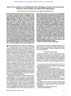
From www.bloodjournal.org by guest on February 6, 2015. For personal use only.
COMMENTARY
Commentary: A New Classification for von Willebrand Disease
By J. Evan Sadler and Harvey R. Gralnick
V
ON WILLEBRAND DISEASE (VWD) is the most
common inherited human bleeding disorder. Approximately 125 individuals per million population have symptomatic VWD, and this is roughly twice the prevalence of
hemophilia A. Asymptomatic inherited defects in von Willebrand factor (VWF) function are extremely common, and
are detectable in nearly 1% of unselected persons. Within
the last decade dozens of new variants of VWD have been
identified, based on abnormalities in either the biochemical
properties of the protein or the nucleotide sequence of the
gene. As a result, the evolving nomenclature for VWD has
become unwieldy. At last count, there were more than 14
reported varieties of type I VWD (having a normal-appearing
distribution of VWF multimers) and more than 13 reported
varieties of type I1 VWD (characterized by the absence
of large VWF multimers). The remaining category, type
111 VWD, is associated with the virtual absence of detectable VWF.
Recent progress in the characterization of mutations that
cause VWD has provided the necessary stimulus to readdress
the issue ofhow VWDis classified. In response to this
situation, a revised classification was developed and endorsed last year by the Subcommittee onvon Willebrand
Factor of the International Society on Thrombosis and Haemostasis (ISTH).’ This simplified scheme is based primarily
on the phenotype of the VWF protein that is present in
patient plasma and platelets. In this commentary we will
discuss the conceptual framework and goals of the new classification. The major features of the classification are listed
in Table 1. A more detailed discussion, as well as a correspondence table that relates the new classification to previous
nomenclature, can be found in the Subcommittee report.’
The principles underlying the new classification derive
from several sources, beginning with the first splitting of
VWD into quantitative (type I) and qualitative (type 11) variants by the behavior of VWF on crossed immunoelectrophoresis.’ Improved methods for electrophoretic analysis later
showed that VWF is a multimeric protein with a characteristic normal distribution of multimer sizes inplasma’,4 (Fig
1). In type I VWD the multimers appeared to be normal in
structure and function, but to be decreased in concentration.
In type I1 VWD the larger multimers were absent and function was decreased, usually disproportionately, both in vitro
From the Howard Hughes Medical Institute, the Departments of
Medicine and Biochemistry & MolecularBiophysics, The Jewish
Hospital of St Louis, Washington University School of Medicine, St
Louis, MO; and Hematology Service, National Institutes of Health
Clinical Center, Bethesda, MD.
Submitted May 25, 1994; accepted May 25, 1994.
Address reprint requests to J. Evan Sadler, MD, PhD, Howard
Hughes Medical Institute, Washington University, 660 S Euclid, Box
8022, St Louis, MO 63110.
0 1994 by The American Society of Hematology.
0006-4971/94/8403-0046$3.00/0
676
and in vivo. In 1980, two major subtypes of type I1 VWD
were distinguished. Assays in vitro showed that type IIA
VWD wasassociated with markedly decreased VWF binding
to platelets, whereas type IIB VWD was associated paradoxically with increased VWF binding to platelet^.^
Further refinements in protein electrophoresis led to extensive splitting of both type I and type I1 categories based on
subtle abnormalities of multimer structure, including
changes in the number and intensity of specific major bands
or minor “satellite” bands. More recently, mutations have
been identified that do not detectably affect VWF multimer
distribution, but drastically impair either of the two main
hemostatic functions of VWF: the ability to support platelet
adhesion, and the stabilization of factor vIII.~Thus, type
I VWD has come to include a mixture of qualitative and
quantitative abnormalities (Fig l A), with varying pathophysiology and clinical behavior. Type I1 VWD categories have
multiplied similarly (Fig 1B). A logical classification based
on pathophysiology was suggested by Ruggeri and Zimmerman in 1987,’ and the new classification is an extension of
that proposal.
ISSUES IN THECLASSIFICATION
OF VWD
At first glance, the reclassification of VWD might appear
to be conceptually simple. However, VWF has two characteristics that bedevil any attempt at the classification of inherited defects: it is encoded byan autosomal gene, and the
protein is polymeric. As a consequence, a patient may have
one or two mutant genes, and the phenotype of the resulting
multimeric VWF may be determined not only by the specific
properties of either mutant allele, but also by interactions
between the defective protein products of the alleles. For
example, a small admixture of mutant subunits may prevent
normal multimer assembly, or may mark the associated normal subunits for rapid clearance from the circulation.
VWF shares these problems with hemoglobin: both are
multisubunit proteins encoded by autosomal loci. Therefore,
the extensive experience with hemoglobinopathies and thalassemias is relevant to the classification of VWD. In general, the clinically useful disease categories are based on
pathophysiology. Specific molecular defects are important
but subsidiary to these features. When appropriate, the
unique and potentially significant features of individual patients are recognized by the use of geographical names
(“toponyms”) or other descriptors that may include the molecular defect. To accommodate the requirements of computerized databases and electronic mail, the categories are case
insensitive (capital letters preferred), use Arabic numerals,
and avoid special characters or formats. The key simplifications in the new classification of VWD are the definition of
major categories strictly in terms of quantitative or qualitative VWF defects, and the further division of qualitative
defects in terms of discrete pathophysiologic mechanisms.
Blood, Vol 84, NO3 (August l ) , 1994: pp 676-679
From www.bloodjournal.org by guest on February 6, 2015. For personal use only.
CLASSIFICATION OF VWD
677
Table 1. A Revised Classification of VWD
1. All VWD is caused by mutations at the VWF locus.
2. Type 1 VWD refers to partial quantitative deficiency of VWF.
Type 2 VWD refers to qualitativedeficiency of VWF. Type 3 VWD
refers to virtually complete deficiency of VWF.
3. Type 2A VWD refers to qualitativevariants with decreased
platelet-dependent function that isassociated with the absence
of high-molecular-weight VWF multimers.
4. Type 28 VWD refers to qualitative variants with increased affinity
for platelet glycoprotein lb.
5. Type 2M VWD refers to qualitative variants with decreased
platelet-dependent function that is notcaused by the absence of
high-molecular-weight VWF multimers.
6. Type 2N VWD refers to qualitative variants with markedly
decreased affinity for factor VIII.
7. When recognized, a mixed phenotype caused by compound
heterozygosity is indicated by separate classification of each
allele separated by a slash (h.
8. For the description of mutations, numbering systems are
suggested for amino acids and nucleotides.
A
1
2
3
4
THE REVISED CLASSIFICATION
A basic tenet of the new classification is that VWD is
caused only by mutations at the VWF locus. Potential mimics of VWD that arise through some other mechanism can
be designated asa particular form of “pseudo-VWD” if
they are inherited,*.’ or as “von Willebrand syndrome” if
they are acquired. The revised classification distinguishes
partial quantitative (type l), qualitative (type 2). and total
quantitative (type 3) deficiency. The minimal change in the
nomenclature from Roman to Arabic numerals differentiates
the new categories from those of previous classifications,
but preserves the oldest and most useful distinctions made
by earlier classifications of VWD, starting with Holmberg
and Nilsson.*Type 1 VWD is often dominant, type 2 VWD
may be dominant or recessive, and type 3 VWD is recessive.
In general, quantitative deficiencies will correlate with promoter, nonsense, and frameshift mutations, and with large
deletions. Qualitative deficiencies will correlate with missense mutations and small in-frame deletions or insertions.
No distinction is made between patients with typical dominantly inherited type 1 VWD and the occasional phenotypically similar, symptomatic relatives of patients with type 3
VWD: both are classified as type 1 VWD.
Type 2 VWD is further subdivided according to the apparent primary cause of the hemostatic defect. Because the
largest VWF multimers per se are required for normal platelet adhesion to sites of vascular injury, any mechanism that
causes a deficiency of large multimers may result in VWD.
Such variants are classified under type 2A, which may be
considered to encompass nonspecific defects in multimer
size. Type 2A corresponds roughly to the previous “type
IIA” variant, but also includes a variety of other subtypes
that have in common the loss of large, hemostatically effective VWF multimers (Fig 1B). Defective platelet adhesion
also may result, paradoxically, from increased binding of
VWF to platelets. This category, designated as type 2B, is
approximately the same as the previous “type IIB,” but
0
N 1 2 3 N 4 5 6 N
Fig 1. Mukimer patterns for plasma VWF from patients with selected variants of VWD. Samples were electrophoresed on a 1.3%
agarose gel in the presence of sodium dodecyl sulfate, and VWF
multimers werevisualized with a radioiodinated anti-VWF antibody.
(AI The type ofVWD is indicated with the terminology
of the revised
classification given first, followed by the previous classification in
parentheses. Lane 1, normal plasma; lane 2, VWD type 1 (formerly
type 11; lane 3, VWD type 2A (formerly typeIBI; lane 4, normal plasma.
(B1 Lanes N contain normalplasma samples. In the revised classification, all of the patterns represented in lanes 1 through 6 are designated VWD type 2A. In the terminologyof the previousclassification
these types of
VWD were designated as follows: lane 1, type IIC; lane
2, type IIG; lane 3, type IIC (sibling of patient of lane 1); lane 4, type
IID; lane 5, type llG (sibling of patient of lane 21; lane 6, type IIA.
From www.bloodjournal.org by guest on February 6, 2015. For personal use only.
SADLER AND GRALNICK
678
includes some patients with relatively normal VWF multimer
distribution.
The two remaining secondary categories reflect the recent
progress in defining the biochemical interactions of VWF in
molecular detail. A few patients exhibit markedly decreased
platelet-dependent VWF function or other evidence of a
qualitative abnormality despite a relatively normal multimer
distribution, and these patients are designated as type 2M
(“M” for “multimer”). Some of these patients have mutations in domains of VWF that are required for binding to
platelet glycoprotein Ib. The last variant, type 2N, is characterized by failure of VWF to bind factor VIII. Because this
interaction is required for the normal survival of factor VI11
in the circulation, these patients have factor VI11 deficiency
associated with normal platelet adhesion. The name “type
2N” reflects the origin in Normandy, France, of one of the
first families to be identified with this disorder.” Recognition
of this autosomal mimic of hemophilia is required for patients to receive appropriate blood component therapy and
genetic counseling.
In addition to defining specific diagnostic categories, the
new classification suggests a nomenclature for compound
heterozygous patients. Inherited defects of VWF function
are extremely common, and the accidental coinheritance of
otherwise “recessive” VWD alleles is proving to be common also. Awareness of the possibility of compound heterozygosity is important because genetic counseling in such
families can be substantially different than for dominant type
l VWD.
Finally, numbering systems are suggested for the description of nucleotide and amino acid substitutions (Table l).’
These recommendations will be useful mainly for the reporting and tabulation of molecular defects in VWD.
DISCUSSION
The revised classification preserves many desirable features of previous classifications, but is substantially simpler. There are two levels of the classification: primary
(types 1, 2, and 3 ) and secondary (A, B, M, N). These are
combined to yield a total of only six diagnostic categories,
compared with more than 28 previously named subtypes.
Additional descriptors may be suggested by the reporting
scientist (ie, toponyms), but are not part of the formal diagnostic name.
As described in the Subcommittee report,’ the classification of a new patient with VWD can be approached by
focusing on pathophysiology. Does the patient bleed because
of a quantitative (type 1 or 3 ) or qualitative (type 2) VWF
defect? If qualitative, does the defect affect platelet function
or factor VI11 binding (type 2N)? If the defect affects platelet
function, is it caused by loss-of-function or, paradoxically,
by gain-of-function (type 2B)? If loss-of-function, is this
caused nonspecifically by the absence of large multimers
(type 2A) or not (type2M)?
Some conceptual problems are necessarily shared by any
practical, concise classification of VWD. No matter how the
categories are defined, some patients will not fit cleanly into
any of them. For example, the distinction between type 1
and type 2M depends on differentiating normal andabnormal
VWF primary, secondary, and tertiary structures. The existing analytical methods are not particularly sensitive, so
errors in classification will occur. Similarly, the distinction
between type 2A and type 2M depends on recognizing the
absence of high-molecular-weight VWF multimers, but how
much of a decrease inmultimer size is required? These
problems are not peculiar to the new classification, however,
because the distinction between the previous categories
“type I” and “type 11” depended on the same assays.
One unavoidable problem with anypathophysiologic classification of VWD is that a mutation could, in principle,
cause a qualitative VWF defect by more than one mechanism. If a predominant mechanism cannot be identified, such
a patient can be classified simply as “type 2.” This is the
sort of patient for whoman associated descriptive name
would be especially useful, to serve as a reminder of the
unique and complex phenotype.
A goal of the new classification is to minimize the number and the clinical significance of hard-to-classify variants,
while maximizing the correlation of subtypes with the response to therapy with l-deamino-8-D-arginine vasopressin
(DDAVP) or plasma fractions. In this respect, it should
succeed at least as well as previous classifications.” Most
responders to DDAVP will have type 1 VWD, some will
have type 2A or 2M VWD, and very few will have type
2N or type 3 VWD. Most clinicians will elect not to use
DDAVP in type 2B VWD, although this apparently has
been useful in some patients. Highly purified factor VI11
preparations may lack sufficient VWF to stabilize their factor VI11 in vivo. Consequently, such preparations may exhibit poor recovery and shortened plasma half-life when
administered to patients with VWD type 2N or type 3.
For these subtypes of VWD, factor VI11 preparations that
contain substantial amounts of normal VWF would be preferred.
The process of developing this classification was instructive for all the participants. As a result of this exercise, the
critical gaps in our knowledge of VWD seem to be better
defined. However, the new scheme will endure only if clinicians actually apply it to patients and also find it useful to
predict clinical behavior. Of course, our hope is that this
will be the case. By working through the Subcommittee on
von Willebrand factor, reaching a consensus was surprisingly easy. Therefore, this ISTH mechanism should provide
a regular, international forum to insure that the classification
of VWD can evolve to reflect new knowledge.
REFERENCES
1. Sadler JE: A revised classification of von Willebrand disease.
Thromb Haemost 71520, 1994
2. Holmberg L, Nilsson I M : Genetic variants of von Willebrand’s
disease. Br Med J 3:317, 1972
3. Hoyer LW, Shainoff JR: Factor VIII-related protein circulates
in normal human plasma as high molecular weight multimers.
Blood
55:1056, 1980
4. Ruggeri ZM, Zimmerman TS: VariantvonWillebrand’s disease. Characterization of two subtypesbyanalysis
of multimeric
From www.bloodjournal.org by guest on February 6, 2015. For personal use only.
CLASSIFICATION OF
VWD
compositionof factor W v o n Willebrandfactorinplasmaand
platelets. J Clin Invest 651318, 1980
5. Ruggeri Z M , Pareti FI, Mannucci PM, CiavarellaN. Zimmerman TS: Heightened interaction between platelets and factor VIW
von Willebrand factor in a new subtype
of von Willebrand's disease.
N Engl J Med 302:1047, 1980
6. Ginsburg D, Sadler E: von Willebrand disease: A database of
point mutations, insertions, and deletions. Thromb Haemost
69:177,
1993
M, Zimmerman TS:von Willebrand factor and von
7. Ruggeri Z
Willebrand disease. Blood 70895, 1987
8. Weiss HJ, Meyer D,Rabinowitz R, Pietu G, Girma JP, Vicic
An intrinsic platelet
WJ, Rogers J: Pseudo-von Willebrand's disease.
679
defect with aggregation by unmodified human
factor VIIVvon Willebrand factor and enhanced adsorption of its high-molecular-weight
multimers. N Engl J Med 306:326, 1982
9. Miller JL, Castella A Platelet-type von Willebrand's disease:
Characterizationof a new bleeding disorder. Blood 60:790, 1982
10. Mazurier C, Dieval J, Jorieux S, Delobel J, Goudemand M
A new von Willebrand factor (vWF) defect in a patientwith factor
VIII (FVIII) deficiency but with normal levels and multimeric patterns of both plasma and platelet
vWF. Characterizationof abnormal
vWFlFVm interaction. Blood7520, 1990
11. Rodeghiero F, Castaman G, Mannucci PM Clinical indications for desmopressin @DAW) in congenital and acquired von
Willebrand disease. Blood Rev 5155, 1991
From www.bloodjournal.org by guest on February 6, 2015. For personal use only.
1994 84: 676-679
Commentary: a new classification for von Willebrand disease
JE Sadler and HR Gralnick
Updated information and services can be found at:
http://www.bloodjournal.org/content/84/3/676.citation.full.html
Articles on similar topics can be found in the following Blood collections
Information about reproducing this article in parts or in its entirety may be found online at:
http://www.bloodjournal.org/site/misc/rights.xhtml#repub_requests
Information about ordering reprints may be found online at:
http://www.bloodjournal.org/site/misc/rights.xhtml#reprints
Information about subscriptions and ASH membership may be found online at:
http://www.bloodjournal.org/site/subscriptions/index.xhtml
Blood (print ISSN 0006-4971, online ISSN 1528-0020), is published weekly by the American
Society of Hematology, 2021 L St, NW, Suite 900, Washington DC 20036.
Copyright 2011 by The American Society of Hematology; all rights reserved.

© Copyright 2026