
Genetic susceptibility to age-related macular degeneration: a
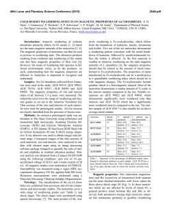
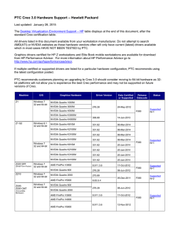
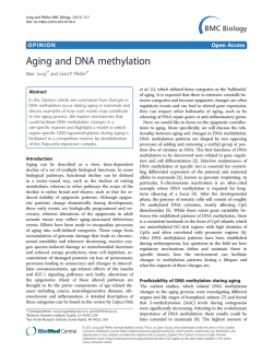
Human Molecular Genetics, 2007, Vol. 16, Review Issue 2
doi:10.1093/hmg/ddm212
R174–R182
Genetic susceptibility to age-related macular
degeneration: a paradigm for dissecting
complex disease traits
Anand Swaroop1,2,*, Kari EH Branham1, Wei Chen3 and Goncalo Abecasis3,*
1
Departments of Ophthalmology and Visual Sciences, 2Department of Human Genetics and 3Department of
Biostatistics, University of Michigan, Ann Arbor, MI 48105, USA
Received July 20, 2007; Revised July 20, 2007; Accepted July 26, 2007
Age-related macular degeneration (AMD) is a progressive neurodegenerative disease, which affects quality of
life for millions of elderly individuals worldwide. AMD is associated with a diverse spectrum of clinical phenotypes, all of which include the death of photoreceptors in the central part of the human retina (called the
macula). Tremendous progress has been made in identifying genetic susceptibility variants for AMD.
Variants at chromosome 1q32 (in the region of CFH ) and 10q26 (LOC387715/ARMS2) account for a large
part of the genetic risk to AMD and have been validated in numerous studies. In addition, susceptibility variants at other loci, several as yet unidentified, make substantial cumulative contribution to genetic risk for
AMD; among these, multiple studies support the role of variants in APOE and C2/BF genes. Genome-wide
association and re-sequencing projects, together with gene-environment interaction studies, are expected
to further define the causal relationships that connect genetic variants to AMD pathogenesis and should
assist in better design of prevention and intervention.
INTRODUCTION
A vast majority of common diseases are complex and
multi-factorial, resulting from the interplay of genetic components and environmental factors. As no single gene or
genetic variant can on its own cause the disease pathology, complexities associated with common diseases present unique challenges for management and therapy. While genetic defects have
been identified for over 2000 Mendelian diseases (generally rare
and affecting small population subsets) during the last
two decades (Online Mendalian Inheritance in Man, http://
www.ncbi.nlm.nih.gov/sites/entrez?db ¼ OMIM), the progress
towards understanding more frequent multi-factorial diseases
has been painfully slow until recently. Completion of the
human genome sequence and cataloging of millions of
common single nucleotide polymorphisms (SNPs) (1 –3) have
led to rapid and unprecedented advances in uncovering
genetic contributions of disease susceptibility for several
complex diseases, including diabetes, coronary artery disease
and macular degeneration (4 –9).
Age-related macular degeneration (AMD) is an ideal prototype of a complex and common disease trait, and genetic
studies of AMD illustrate both the promise and challenges of
new gene-mapping approaches. Early-onset maculopathies,
such as Stargardt’s disease, have long been known to have hereditary basis. In contrast, by mid-1990s, only a handful of
publications suggested role of genes in determining AMD risk
(10–12), and it was difficult to convince most clinicians and
scientists about the value of genetic studies. Recent investigations have not only unambiguously established the role of
genetic variants in AMD pathogenesis, but have also made it
feasible to uncover the role of gene–gene and gene–environment interactions for this debilitating blinding disease. In this
review, we will summarize the progress in unraveling the
genetic basis of AMD and present directions for future
studies that can be applicable to other multi-factorial disorders.
PREVALENCE, RISK FACTORS AND CLINICAL
PHENOTYPES
AMD can be defined as aging-associated progressive degeneration of photoreceptors and/or retinal pigment epithelium (RPE)
in the central part of the human retina (called the macula),
*To whom correspondence should be addressed. Email: [email protected]; [email protected]
# 2007 The Author(s)
This is an Open Access article distributed under the terms of the Creative Commons Attribution Non-Commercial License (http://creativecommons.org/
licenses/by-nc/2.0/uk/) which permits unrestricted non-commercial use, distribution, and reproduction in any medium, provided the original work is
properly cited.
Human Molecular Genetics, 2007, Vol. 16, Review Issue 2
eventually leading to the loss of central vision. It is the major
cause of uncorrectable visual impairment in the elderly population of the developed countries (13 –15). AMD affects over
1.7 million people in US alone, and this number is expected
to reach three million by the year 2020 (13). With increased
life expectancy, this devastating disease will continue to
have a significant public health impact on the quality of life
worldwide (16,17).
Clinical presentation of AMD is rather diverse (Fig. 1).
Small and hard macular drusen occur as part of the normal
aging process and do not necessarily predict the disease
(18). Early stages of AMD include atrophy of the RPE (18).
Large and soft drusen in the macula are identified as strong
risk factors for the development of advanced forms of
AMD, which include central geographic atrophy (GA) and
choroidal neovascularization (CNV) (19). Advanced age and
family history are the two major risk factors (20). However,
a number of environmental factors can also contribute to clinical manifestations of AMD; these include smoking, vascular
disease, UV exposure and nutritional status (15,20).
For genetic studies, a clear definition of the disease status
and/or phenotype is an essential aspect of the study design.
The phenotypic determination of affected status of controls
is equally important to the success of genetic studies. Since
AMD is a widespread late-onset disease, the most effective
controls are older than the typical age of onset for the
disease (21). No consensus has emerged on an optimal
disease classification and grading scheme. Instead, several
grading systems are currently being used in different
population-based and cohort-based studies (22 – 25). As susceptibility to distinct AMD subtypes appears to be driven by
different genetic risk factors, we expect that attention to
disease classification will be an important aspect of future
genetic studies of AMD. In particular, we expect it will help
to elucidate the molecular switches that ultimately result in
distinct phenotypes for different individuals.
Population-based studies have revealed significant differences in the incidence and prevalence of AMD subtypes
among different ethnic/racial groups in US (13,26 –29),
Europe (30,31), and Australia (32,33). Specifically, pooled
results from several studies reveal that white individuals of
European ancestry have a higher age-adjusted risk of developing AMD than individuals of more recent African ancestry
(13). A lower prevalence of AMD is also suggested in populations from China and Japan compared to whites (34,35).
However, a pilot study has demonstrated similar prevalence
of late AMD in populations from India and Europe (36). At
this stage, it is unclear if these population differences are
due to genetic or environmental factors, or both. Nevertheless,
clear differences in disease prevalence between ethnic groups
emphasize the importance of design of genetic studies (37,38).
GENETIC STUDIES
Dissecting the genetic basis of AMD using linkage analysis
has presented several challenges, particularly, because of the
difficulty in collecting large multi-generational families or
even small pedigrees with multiple affected sib-pairs that
are more readily available for the study of diseases with an
R175
earlier age of onset. Some of the early suggestions of
genetic predisposition originated from familial aggregation
studies. For example, a high concordance of disease phenotype
was observed in monozygotic twins (11,39 –41). A higher
incidence of AMD was also reported in first-degree relatives
of affected probands versus controls (10,42). Overall, siblings
of individuals with AMD have a three to six-fold increase in
disease risk (10,42,43). These initial studies established the
need for further genetic evaluation of AMD.
Linkage analysis
Genetic linkage studies search for regions of chromosome that
are shared between closely related affected individuals. Since
close relatives share relatively long stretches of chromosome,
linkage studies can survey the whole genome with only a few
hundred micro-satellite markers or a few thousand SNPs. Such
analyses have proven extremely effective in the dissection of
Mendelian traits (44). Although the results of linkage studies
for other complex diseases have been relatively disappointing
(45), such investigations have overall been quite successful for
AMD. The first report of linkage in a large family (10 affected
individuals) with the dry form of AMD exhibiting autosomal
dominant inheritance identified a genetic locus (ARMD1) at
chromosome 1q25 –q31 (LOD score 3.0) (46). As large
AMD families are difficult to obtain due to the late age of
disease-onset and their analysis may be complicated by phenocopy effects, the majority of subsequent linkage studies utilized the affected sib-pair or relative-pair design; these
investigations have implicated several regions of the genome
as harboring susceptibility loci with potentially major or
minor contributions to AMD (Fig. 2) (47 –55). The chromosomal regions at 1q31 –32 and 10q26 were identified in several
independent studies and confirmed by meta-analysis of six
datasets (56). As noted below, these regions harbor the
largest effect susceptibility variants for AMD, identified to
date.
Association studies
Contrary to linkage studies, which entail only a coarse
measurement of genetic variation, association analysis
requires more detailed measurements with hundreds of thousands of markers to cover the genome (1,57,58). Due to technical limitations, until recently, genetic association studies
have been limited to the study of candidate genes. Even
within these limited regions, most association studies generally examined only a subset of all genetic variants, making
it difficult to interpret negative results.
For macular degeneration, the first association studies
explored the genes associated with rare monogenic macular
diseases, whose clinical phenotypes overlap with AMD but
typically have an earlier age of onset (59). These genes
included RDS/peripherin (associated with retinitis pigmentosa, adult-onset vitelliform macular dystrophy, butterfly
dystrophy, and bulls-eye maculopathy) (60), TIMP3
(mutated in Sorsby’s Fundus Dystrophy) (61), EFEMP1
(responsible for Doyne Honeycomb retinal dystrophy) (62),
VMD2 (for Best’s Macular Degeneration) (63,64), and
ELOVL4 (associated with Stargardt-like macular dystrophy)
R176
Human Molecular Genetics, 2007, Vol. 16, Review Issue 2
Figure 1. Fundus pictures of: (A) Normal, 74 year old man, (B) 66 year old woman with few small drusen and RPE changes, (C) 71 year old male with extensive
soft drusen, (D) 87 year old woman with GA of the RPE, (E) 75 year old woman with disciform scar due to previous neovascularization, and (F) 15 year old
female with pisciform flecks associated with Stargardt’s macular degeneration.
Figure 2. Compilation of results of previous linkage studies, showing chromosomal regions harboring potential AMD susceptibility loci.
(65). To date, these studies have not yielded any striking
genetic associations with AMD. However, with advances in
genotype and re-sequencing technologies that allow genetic
association studies to examine larger numbers of individuals
in greater detail, we may need to re-assess the impact of
these genes on AMD.
Human Molecular Genetics, 2007, Vol. 16, Review Issue 2
Motivated by phenotypic similarities between Stargardt
disease and AMD (Fig. 1F), multiple association studies
have been performed on the gene responsible for the majority
of cases of Stargardt disease, the ABCA4 gene. Several of
these have indicated an association of AMD with certain missense changes in ABCA4 (notably D2177N and G1961E)
(66,67), although there are also negative reports (68 –70). At
this stage, the ABCA4 variants do not seem to make a major
contribution to AMD susceptibility.
Among candidates examined in single gene studies, APOE
exhibits the clearest association with AMD. APOE is involved
in transport and metabolism of lipid and cholesterol and in the
response to neuronal injury (reviewed by Mahley and Rall,
71). APOE has three common alleles, 12, 13 and 14. Initially,
two studies reported a reduction in the frequency of the 14
allele in patients with AMD compared to controls, suggesting
a protective effect (72,73). In addition, 12 allele frequency was
increased in AMD patients compared to controls (72). The
association between APOE and AMD has now been replicated
by several independent reports (74 –76) though at least in one
instance no association was obtained (77). Our meta-analysis
of published studies provides relatively strong evidence of
association between APOE 14 allele and AMD susceptibility
(Table 1). These findings differ somewhat from those of a previous meta-analysis, which analyzed pooled data across
studies (78). Meta-analysis, rather than analysis of pooled
data, is more appropriate when allele frequencies differ
across populations.
Chromosomal regions at 1q32 and 10q26
A pioneering discovery was made by Josephine Hoh and colleagues when they executed a genome-wide association study
with 100 000 SNPs in a small case-control sample; the
results of this initial scan and their subsequent follow-up
identified strong association between Y402H variant in the
Complement Factor H (CFH ) gene and increased risk of
AMD (9). Concurrently, other groups obtained similar
results using distinct approaches and replicated the findings
(9,79 – 82). A meta-analysis combining results from multiple
association studies of CFH and AMD indicate that heterozygote carriers of the risk allele have a 2.5-fold increase in
developing AMD and homozygous carriers have a six-fold
increase in developing AMD compared to the non-risk allele
(83). Again, as noted in Table 1, the consensus of studies published to date provides compelling evidence for association
between this SNP and AMD in populations of European
descent.
While initial studies of the association between CFH and
macular degeneration focused on the Y402H variant, recent
investigations have examined the region more broadly. For
example, Li et al. (84). evaluated 84 polymorphisms in and
around CFH and demonstrated that 20 of these exhibited
stronger association with disease susceptibility than the
Y402H variant. Furthermore, no single SNP itself accounted
for the contribution of the CFH locus to AMD. Instead, multiple polymorphisms defined a set of four common haplotypes
(two associated with disease susceptibility and two protective)
and multiple rare haplotypes (associated with increased
susceptibility in aggregate). These results suggest multiple
R177
susceptibility alleles in the region, with non-coding CFH variants playing a key role in determining disease risk. These
studies were confirmed in a companion paper, which also
showed that non-coding variants in and around CFH appear
to impact AMD independently of the Y402H variant (85).
Further complexity came from the association of a deletion
in CFH-like nearby genes (CFHR1 and CFHR3) with
disease susceptibility (86). Overall, the current data suggest
that identifying causal variants in a complex disease may be
quite challenging and that, potentially, regulatory variants
may have effects that are as important—or perhaps even
more important—than the coding variants or loss of function
mutations that are generally associated with Mendelian
disorders.
The strong association between CFH and AMD sparked
renewed interest in the complement pathway. It is now
clear that polymorphism in the C2 and BF genes are also
associated with disease (85,87) (unpublished data from our
lab; Table 1) and it is tempting to speculate that careful
assessment of other complement genes will uncover further
novel associations.
Similar to the 1q32 region, chromosome 10q26 shows
convincing evidence of linkage both in individual studies
(54) and in a meta-analysis of several published reports (56).
Fine-mapping efforts by a number of groups have yielded
convincing evidence of association at two neighboring
genes, PLEKHA1 and LOC387715 (88 –90), which has been
replicated (85). More recently, a genome-wide association
scan also provides evidence of association between AMD susceptibility and this region (91,92), and implicates another
gene, HTRA1, in AMD pathogenesis. These three genes are
in linkage disequilibrium with each other, and association
to this cluster has now been replicated in several studies
(93 – 97) (Table 1). In an attempt to disentangle the relationship between polymorphisms in the region and AMD susceptibility, we recently examined 45 SNPs in the region (98). In
contrast to fine-mapping efforts within the CFH locus (84)
suggesting multiple susceptibility variants, our data show
that a single coding variant in the LOC387715 gene (now
called ARMS2)—rs10490924—can account for the association between other SNPs in the region and macular degeneration. All other examined variants revealed significantly
weaker association and could not account for the effect of
rs10490924. The data illustrate the challenges in interpreting
association study results when the implicated region includes
multiple genes in linkage disequilibrium or multiple functional candidates. It is tempting to speculate that the identity
of the gene responsible in the 10q26 region will be further
elucidated by future association studies that implicate
PLEKHA1-like, LOC387715-like or HTRA1-like genes in
disease susceptibility.
To date, variants in the CFH region at chromosome 1q32
and in the PLEKHA1 / LOC387715 / HTRA1 region at
10q26 have demonstrated the strongest replicable association
with AMD. Variants in the APOE, C2 and BF genes also
show replicable, but smaller, association with AMD across
studies and contribute to disease susceptibility. The results
of several additional reports [e.g. CST3 (99), CX3CR1 (100),
TLR4 (101), fibulin 5 (102), VEGF (103)] are encouraging;
however, in our opinion, none of these have achieved the
R178
Human Molecular Genetics, 2007, Vol. 16, Review Issue 2
Table 1. A summary of conclusions from meta-analysis of established associations (three or more published reports) between AMD and genetic variants
Gene
CFH
LOC387715
C2
C2
BF
APOE
Polymorphism
rs1061170 (C/T)
rs10490924 (G/T)
rs9332739 (C/G)
rs547154 (A/C)
rs4151667 (A/T)
–
Total
studies
Total (N )
14
8
4
4
4
8
10 930
8473
4184
4162
4197
4290
Allele frequencies
Allele
Cases
Controls
T
T
G
C
T
12
13
14
0.435
0.420
0.977
0.949
0.974
0.097
0.808
0.095
0.639
0.207
0.943
0.892
0.942
0.076
0.784
0.152
level of evidence required to produce broad scientific
consensus.
FUTURE DIRECTIONS
A complete dissection of the genetic basis of AMD
Siblings of individuals with AMD have three to six-fold higher
risk of disease compared to individuals from the population at
large. Individually, the genetic variants at the chromosome
1q32 (CFH ) and 10q26 (LOC387715/ARMS2) regions
correspond to an increase in risk to siblings of only 1.2 to
1.6-fold (84,98). It is very likely that additional susceptibility
loci and variants remain to be identified, and several promising
approaches are now available to do so. Using new highthroughput re-sequencing platforms, it will be possible to
comprehensively search for new susceptibility variants in
previously identified loci (104). Additional genome-wide
association scans that examine larger numbers of individuals
are also likely to point to new loci that were missed in
earlier smaller scans—as happened in the case of Crohn’s
disease (6,105,106) and diabetes (4,5,8,107). Candidate gene
studies that focus on those identified in the initial studies
(e.g. additional genes in the complement pathway) are likely
to be promising and have already resulted in some success
(87). Finally, it will be possible to take advantage of the
ability to examine large numbers of individuals to carefully
evaluate the role of gene – gene and gene – environment interactions and examine their contributions to disease risk.
Genome-wide association
In our view, one step that is likely to lead to additional AMD
susceptibility variants will be to undertake genome-wide
association studies using higher density panels that provide
better coverage of the genome (108) and that examine larger
number of individuals. Although larger sample sizes will
always provide more power and improve the chance of new
discoveries, one cost-effective option is to use a few large
case-control cohorts for initial screening of 100 000s of variants, followed by detailed examination of the positive
genomic regions in additional cohorts (109). Such large
genome-wide scans will have profound impact on revealing
a more complete genetic picture of AMD susceptibility, as
demonstrated recently by 500K SNP examination of 17 000
genomes evaluated for seven common diseases (6).
Odds ratio
Meta-analysis
(P-value)
References
2.00
2.62
2.42
2.20
2.20
1.33
1.28
0.60
,102100
,102100
1.0 10212
6.8 10210
9.5 10211
0.042
0.00024
1.7 10211
(9,79,82,85,90,96,117–123)
(85,88,97,98,117,124,125)
(85,87) þ our own unpublished data
(85,87) þ our own unpublished data
(85,87) þ our own unpublished data
(72,73,75–77,126–129)
Re-sequencing of selected regions
Genome-wide association studies are designed to identify
common SNPs that may only increase the risk of disease by
a modest amount. Since they are common, these variants
can nevertheless explain a substantial fraction of disease risk
in the population. However, many variations will be missed
in such scans, especially rare ones that are not well tagged
by the common variants, which account for the bulk of
SNPs in commercial genotyping panels. Identification of rare
variants with impact on disease susceptibility can be accomplished by re-sequencing the associated gene regions in a
large population. This approach has been successfully used
for finding coding variants in the adipokine gene that is associated with plasma triglyceride levels (110). The targeted population re-sequencing strategy could help identify genetic
variants associated with AMD if it is applied to known susceptibility loci, such as the regions surrounding CFH and
LOC387715/ARMS2, and other promising genomic regions
(such as the genes in the complement pathway or HLA
region).
CONCLUSIONS
Further dissection of genetic susceptibility and the possible
interactions between variants and environmental factors
(such as smoking and nutrition) will be essential for elucidating mechanisms of disease pathology. Association studies
have provided valuable insights into the general location of
genetic differences that influence susceptibility to AMD. Currently, we can confidently point to variants in the CFH region
(at chromosome 1q32) and in LOC387715/ARMS2 (at
chromosome 10q26) among the major contributors. A role in
disease susceptibility is also strongly supported by the available evidence for variants in the APOE and C2/BF genes.
Many other genetic variants have been implicated but only
in a small number reports, and a definite conclusion about
their impact on disease susceptibility is not yet possible.
Nevertheless, with all the genetic findings, it may soon be
possible to provide pre-symptomatic diagnosis with reasonable accuracy, leading to better disease management strategies
for high-risk individuals.
We must, however, mention three points of caution and/or
consideration. (i) We should be aware of the difference
between susceptibility and causality. For complex diseases,
association (even strong) of gene variants to disease does
Human Molecular Genetics, 2007, Vol. 16, Review Issue 2
not represent a causal relationship as the presence of no single
sequence variation can lead to clinical pathology. A number of
individuals carrying high-risk Y402H allele of CFH and/or
other haplotypes may never develop macular degeneration
phenotype. (ii) We still have no clear understanding of the
early steps in disease manifestation. Numerous studies indicate RPE as the primary site of disease initiation, with death
of photoreceptors caused by RPE dysfunction selectively in
the macula (111). Oxidative stress, inflammatory pathways,
mitochondria-associated cellular processes have also been
implicated in AMD pathogenesis (112 –114). Animal models
are required to delineate the underlying pathway(s) and discover treatments; several mouse models manifesting at least
a part of the AMD disease spectrum have been reported
(115,116). Further investigations are necessary to correlate
risk or protective variants in associated genes to AMD pathology. (iii) Like other complex diseases, the environment is
expected to play a key role in triggering AMD. We are beginning to decipher the role of some of the factors (such as
smoking); yet, much work remains. A better assessment of
gene – environment interaction will require larger wellphenotyped AMD population cohorts and allow opportunities
for comprehensive planning of disease prevention and
treatment.
ACKNOWLEDGEMENTS
We thank John Heckenlively and Stephen Saxe for clinical
insights and study subject photographs; Mohammad Othman
and Atsuhiro Kanda for productive discussions; Ritu Khanna
for assistance with figures and databases; and Sharyn Ferrara
for administrative support. This research was supported by
grants from the National Institutes of Health, The Foundation
Fighting Blindness (FFB), the Elmer and Sylvia Sramek
Foundation, Thompson Foundation, and Research to Prevent
Blindness (RPB). A.S. is Harold F. Falls Collegiate Professor
and a recipient of RPB Senior Scientific Investigator award.
G.R.A. is a Pew Scholar for the Biomedical Sciences.
Funding to pay the Open Access publication charges for this
article was provided by a grant from National Institutes of
Health, EY 016862.
Conflict of Interest statement. None declared.
REFERENCES
1. The International HapMap Consortium (2005) A haplotype map of the
human genome. Nature, 437, 1299– 1320.
2. Lander, E.S., Linton, L.M., Birren, B., Nusbaum, C., Zody, M.C.,
Baldwin, J., Devon, K., Dewar, K., Doyle, M., FitzHugh, W. et al.
(2001) Initial sequencing and analysis of the human genome. Nature,
409, 860–921.
3. Venter, J.C., Adams, M.D., Myers, E.W., Li, P.W., Mural, R.J., Sutton,
G.G., Smith, H.O., Yandell, M., Evans, C.A., Holt, R.A. et al. (2001)
The sequence of the human genome. Science, 291, 1304–1351.
4. Saxena, R., Voight, B.F., Lyssenko, V., Burtt, N.P., de Bakker, P.I.,
Chen, H., Roix, J.J., Kathiresan, S., Hirschhorn, J.N., Daly, M.J. et al.
(2007) Genome-wide association analysis identifies loci for type 2
diabetes and triglyceride levels. Science, 316, 1331–1336.
5. Scott, L.J., Mohlke, K.L., Bonnycastle, L.L., Willer, C.J., Li, Y., Duren,
W.L., Erdos, M.R., Stringham, H.M., Chines, P.S., Jackson, A.U. et al.
(2007) A genome-wide association study of type 2 diabetes in Finns
detects multiple susceptibility variants. Science, 316, 1341–1345.
R179
6. The Welcome Trust Case Control Consortium (2007) Genome-wide
association study of 14,000 cases of seven common diseases and 3,000
shared controls. Nature, 447, 661–678.
7. McPherson, R., Pertsemlidis, A., Kavaslar, N., Stewart, A., Roberts, R.,
Cox, D.R., Hinds, D.A., Pennacchio, L.A., Tybjaerg-Hansen, A.,
Folsom, A.R. et al. (2007) A common allele on chromosome 9
associated with coronary heart disease. Science, 316, 1488–1491.
8. Zeggini, E., Weedon, M.N., Lindgren, C.M., Frayling, T.M., Elliott,
K.S., Lango, H., Timpson, N.J., Perry, J.R., Rayner, N.W., Freathy, R.M.
et al. (2007) Replication of genome-wide association signals in UK
samples reveals risk loci for type 2 diabetes. Science, 316, 1336–1341.
9. Klein, R.J., Zeiss, C., Chew, E.Y., Tsai, J.Y., Sackler, R.S., Haynes, C.,
Henning, A.K., Sangiovanni, J.P., Mane, S.M., Mayne, S.T. et al. (2005)
Complement factor H polymorphism in age-related macular
degeneration. Science, 308, 385–389.
10. Seddon, J.M., Ajani, U.A. and Mitchell, B.D. (1997) Familial
aggregation of age-related maculopathy. Am. J. Ophthalmol., 123,
199–206.
11. Meyers, S.M. (1994) A twin study on age-related macular degeneration.
Trans. Am. Ophthalmol. Soc., 92, 775– 843.
12. Silvestri, G., Johnston, P.B. and Hughes, A.E. (1994) Is genetic
predisposition an important risk factor in age-related macular
degeneration? Eye, 8 ( Pt 5), 564–568.
13. Friedman, D.S., O’Colmain, B.J., Munoz, B., Tomany, S.C., McCarty,
C., de Jong, P.T., Nemesure, B., Mitchell, P. and Kempen, J. (2004)
Prevalence of age-related macular degeneration in the United States.
Arch. Ophthalmol., 122, 564–572.
14. Klaver, C.C., Assink, J.J., van Leeuwen, R., Wolfs, R.C., Vingerling,
J.R., Stijnen, T., Hofman, A. and de Jong, P.T. (2001) Incidence and
progression rates of age-related maculopathy: the Rotterdam Study.
Invest. Ophthalmol. Vis. Sci., 42, 2237–2241.
15. Klein, R., Peto, T., Bird, A. and Vannewkirk, M.R. (2004) The
epidemiology of age-related macular degeneration. Am. J. Ophthalmol.,
137, 486–495.
16. Hassell, J.B., Lamoureux, E.L. and Keeffe, J.E. (2006) Impact of age
related macular degeneration on quality of life. Br. J. Ophthalmol., 90,
593–596.
17. Lotery, A.J., Xu, X., Zlatava, G. and Loftus, J. (2007) Burden of illness,
visual impairment, and health resource utilization of patients with
neovascular age-related macular degeneration: results from the united
kingdom cohort of a five-country cross-sectional study.
Br. J. Ophthalmol. doi: 10.1136/bjo.2007.116939.
18. Bressler, S. and Rosberger, D. (1999) Nonneovascular (nonexudative)
age-related macular degeneration. In Guyer, D., Yannuzzi, L., Chang, S.,
Shields, J., Green, W.R. (eds), Retina-Vitreous-Macula, WB Saunders
Co., Vol. 1, pp. 79 –93.
19. Klein, R., Klein, B.E., Jensen, S.C. and Meuer, S.M. (1997) The
five-year incidence and progression of age-related maculopathy: the
Beaver Dam Eye Study. Ophthalmology, 104, 7–21.
20. Evans, J.R. (2001) Risk factors for age-related macular degeneration.
Prog. Retin. Eye Res., 20, 227 –253.
21. Morton, N.E. and Collins, A. (1998) Tests and estimates of allelic
association in complex inheritance. Proc. Natl Acad. Sci. USA, 95,
11389–11393.
22. Age-Related Eye Disease Study Research Group (2001) A randomized,
placebo-controlled, clinical trial of high-dose supplementation with
vitamins C and E and beta carotene for age-related cataract and vision
loss: AREDS report no. 9. Arch. Ophthalmol., 119, 1439–1452.
23. Bird, A.C., Bressler, N.M., Bressler, S.B., Chisholm, I.H., Coscas, G.,
Davis, M.D., de Jong, P.T., Klaver, C.C., Klein, B.E., Klein, R. et al.
(1995) An international classification and grading system for age-related
maculopathy and age-related macular degeneration. The International
ARM Epidemiological Study Group. Surv. Ophthalmol., 39, 367–374.
24. Klein, R., Davis, M.D., Magli, Y.L., Segal, P., Klein, B.E. and Hubbard,
L. (1991) The Wisconsin age-related maculopathy grading system.
Ophthalmology, 98, 1128–1134.
25. Seddon, J.M., Sharma, S. and Adelman, R.A. (2006) Evaluation of the
clinical age-related maculopathy staging system. Ophthalmology, 113,
260–266.
26. Klein, R., Klein, B.E., Jensen, S.C., Mares-Perlman, J.A., Cruickshanks,
K.J. and Palta, M. (1999) Age-related maculopathy in a multiracial
United States population: the national health and nutrition examination
survey III. Ophthalmology, 106, 1056–1065.
R180
Human Molecular Genetics, 2007, Vol. 16, Review Issue 2
27. Klein, R., Klein, B.E. and Cruickshanks, K.J. (1999) The prevalence of
age-related maculopathy by geographic region and ethnicity. Prog.
Retin. Eye Res., 18, 371–389.
28. Klein, R., Klein, B.E., Knudtson, M.D., Wong, T.Y., Cotch, M.F., Liu,
K., Burke, G., Saad, M.F. and Jacobs, D.R., Jr. (2006) Prevalence of
age-related macular degeneration in 4 racial/ethnic groups in the
multi-ethnic study of atherosclerosis. Ophthalmology, 113, 373 –380.
29. Klein, R., Klein, B.E., Marino, E.K., Kuller, L.H., Furberg, C., Burke,
G.L. and Hubbard, L.D. (2003) Early age-related maculopathy in the
cardiovascular health study. Ophthalmology, 110, 25 –33.
30. Klaver, C.C., Wolfs, R.C., Vingerling, J.R., Hofman, A. and de Jong,
P.T. (1998) Age-specific prevalence and causes of blindness and visual
impairment in an older population: the Rotterdam Study. Arch.
Ophthalmol., 116, 653–658.
31. Vingerling, J.R., Dielemans, I., Hofman, A., Grobbee, D.E., Hijmering,
M., Kramer, C.F. and de Jong, P.T. (1995) The prevalence of age-related
maculopathy in the Rotterdam Study. Ophthalmology, 102, 205 –210.
32. Mitchell, P., Smith, W., Attebo, K. and Wang, J.J. (1995) Prevalence of
age-related maculopathy in Australia. The Blue Mountains Eye Study.
Ophthalmology, 102, 1450–1460.
33. VanNewkirk, M.R., Nanjan, M.B., Wang, J.J., Mitchell, P., Taylor, H.R.
and McCarty, C.A. (2000) The prevalence of age-related maculopathy:
the visual impairment project. Ophthalmology, 107, 1593–1600.
34. Li, Y., Xu, L., Jonas, J.B., Yang, H., Ma, Y. and Li, J. (2006) Prevalence
of age-related maculopathy in the adult population in China: the Beijing
eye study. Am. J. Ophthalmol., 142, 788 –793.
35. Oshima, Y., Ishibashi, T., Murata, T., Tahara, Y., Kiyohara, Y. and
Kubota, T. (2001) Prevalence of age related maculopathy in a
representative Japanese population: the Hisayama study.
Br. J. Ophthalmol., 85, 1153–1157.
36. Gupta, S.K., Murthy, G.V., Morrison, N., Price, G.M., Dherani, M.,
John, N., Fletcher, A.E. and Chakravarthy, U. (2007) Prevalence of early
and late age-related macular degeneration in a rural population in
northern India: the INDEYE feasibility study. Inves. Ophthalmol. Vis.
Sci., 48, 1007–1011.
37. Pritchard, J.K. and Rosenberg, N.A. (1999) Use of unlinked genetic
markers to detect population stratification in association studies.
Am. J. Hum. Genet., 65, 220 –228.
38. Devlin, B. and Roeder, K. (1999) Genomic control for association
studies. Biometrics, 55, 997– 1004.
39. Grizzard, S.W., Arnett, D. and Haag, S.L. (2003) Twin study of
age-related macular degeneration. Ophthalmic Epidemiol., 10, 315–322.
40. Hammond, C.J., Webster, A.R., Snieder, H., Bird, A.C., Gilbert, C.E.
and Spector, T.D. (2002) Genetic influence on early age-related
maculopathy: a twin study. Ophthalmology, 109, 730– 736.
41. Klein, M.L., Mauldin, W.M. and Stoumbos, V.D. (1994) Heredity and
age-related macular degeneration. Observations in monozygotic twins.
Arch. Ophthalmol., 112, 932–937.
42. Klaver, C.C., Wolfs, R.C., Assink, J.J., van Duijn, C.M., Hofman, A. and
de Jong, P.T. (1998) Genetic risk of age-related maculopathy.
Population-based familial aggregation study. Arch. Ophthalmol., 116,
1646–1651.
43. Heiba, I.M., Elston, R.C., Klein, B.E. and Klein, R. (1994) Sibling
correlations and segregation analysis of age-related maculopathy: the
Beaver Dam Eye Study. Genet. Epidemiol., 11, 51–67.
44. Botstein, D. and Risch, N. (2003) Discovering genotypes underlying
human phenotypes: past successes for mendelian disease, future
approaches for complex disease. Nat. Genet., 33, 228 –237.
45. Lernmark, A. and Ott, J. (1998) Sometimes it’s hot, sometimes it’s not.
Nat. Genet., 19, 213 –214.
46. Klein, M.L., Schultz, D.W., Edwards, A., Matise, T.C., Rust, K.,
Berselli, C.B., Trzupek, K., Weleber, R.G., Ott, J., Wirtz, M.K. et al.
(1998) Age-related macular degeneration. Clinical features in a large
family and linkage to chromosome 1q. Arch. Ophthalmol., 116,
1082–1088.
47. Majewski, J., Schultz, D.W., Weleber, R.G., Schain, M.B., Edwards,
A.O., Matise, T.C., Acott, T.S., Ott, J. and Klein, M.L. (2003)
Age-related macular degeneration – a genome scan in extended families.
Am. J. Hum. Genet., 73, 540 –550.
48. Schick, J.H., Iyengar, S.K., Klein, B.E., Klein, R., Reading, K., Liptak,
R., Millard, C., Lee, K.E., Tomany, S.C., Moore, E.L. et al. (2003) A
whole-genome screen of a quantitative trait of age-related maculopathy
49.
50.
51.
52.
53.
54.
55.
56.
57.
58.
59.
60.
61.
62.
63.
64.
65.
in sibships from the Beaver Dam Eye Study. Am. J. Hum. Genet., 72,
1412–1424.
Seddon, J.M., Santangelo, S.L., Book, K., Chong, S. and Cote, J. (2003)
A genomewide scan for age-related macular degeneration provides
evidence for linkage to several chromosomal regions. Am. J. Hum.
Genet., 73, 780–790.
Abecasis, G.R., Yashar, B.M., Zhao, Y., Ghiasvand, N.M., Zareparsi, S.,
Branham, K.E., Reddick, A.C., Trager, E.H., Yoshida, S., Bahling, J.
et al. (2004) Age-related macular degeneration: a high-resolution
genome scan for susceptibility loci in a population enriched for late-stage
disease. Am. J. Hum. Genet., 74, 482 –494.
Iyengar, S.K., Song, D., Klein, B.E., Klein, R., Schick, J.H., Humphrey,
J., Millard, C., Liptak, R., Russo, K., Jun, G. et al. (2004) Dissection of
genomewide-scan data in extended families reveals a major locus and
oligogenic susceptibility for age-related macular degeneration.
Am. J. Hum. Genet., 74, 20–39.
Kenealy, S.J., Schmidt, S., Agarwal, A., Postel, E.A., De La Paz, M.A.,
Pericak-Vance, M.A. and Haines, J.L. (2004) Linkage analysis for
age-related macular degeneration supports a gene on chromosome
10q26. Mol. Vis., 10, 57–61.
Schmidt, S., Scott, W.K., Postel, E.A., Agarwal, A., Hauser, E.R., De La
Paz, M.A., Gilbert, J.R., Weeks, D.E., Gorin, M.B., Haines, J.L. et al.
(2004) Ordered subset linkage analysis supports a susceptibility locus for
age-related macular degeneration on chromosome 16p12. BMC Genet.,
5, 18.
Weeks, D.E., Conley, Y.P., Tsai, H.J., Mah, T.S., Schmidt, S., Postel,
E.A., Agarwal, A., Haines, J.L., Pericak-Vance, M.A., Rosenfeld, P.J.
et al. (2004) Age-related maculopathy: a genomewide scan with
continued evidence of susceptibility loci within the 1q31, 10q26, and
17q25 regions. Am. J. Hum. Genet., 75, 174– 189.
Barral, S., Francis, P.J., Schultz, D.W., Schain, M.B., Haynes, C.,
Majewski, J., Ott, J., Acott, T., Weleber, R.G. and Klein, M.L. (2006)
Expanded genome scan in extended families with age-related macular
degeneration. Invest. Ophthalmol. Vis. Sci., 47, 5453–5459.
Fisher, S.A., Abecasis, G.R., Yashar, B.M., Zareparsi, S., Swaroop, A.,
Iyengar, S.K., Klein, B.E., Klein, R., Lee, K.E., Majewski, J. et al.
(2005) Meta-analysis of genome scans of age-related macular
degeneration. Hum. Mol. Genet., 14, 2257–2264.
The International HapMap Consortium (2003) The International
HapMap Project. Nature, 426, 789–796.
Risch, N. and Merikangas, K. (1996) The future of genetic studies of
complex human diseases. Science, 273, 1516–1517.
Stone, E.M., Sheffield, V.C. and Hageman, G.S. (2001) Molecular
genetics of age-related macular degeneration. Hum. Mol. Genet., 10,
2285–2292.
Baird, P.N., Richardson, A., Islam, A., Lim, L. and Guymer, R. (2007)
Analysis of the RDS/peripherin gene in age-related macular
degeneration. Clin. Experiment. Ophthalmol., 35, 194–195.
De La Paz, M.A., Pericak-Vance, M.A., Lennon, F., Haines, J.L. and
Seddon, J.M. (1997) Exclusion of TIMP3 as a candidate locus in
age-related macular degeneration. Invest. Ophthalmol. Vis. Sci., 38,
1060–1065.
Stone, E.M., Lotery, A.J., Munier, F.L., Heon, E., Piguet, B., Guymer,
R.H., Vandenburgh, K., Cousin, P., Nishimura, D., Swiderski, R.E. et al.
(1999) A single EFEMP1 mutation associated with both Malattia
Leventinese and Doyne honeycomb retinal dystrophy. Nat. Genet., 22,
199–202.
Lotery, A.J., Munier, F.L., Fishman, G.A., Weleber, R.G., Jacobson,
S.G., Affatigato, L.M., Nichols, B.E., Schorderet, D.F., Sheffield, V.C.
and Stone, E.M. (2000) Allelic variation in the VMD2 gene in best
disease and age-related macular degeneration. Invest. Ophthalmol. Vis.
Sci., 41, 1291– 1296.
Allikmets, R., Seddon, J.M., Bernstein, P.S., Hutchinson, A., Atkinson,
A., Sharma, S., Gerrard, B., Li, W., Metzker, M.L., Wadelius, C. et al.
(1999) Evaluation of the Best disease gene in patients with age-related
macular degeneration and other maculopathies. Hum. Genet., 104,
449–453.
Ayyagari, R., Zhang, K., Hutchinson, A., Yu, Z., Swaroop, A., Kakuk,
L.E., Seddon, J.M., Bernstein, P.S., Lewis, R.A., Tammur, J. et al.
(2001) Evaluation of the ELOVL4 gene in patients with age-related
macular degeneration. Ophthalmic. Genet., 22, 233–239.
Human Molecular Genetics, 2007, Vol. 16, Review Issue 2
66. Allikmets, R. (2000) Further evidence for an association of ABCR
alleles with age-related macular degeneration. The International ABCR
Screening Consortium. Am. J. Hum. Genet., 67, 487– 491.
67. Allikmets, R., Shroyer, N.F., Singh, N., Seddon, J.M., Lewis, R.A.,
Bernstein, P.S., Peiffer, A., Zabriskie, N.A., Li, Y., Hutchinson, A. et al.
(1997) Mutation of the Stargardt disease gene (ABCR) in age-related
macular degeneration. Science, 277, 1805–1807.
68. Rivera, A., White, K., Stohr, H., Steiner, K., Hemmrich, N., Grimm, T.,
Jurklies, B., Lorenz, B., Scholl, H.P., Apfelstedt-Sylla, E. et al. (2000) A
comprehensive survey of sequence variation in the ABCA4 (ABCR)
gene in Stargardt disease and age-related macular degeneration.
Am. J. Hum. Genet., 67, 800 –813.
69. Schmidt, S., Postel, E.A., Agarwal, A., Allen, I.C., Jr., Walters, S.N., De
la Paz, M.A., Scott, W.K., Haines, J.L., Pericak-Vance, M.A. and
Gilbert, J.R. (2003) Detailed analysis of allelic variation in the ABCA4
gene in age-related maculopathy. Invest. Ophthalmol. Vis. Sci., 44,
2868–2875.
70. Stone, E.M., Webster, A.R., Vandenburgh, K., Streb, L.M., Hockey,
R.R., Lotery, A.J. and Sheffield, V.C. (1998) Allelic variation in ABCR
associated with Stargardt disease but not age-related macular
degeneration. Nat. Genet., 20, 328 –329.
71. Mahley, R.W. and Rall, S.C., Jr. (2000) Apolipoprotein E: far more than
a lipid transport protein. Annu. Rev. Genomics Hum. Genet., 1, 507–537.
72. Klaver, C.C., Kliffen, M., van Duijn, C.M., Hofman, A., Cruts, M.,
Grobbee, D.E., van Broeckhoven, C. and de Jong, P.T. (1998) Genetic
association of apolipoprotein E with age-related macular degeneration.
Am. J. Hum. Genet., 63, 200 –206.
73. Souied, E.H., Benlian, P., Amouyel, P., Feingold, J., Lagarde, J.P.,
Munnich, A., Kaplan, J., Coscas, G. and Soubrane, G. (1998) The
epsilon4 allele of the apolipoprotein E gene as a potential protective
factor for exudative age-related macular degeneration.
Am. J. Ophthalmol., 125, 353 –359.
74. Schmidt, S., Klaver, C., Saunders, A., Postel, E., De La Paz, M.,
Agarwal, A., Small, K., Udar, N., Ong, J., Chalukya, M. et al. (2002) A
pooled case-control study of the apolipoprotein E (APOE) gene in
age-related maculopathy. Ophthalmic. Genet., 23, 209 –223.
75. Baird, P.N., Guida, E., Chu, D.T., Vu, H.T. and Guymer, R.H. (2004)
The epsilon2 and epsilon4 alleles of the apolipoprotein gene are
associated with age-related macular degeneration. Invest. Ophthalmol.
Vis. Sci., 45, 1311–1315.
76. Zareparsi, S., Reddick, A.C., Branham, K.E., Moore, K.B., Jessup, L.,
Thoms, S., Smith-Wheelock, M., Yashar, B.M. and Swaroop, A. (2004)
Association of apolipoprotein E alleles with susceptibility to age-related
macular degeneration in a large cohort from a single center. Invest.
Ophthalmol. Vis. Sci., 45, 1306–1310.
77. Schultz, D.W., Klein, M.L., Humpert, A., Majewski, J., Schain, M.,
Weleber, R.G., Ott, J. and Acott, T.S. (2003) Lack of an association of
apolipoprotein E gene polymorphisms with familial age-related macular
degeneration. Arch. Ophthalmol., 121, 679–683.
78. Thakkinstian, A., Bowe, S., McEvoy, M., Smith, W. and Attia, J. (2006)
Association between apolipoprotein E polymorphisms and age-related
macular degeneration: A HuGE review and meta-analysis.
Am. J. Epidemiol., 164, 813 –822.
79. Edwards, A.O., Ritter, R.,III, Abel, K.J., Manning, A., Panhuysen, C. and
Farrer, L.A. (2005) Complement factor H polymorphism and age-related
macular degeneration. Science, 308, 421 –424.
80. Haines, J.L., Hauser, M.A., Schmidt, S., Scott, W.K., Olson, L.M.,
Gallins, P., Spencer, K.L., Kwan, S.Y., Noureddine, M., Gilbert, J.R.
et al. (2005) Complement factor H variant increases the risk of
age-related macular degeneration. Science, 308, 419–421.
81. Hageman, G.S., Anderson, D.H., Johnson, L.V., Hancox, L.S., Taiber,
A.J., Hardisty, L.I., Hageman, J.L., Stockman, H.A., Borchardt, J.D.,
Gehrs, K.M. et al. (2005) A common haplotype in the complement
regulatory gene factor H (HF1/CFH) predisposes individuals to
age-related macular degeneration. Proc. Natl Acad. Sci. USA, 102,
7227–7232.
82. Zareparsi, S., Branham, K.E., Li, M., Shah, S., Klein, R.J., Ott, J., Hoh,
J., Abecasis, G.R. and Swaroop, A. (2005) Strong association of the
Y402H variant in complement factor H at 1q32 with susceptibility to
age-related macular degeneration. Am. J. Hum. Genet., 77, 149–153.
83. Thakkinstian, A., Han, P., McEvoy, M., Smith, W., Hoh, J., Magnusson,
K., Zhang, K. and Attia, J. (2006) Systematic review and meta-analysis
of the association between complement factor H Y402H polymorphisms
84.
85.
86.
87.
88.
89.
90.
91.
92.
93.
94.
95.
96.
97.
98.
99.
100.
R181
and age-related macular degeneration. Hum. Mol. Genet., 15,
2784–2790.
Li, M., Atmaca-Sonmez, P., Othman, M., Branham, K.E., Khanna, R.,
Wade, M.S., Li, Y., Liang, L., Zareparsi, S., Swaroop, A. et al. (2006)
CFH haplotypes without the Y402H coding variant show strong
association with susceptibility to age-related macular degeneration. Nat.
Genet., 38, 1049–1054.
Maller, J., George, S., Purcell, S., Fagerness, J., Altshuler, D., Daly, M.J.
and Seddon, J.M. (2006) Common variation in three genes, including a
noncoding variant in CFH, strongly influences risk of age-related
macular degeneration. Nat. Genet., 38, 1055–1059.
Hughes, A.E., Orr, N., Esfandiary, H., Diaz-Torres, M., Goodship, T. and
Chakravarthy, U. (2006) A common CFH haplotype, with deletion of
CFHR1 and CFHR3, is associated with lower risk of age-related macular
degeneration. Nat. Genet., 38, 1173–1177.
Gold, B., Merriam, J.E., Zernant, J., Hancox, L.S., Taiber, A.J., Gehrs,
K., Cramer, K., Neel, J., Bergeron, J., Barile, G.R. et al. (2006) Variation
in factor B (BF) and complement component 2 (C2) genes is associated
with age-related macular degeneration. Nat. Genet., 38, 458 –462.
Rivera, A., Fisher, S.A., Fritsche, L.G., Keilhauer, C.N., Lichtner, P.,
Meitinger, T. and Weber, B.H. (2005) Hypothetical LOC387715 is a
second major susceptibility gene for age-related macular degeneration,
contributing independently of complement factor H to disease risk. Hum.
Mol. Genet., 14, 3227–3236.
Jakobsdottir, J., Conley, Y.P., Weeks, D.E., Mah, T.S., Ferrell, R.E. and
Gorin, M.B. (2005) Susceptibility genes for age-related maculopathy on
chromosome 10q26. Am. J. Hum. Genet., 77, 389 –407.
Conley, Y.P., Jakobsdottir, J., Mah, T., Weeks, D.E., Klein, R., Kuller,
L., Ferrell, R.E. and Gorin, M.B. (2006) CFH, ELOVL4, PLEKHA1 and
LOC387715 genes and susceptibility to age-related maculopathy:
AREDS and CHS cohorts and meta-analyses. Hum. Mol. Genet., 15,
3206–3218.
Dewan, A., Liu, M., Hartman, S., Zhang, S.S., Liu, D.T., Zhao, C., Tam,
P.O., Chan, W.M., Lam, D.S., Snyder, M. et al. (2006) HTRA1 promoter
polymorphism in wet age-related macular degeneration. Science, 314,
989–992.
Yang, Z., Camp, N.J., Sun, H., Tong, Z., Gibbs, D., Cameron, D.J., Chen,
H., Zhao, Y., Pearson, E., Li, X. et al. (2006) A variant of the HTRA1
gene increases susceptibility to age-related macular degeneration.
Science, 314, 992 –993.
Seddon, J.M., Francis, P.J., George, S., Schultz, D.W., Rosner, B. and
Klein, M.L. (2007) Association of CFH Y402H and LOC387715 A69S
with progression of age-related macular degeneration. JAMA, 297,
1793–1800.
Shastry, B.S. (2006) Further support for the common variants in
complement factor H (Y402H) and LOC387715 (A69S) genes as major
risk factors for the exudative age-related macular degeneration.
Ophthalmologica, 220, 291 –295.
Shuler, R.K., Jr., Hauser, M.A., Caldwell, J., Gallins, P., Schmidt, S.,
Scott, W.K., Agarwal, A., Haines, J.L., Pericak-Vance, M.A. and Postel,
E.A. (2007) Neovascular age-related macular degeneration and its
association with LOC387715 and complement factor H polymorphism.
Arch. Ophthalmol., 125, 63–67.
Schaumberg, D.A., Christen, W.G., Kozlowski, P., Miller, D.T., Ridker,
P.M. and Zee, R.Y. (2006) A prospective assessment of the Y402H
variant in complement factor H, genetic variants in C-reactive protein,
and risk of age-related macular degeneration. Invest. Ophthalmol. Vis.
Sci., 47, 2336– 2340.
Schmidt, S., Hauser, M.A., Scott, W.K., Postel, E.A., Agarwal, A.,
Gallins, P., Wong, F., Chen, Y.S., Spencer, K., Schnetz-Boutaud, N.
et al. (2006) Cigarette smoking strongly modifies the association of
LOC387715 and age-related macular degeneration. Am. J. Hum. Genet.,
78, 852–864.
Kanda, A., Chen, W., Othman, M., Branham, K.E.H., Brooks, M.,
Khanna, R., He, S., Lyons, R., Abecasis, G.R. and Swaroop, A. (2007) A
variant of mitochondrial protein LOC387715/ARMS2, not HTRA1, is
strongly associated with age-related macular degeneration. PNAS. In press
Zurdel, J., Finckh, U., Menzer, G., Nitsch, R.M. and Richard, G. (2002)
CST3 genotype associated with exudative age related macular
degeneration. Br. J. Ophthalmol., 86, 214–219.
Tuo, J., Smith, B.C., Bojanowski, C.M., Meleth, A.D., Gery, I., Csaky,
K.G., Chew, E.Y. and Chan, C.C. (2004) The involvement of sequence
R182
101.
102.
103.
104.
105.
106.
107.
108.
109.
110.
111.
112.
113.
114.
115.
116.
117.
Human Molecular Genetics, 2007, Vol. 16, Review Issue 2
variation and expression of CX3CR1 in the pathogenesis of age-related
macular degeneration. FASEB J, 18, 1297–1299.
Zareparsi, S., Buraczynska, M., Branham, K.E., Shah, S., Eng, D., Li,
M., Pawar, H., Yashar, B.M., Moroi, S.E., Lichter, P.R. et al. (2005)
Toll-like receptor 4 variant D299G is associated with susceptibility to
age-related macular degeneration. Hum. Mol. Genet., 14, 1449–1455.
Stone, E.M., Braun, T.A., Russell, S.R., Kuehn, M.H., Lotery, A.J.,
Moore, P.A., Eastman, C.G., Casavant, T.L. and Sheffield, V.C. (2004)
Missense variations in the fibulin 5 gene and age-related macular
degeneration. N. Engl. J. Med., 351, 346– 353.
Haines, J.L., Schnetz-Boutaud, N., Schmidt, S., Scott, W.K., Agarwal,
A., Postel, E.A., Olson, L., Kenealy, S.J., Hauser, M., Gilbert, J.R. et al.
(2006) Functional candidate genes in age-related macular degeneration:
significant association with VEGF, VLDLR, and LRP6. Invest.
Ophthalmol. Vis. Sci., 47, 329 –335.
Bentley, D.R. (2006) Whole-genome re-sequencing. Curr. Opin. Genet.
Dev., 16, 545–552.
Libioulle, C., Louis, E., Hansoul, S., Sandor, C., Farnir, F., Franchimont,
D., Vermeire, S., Dewit, O., de Vos, M., Dixon, A. et al. (2007) Novel
Crohn disease locus identified by genome-wide association maps to a
gene desert on 5p13.1 and modulates expression of PTGER4. PLoS
Genet., 3, e58.
Duerr, R.H., Taylor, K.D., Brant, S.R., Rioux, J.D., Silverberg, M.S.,
Daly, M.J., Steinhart, A.H., Abraham, C., Regueiro, M., Griffiths,
A. et al. (2006) A genome-wide association study identifies IL23R as an
inflammatory bowel disease gene. Science, 314, 1461–1463.
Sladek, R., Rocheleau, G., Rung, J., Dina, C., Shen, L., Serre, D., Boutin,
P., Vincent, D., Belisle, A., Hadjadj, S. et al. (2007) A genome-wide
association study identifies novel risk loci for type 2 diabetes. Nature,
445, 881–885.
Barrett, J.C. and Cardon, L.R. (2006) Evaluating coverage of
genome-wide association studies. Nat. Genet., 38, 659–662.
Skol, A.D., Scott, L.J., Abecasis, G.R. and Boehnke, M. (2006) Joint
analysis is more efficient than replication-based analysis for two-stage
genome-wide association studies. Nat. Genet., 38, 209–213.
Romeo, S., Pennacchio, L.A., Fu, Y., Boerwinkle, E., Tybjaerg-Hansen,
A., Hobbs, H.H. and Cohen, J.C. (2007) Population-based resequencing
of ANGPTL4 uncovers variations that reduce triglycerides and increase
HDL. Nat. Genet., 39, 513–516.
Zarbin, M.A. (2004) Current concepts in the pathogenesis of age-related
macular degeneration. Arch. Ophthalmol., 122, 598–614.
Donoso, L.A., Kim, D., Frost, A., Callahan, A. and Hageman, G. (2006)
The role of inflammation in the pathogenesis of age-related macular
degeneration. Surv. Ophthalmol., 51, 137–152.
Beatty, S., Koh, H., Phil, M., Henson, D. and Boulton, M. (2000) The
role of oxidative stress in the pathogenesis of age-related macular
degeneration. Surv. Ophthalmol., 45, 115–134.
Seddon, J.M., Gensler, G., Milton, R.C., Klein, M.L. and Rifai, N. (2004)
Association between C-reactive protein and age-related macular
degeneration. JAMA, 291, 704–710.
Edwards, A.O. and Malek, G. (2007) Molecular genetics of AMD and
current animal models. Angiogenesis, 10, 119–132.
Elizabeth Rakoczy, P., Yu, M.J., Nusinowitz, S., Chang, B. and
Heckenlively, J.R. (2006) Mouse models of age-related macular
degeneration. Exp. Eye Res., 82, 741– 752.
Fisher, S.A., Rivera, A., Fritsche, L.G., Babadjanova, G., Petrov, S. and
Weber, B.H. (2007) Assessment of the contribution of CFH and
118.
119.
120.
121.
122.
123.
124.
125.
126.
127.
128.
129.
chromosome 10q26 AMD susceptibility loci in a Russian population
isolate. Br. J. Ophthalmol., 91, 576–578.
Simonelli, F., Frisso, G., Testa, F., di Fiore, R., Vitale, D.F., Manitto,
M.P., Brancato, R., Rinaldi, E. and Sacchetti, L. (2006) Polymorphism
p.402Y.H in the complement factor H protein is a risk factor for age
related macular degeneration in an Italian population.
Br. J. Ophthalmol., 90, 1142–1145.
Seitsonen, S., Lemmela, S., Holopainen, J., Tommila, P., Ranta, P.,
Kotamies, A., Moilanen, J., Palosaari, T., Kaarniranta, K., Meri, S. et al.
(2006) Analysis of variants in the complement factor H, the elongation
of very long chain fatty acids-like 4 and the hemicentin 1 genes of
age-related macular degeneration in the Finnish population. Mol. Vis.,
12, 796–801.
Seddon, J.M., George, S., Rosner, B. and Klein, M.L. (2006) CFH gene
variant, Y402H, and smoking, body mass index, environmental
associations with advanced age-related macular degeneration. Hum.
Hered., 61, 157 –165.
Souied, E.H., Leveziel, N., Richard, F., Dragon-Durey, M.A., Coscas,
G., Soubrane, G., Benlian, P. and Fremeaux-Bacchi, V. (2005) Y402H
complement factor H polymorphism associated with exudative
age-related macular degeneration in the French population. Mol. Vis., 11,
1135–1140.
Magnusson, K.P., Duan, S., Sigurdsson, H., Petursson, H., Yang, Z.,
Zhao, Y., Bernstein, P.S., Ge, J., Jonasson, F., Stefansson, E. et al.
(2006) CFH Y402H confers similar risk of soft drusen and both forms of
advanced AMD. PLoS Med., 3, e5.
Baird, P.N., Islam, F.M., Richardson, A.J., Cain, M., Hunt, N. and
Guymer, R. (2006) Analysis of the Y402H variant of the complement
factor H gene in age-related macular degeneration. Invest. Ophthalmol.
Vis. Sci., 47, 4194–4198.
Ross, R.J., Bojanowski, C.M., Wang, J.J., Chew, E.Y., Rochtchina, E.,
Ferris, F.L.,III, Mitchell, P., Chan, C.C. and Tuo, J. (2007) The
LOC387715 polymorphism and age-related macular degeneration:
replication in three case-control samples. Invest. Ophthalmol. Vis. Sci.,
48, 1128–1132.
Francis, P.J., George, S., Schultz, D.W., Rosner, B., Hamon, S., Ott, J.,
Weleber, R.G., Klein, M.L. and Seddon, J.M. (2007) The LOC387715
gene, smoking, body mass index, environmental associations with
advanced age-related macular degeneration. Hum. Hered., 63, 212–218.
Bojanowski, C.M., Shen, D., Chew, E.Y., Ning, B., Csaky, K.G., Green,
W.R., Chan, C.C. and Tuo, J. (2006) An apolipoprotein E variant may
protect against age-related macular degeneration through cytokine
regulation. Environ. Mol. Mutagen., 47, 594–602.
Simonelli, F., Margaglione, M., Testa, F., Cappucci, G., Manitto, M.P.,
Brancato, R. and Rinaldi, E. (2001) Apolipoprotein E polymorphisms in
age-related macular degeneration in an Italian population. Ophthalmic
Res., 33, 325–328.
Schmidt, S., Saunders, A.M., De La Paz, M.A., Postel, E.A., Heinis,
R.M., Agarwal, A., Scott, W.K., Gilbert, J.R., McDowell, J.G., Bazyk,
A. et al. (2000) Association of the apolipoprotein E gene with
age-related macular degeneration: possible effect modification by family
history, age, and gender. Mol. Vis., 6, 287– 293.
Gotoh, N., Kuroiwa, S., Kikuchi, T., Arai, J., Arai, S., Yoshida, N. and
Yoshimura, N. (2004) Apolipoprotein E polymorphisms in Japanese
patients with polypoidal choroidal vasculopathy and exudative
age-related macular degeneration. Am. J. Ophthalmol., 138, 567–573.

© Copyright 2026