
Laboratory Techniques in Microbiology
LABORATORYTECHNIQUES IN .# R PTIWARI G S HOONDAL RTEWARI LABORATORYTECHNIQUES IN MICROBIOLOGY & BIOTECHNOLOGY • R. P. TIWARI • G. S. HOONDAL • R. TEWARI Departments of Microbiology and Biotechnology, Pan jab University, Chandigarh I ~ ABHISHEK PUBLICATIONS CHANDIGARH (INDIA) All nghts reserved No part of this book may be reproduce or transmitted in any form or by any means, electronic or mechanical, including photocoPYing, recording or by any information storage and retrieval system, without permission In wntlng from the publisher. Cover Photographs: Sh. M L Sharma In-charge (Electron Microscope) PU. Chandlgarh- 160014 ISBN :978-81-8247-077-4 ISBN: 81-8247-077-3 Authors First Edition: 2004 Published by : Bharat Shushan Abhlshek Publications 57-59, Sector 17-C, Chandlgarh-17 Ph. : 2707562, Fax: 2704668, Emall:[email protected] Laser Type by : Outline GraphiCS, Chandigarh Cover Design. R P Verma Printed at . Mehra Offset Press, DaryaganJ, New Delhi ·t~ i CONTENTS Ex. No Title Page Preface Introduction Bacteriological laboratory safety rules Unit I: Observation and study of structure of bacteria 2 3 4 5 6 7 8 9 10 II 12 13 14 15 16 17 18 19 20 21 22 23 24 25 26 27 Demonstration of omnipresence of microbes Microscopy: Study, use and care of microscope Examination of microorganisms in live preparations i. Hay infusion examination ii. Examination of protozoa iii. Hanging drop technique iv. Motility in semi ~olid agar Examination of blue green algae (BGA) Preparation of bacterial smear and simple staining Study of morphology of bacteria Observation of capsule and bacteria using negative staining Differential staining of bacteria (Gram's staining) Demonstration of pleomorphism in microorganisms Exam ination of spirochetes Slide culture technique for fungi (microscopic examination) Lacto phenol cotton blue staining for fungi Acid fast staining Staining ofbactcrial spores Capsule staining Demonstration of bacterial cell \vall Demonstration of tlagella in bacteria Demonstration of metachromatic granules in bacteria Demonstration of fat storage globules in bacteria Nucleic acid staining in bacteria Detcrm ination of size of bacteria Differentiation between live and dead microorganisms Unit II: Microbial physiology: growth and metabolism Preparation of nutrient media Aseptic transfer of culture Isolation of pure culture of bacteria I solation of bactcriophages from sewage Determ ination of viable counts of bacteria Pour plating technique Spread plate method (surface viable counts) Drop method (Milcs and Misra method) MPN method Roll tube technique 9 10 13 16 17 20 22 24 26 27 29 30 32 34 36 38 40 42 44 45 46 48 51 56 59 62 63 28 29 30 31 32 33 34 Study of bacterial growth cycle by determining viable counts Study of bacterial growth cycle by measuring turbidity and biomass determ ination Demonstration of catabolic repression in bacterial culture Effect of oxygen on the growth of bacteria Effect of physical factors (pH and temperature) on growth Study of biochemical characteristics of bacteria Identification of unknown bacteria. 35 36 37 38 39 40 41 42 43 44 Unit III: Bacterial genetics and molecular biology Iso Iation of bacterial mutants Study of mutagens by Ames test Genomic DNA extraction from bacteria Agarose gel electrophoresis for DNA Plasm ids DNA isolation Bacterial transformation Bacterial transduction Bacterial conjugation Estimation of protein by biuret method Estimation of protein by Lowry's method 45 46 47 48 49 50 51 52 53 54 55 56 57 58 59 60 61 62 63 Unit IV: Environment microbiology Study of micro flora of soil Isolation of nitrogen fixing organisms (a) Azotobacter from soil and (b) Rhizobium sp from root nodules Preparation of Rhizobium inoculants and inoculation of seeds Isolation of phosphate solubilizing microbes (PSMs) from soil Isolation of bacteria from soil (a) Saccharolytic microorganisms (b) Proteolytic microorganisms (c) Lipolytic microorganisms Determination of dissolved oxygen of water Biochemical oxygen demand (BOD) of water Chemical oxygen demand (COD) of water Microbiological testing of water for its potability Coliforms counts using membrane filter method Demonstration of associative activities of bacteria Study of micro flora of air Direct microscopic count (breed count of milk) Methylene blue dye reduction test Resazurin reduction test. Phosphatase test for milk. Sterility test of milk Stormy clot fermentation test Microbes in food 67 69 70 71 73 75 84 89 91 95 97 99 101 102 103 104 lOS 109 110 112 113 114 115 116 119 120 124 125 126 127 129 130 132 133 134 135 64 65 66 67 68 69 70 71 72 73 74 75 76 77 78 79 80 81 82 83 84 85 86 87 88 89 90 Unit V: Medical microbiology & immunology Koch postulates Study of normal micro flora of skin Laboratory diagnosis of urinary tract infection Animal bleeding Preparation and preservation of plasma and serum Separation of immunoglobulins Agglutination reaction. Haemagglutination and blood grouping Ouchterlony's immunodiffusion technique Electrophoretic separation of serum proteins Counter current immnoelectrophoresis (CIEP) Enzyme linked immunosorbant assay (ELISA) Route of immunization Differential leukocyte counts of blood Use of hemocytometer for counting blood cells Determ ination of total leukocyte count in blood Determination of red blood cells counts in blood. Separation of lymphocytes from peripheral blood Determination ofviabilit) of lymphocyte preparation. Production of antibody against soluble and particulate antigen Isolation of pathogens from sore throat Study of biochem ical properties of Staphylococcus Isolation of pathogcn" from stool sam pIc Unit VI: Control of mic.·obial activities Phenol coefficient determ ination Sterilization and disinfection Antibiotic sensitivity test Lethal effects of ultra violet radiations (UV) on microbes Appendix-I ( Reagents/Stains) Appendix-II (Media) Appendix-III (Buffers and Solutions) 139 140 141 143 145 147 148 149 150 152 153 154 156 158 159 160 161 162 163 164 166 168 169 173 175 178 180 181 185 189 Preface' The pre-:;ent manual comes from the teachers who have more than twenty-five ) cars experience PI' teaching and research in microhi,)logy and hiotechnology to undergraduate and postgraduate classes. I'he manual has been \\ ritten for l'ni\ ersity undergraduates (Medical and Engineering streams) having interest in Microbiolng) and Biotechnolog). Students joining these disciplines are primarily the science students who ma) ha\ e only a hrief exposure to microhiology heing taught as small component along \\ ith hiology. Much emphasis is on the exercises in hotany and zoology and that through models. Some students from non-medical stream who even lack the hasics of hiology or have forgotten what they have learnt up to matriculation join B.E Biotechnology and Food Processing Technology courses. Students often lack the knowledge about the handling of microbes despite the fact they have the information ahout their potential as foes and friends. Hence it becomes obligatory to impart them good training comprising basics of biosciences, microbiology and biotechnology at least at the uni\ ersity or college Ie\ d to comprehend the concept of sterile techniques, cross contamination, decontamination in laboratory, aseptic transfer techniques form one culture medium to another especially during cultivation and examination of microbial and cellular forms. Although there has been a sea change in the research activities with the advent of, molecular biology and hiotechnology but it is not possible to make progress unless one is conversant \\ ith the microbiological techniques pertaining to handling of microhes which has not changed over years. In recent years, very few hooks have been puhlished pertaining to basic techniques, which have heen neglected hecause of hype created with the emergence of relatively newer branches like hiotechnology that also require one to efticient and proficient in handling of microhes to he a good hiotechnologist. We haye the pleasure in hringing ahout the manual of techniques in microbiology and biotechnology that \\Olild he highly useful for the students of hiosciences, microbiology and biotechnology. The manual has been divided in six sections pertaining to exercises in GE\:ERAL MICROBIOLOGY, ApPLIED 1\IICROBIOLOGY, BACTERIAL PHYSIOLOGY, MEDICAL MICROBIOLOGY, I~1MUNOLOGY, BACTERIAL GEi\ETICS A~[) BIOTECHNOLOGY. The experiments have been given in chronological orders and most of these can be performed with materials available in most biological sciences laboratories. For some of the experiments even alternate procedures have been suggested. The experiments covered in this manual are easy to perform, and have been used over the years in our laboratory for imparting practical training regularly to undergraduates and postgraduate students and summer trainees. In the beginning of each experiment, a brief introduction of the exercise familiarizes the students about the nature of experiment and its objective. It is followed by a detailed stepwise protocol. Exercises have been explained in easy and explicit language. Suggestions from readers and users are welcome for its improvement in future . (Email: ramptiwari(clivahoo.com). Authors Introduction Microbiolog) and biotechnology are not mere facts, terms, and concepts. In the present era of biological research. these two applied subjects are making significant contributions towards scientific knowledge and solving human problems. In order to be an accomplished biotechnologist. a thorough knowledge of Microbiology is essential. Microorganisms were probably the first living forms to appear on the earth. Microorganisms are omnipresent in biosphere. Most microorganisms are free living and perform useful activities. Biosphere in general comprises of two categories: animate or living and inanimate or non-living beings. Biology is the science that deals with the li\'ing organisms. The branch of biology that deals with microscopic forms of living organisms (microbes) is termed as the "microbiology". The emergence and development of microbiology has been highly erratic in the heginning. Credit for introduction to microbial world goes to Antony van Leeuwenhoek and to \ istas of microhial activities to Louis Pasteur and Robert Koch. This period is kn()\\ n as the "Golden Era" of microbiology because of the significant discoveries on \ aricl-us aspects of microbiology: microbial fermentations, discovery of various disease causing agents. microbes cultivations, differences in metabolic activities. development of attenuated strains and their use as immunoprophylactic agents. Microbial kingdom includes the study of bacteria, rickettsiae, viruses, fungi, algae and protozoa. Microbiology is further subdivided into bacteriology dealing with bacteria and rickettsiae. virology the study of viruses. mycology study of fungi and protozoology deals \vith protozoa. Microbiology considers the microscopic forms of life as to their occurrence in nature. their reproduction and physiology, their harmful and beneficial relationship \\ith other living things and their significance in science and industry. Microhiologists study bacteria in many \\a) s. The methods used to study bacteria and other l1licrobes include direct microscopic examination, cultivation, biochemical tests. animal inoculation. serological reactivity and recent molecular biological techniques. Currently because of their high \'t.~rsatility and rapid turn over microorganisms are used extenslvel) in genetic engineering and biotechnological processes and new processes are heing de\ eloped to produce variety of enzymes of industrial importance, vaccines. insecticides. pharmaceuticals and other biological products of interest to mankind. animals and plants. Laboratory Safety Rules 1. Always. mop the bench with disinfectant such as 2% phenol or polysan before and after use. 2. Always, wash your hands thoroughly with soap and water at the beginning and at the end of each laboratory period. 3. Keep windows and doors closed to reduce air borne contamination. 4. Be systematic and logical. Keep a faithful record of all the experiments and observations. Update it regularly and submit it for evaluation at the end of each exercise. 5. Always. wear overcoat/apron while working in laboratory. It should be washed at least once a week. Keep the ha~rs and loose garments under check. 6. AI\'\'ays. wear gloves if there are cuts on hands or working with hazardous chemicals. 7. Be familiar about the working of instrument prior to handling it independently. Keep all the laboratory equipments at their respective places after use. 8. Do not remove any culture or any other article from the laboratory. 9. In case of any accident or spillage of culture and stain, immediately report to the instructor or laboratory in-charge for its proper care and disposal. 10. Always. discard the disposables and used cotton, matchstick, paper pieces etc. to trash cans and never into the sink. 11. Always keep disinfectant solution jar on the working bench for disposal of refuse. 12. Do not bring and consume eatables in the laboratory. 13. Dispose of infectious material, cultures and contaminated material carefully in container of disinfectant and check its disinfecting activity regularly. 14. Dispose of all cultures after autoclaving and used pipettes should be placed in disinfectant after use. 15. Always flame the inoculating loops before and after use. Similarly sterilize the necks of all tubes and flasks by passing it through the burner flame before and after each operation. 16. Arrange the cultures and bench equipment at respective place on the desk. Do not allow unused articles to accumulate in your work area. 17. Always, follow the instructions sincerely. 18. Work either using laminar flow chamber or light the burner at least five min prior to making any inoculations and work near the burner. 19. Avoid horseplay and vocalization in the laboratory Unit one Observations and study of structure of microbes "This page is Intentionally Left Blank" Exercise 1: Demonstration of omnipresence of microbes Microorganisms are present everywhere in the universe. They are part and parcel of our life. These are omnipresent i.e. present in air we breathe, food we eat and water we drink as \vell as on our body surface. Some of these comprise the resident flora of skin and mucosal surfaces of our body that are in contact with the atmosphere. Distribution and composition of microflora at particular niches depend on the prevailing environmental conditions and availability of nutrients. It is very difficult to dislodge them from these places. They colonize quickly if removed deliberately using cleansing devices, e.g. washing of hands with simple water or soap. However, scrubbing does decrease their number temporarily. It took very long to prove the existence of these microscopic forms until the developments of microscope and culturing techniques of these organisms. Presence of these organisms can be demonstrated by fingerprinting experiment. Each organism transferred to nutrient medium grows and produces a visible growth called colony representing the progeny of single organism. Number of such colonies appearing on nutrient medium on incubation decreases as the same finger is touched again and again at different places subsequently. Requirements a. Nutrient agar plates b. Soap or any other detergent, alcohol c. Incubator. Procedure I. Take a nutrient agar plate. Remove its lid near the Bunsen burner flame. Now, touch the agar surface at 5-6 places with the forefinger. 2. Repeat the same protocol on second plate with soap or alcohol washed hands. 3. I ncubate both the plates in inverted position in the incubator at 37°C. 4. Next day observe both the plates and note the size and number of colonies 111 each fingerprint impression. 5. Study the colonial morphology of different types of colonies in term of size, shape, color, consistency, translucency, elevation etc. Keep these plates to be used to study the morphology of organisms in next period. Questions I. What do you mean by a colony? ') What is the source of organisms fOllnd in air? 3. Why are the plates incubated in invertcd position? 4. How did the invention of agar helped in development of microbiology? 5. Name the procedures used in the study of bacteria. 9 Exercise 2: Microscopy: Study, use and care of microscope Microscope is an important tool for the microbiologist as the microorganisms arc invisible to naked eye unless magnified. Antony van Leeuwenhoek called as "the father of microbiology" depicted the drawings of major forms of microorganisms while looking through a magnifying glass mounted on mechanical device for observing microbes. It was called a simple microscope. It contains a biconvex lens whose movement could be mechanically regulated. Bright field microscope, a compound microscope, incorporates two or more lens system. The arrangement increases the net magnification of the object many folds depending upon the magnification of two lenses. Zacharias Jansen introduced microscope currently used in most laboratories. Quality of microscope depends on its resolution or resolving power of the objective lens. Microscope comprises two main parts, the supporting stand and the optical system. The supporting stand includes: (I) a base to hold the microscope in its position, (2) an arm to support the optical system and house the fine adjustment, (3) a stage or platform on which the object to be examined is placed. It is usually equipped with mechanical device that holds the glass slide firmly and on which the object is mounted so that it can be moved from place to place by setscrews. (4) Condenser and mirror is fitted beneath the stage. Condenser and mirror focus the light through a central opening in the stage on the object to be examined. The optical system consists of a body tube, which supports ocular lens (eye piece) at top and a set of objective lenses attached to revolving nose piece at the other end. Optical system is connected to the arm of the supporting stand by an intermediate slide, which moves up and down on the arm in response to movement of fine adjustment. The intermediate slide contains the rack and pinion for the coarse adjustment, which acts directly on the tube of the optical system. Iris diaphragm controls the intensity of light entering the object through condenser. Ocular lens (5x, lOx or 15x) is placed at the upper end of the tube. Monocular microscope has one-lens while the binocular has two lenses. Set of objective lenses (lOx, 40x and I OOx an oil immersion objective): Above the stage on other end of mechanical tube is a revolving nosepiece holding three/four objectives. These primary lenses magnify the specimen. The objective can be moved away or closer to the object through coarse (low power objective) and fine adjustment knobs (for high power and oil immersion objective) for focusing the image. Reflecting mirror/ light source: Reflecting mirror has two planes, one concave for incandescent or artificial light and other plane for daylight. Magnification of microscope is the multiplication product of the powers of objective and ocular lenses. Magnification of an objective is usually designated by its equivalent focal length (the focal distance/ working distance of a lens). 16 mm objective magnifies 10 times; 4 mm objective magnifies 40-45 times and most 1.8 mm objectives 97 times. A phase contrast microscope converts sl ight differences in refractive index and density into easily detectable variations in light intensity and is an excellent device to observe living cells, as there are little differences in contrast between the cells and water. Microscope condenser has an annular stop and an opaque disk with a thin transparent ring which produces a hollow cone of light so that background formed by non-deviated light is bright while the unstained object appears dark as the light passing through cone/cell. This type of microscope is highly useful for the detection of bacterial components such as endospores and inclusion bodies containing polyf3 hydroxybutyrate, polyphosphates, sulfur or other substances. 10 . .. . - - - Eye piece/ocular lens Coarse adjustment knobb---Fine adjustment knob Noseimmersion piece !~~.;:=== Oil objective Body Arm - - - - I ~~~--- Stage----{-......::;:; Condenser movement knob ij~~~L= High power objective Low power objective Stage clip -7'E~EJ I,~~.......- - - - Condenser with iris daiphragm Base---- Retkcting mirror Compound light mirroscope Fluorescence microscope : Fluorescence is the property of emlttmg rays having wavelength different from that of incident rays. Object becomes luminous against dark background. Fluorescent materials are generally of two kinds: one present naturally in cells and the other include the induced one by staining the object with fluorescent dyes or flurochromes. In fluorescent microscope, an object emits light when examined under UV rays. Radiations exciting the luminosity do not contribute to image formation. Such objects absorb radiant energy and release trapped energy when excited as visible light quickly to return to more stable state. Two kinds of filters are used for filtering harmful rays. Excitation filters, which transmit only the rays of visible range while blocking UV radiations the illuminating beam only excluding the radiations, and Barrier filters prevent passing of excited radiations in microscope to protect eyes from UV rays. The technique has become very important in medical microbiology, microbial ecology and study of bacterial pathogenesis. The objects are identified after staining with fluorescent dyes or flurochromes or specifically labeled fluorescent antibodies using immunofluorescence procedures. Handling and use of microscope I. Always, carry the microscope with both hands, one beneath and other on the arm. 2. Always focus the object by moving the Objective away from the glass slide. Avoid focusing downwards. 3. With coarse adjustment knob move the Objective to be used until it nearly touches the surface upper surface of the mounted specimen. Then focus by moving the coarse knob 11 upwards until the object comes into view. Complete the focusing with Fine adjustm ent knob. 4. Adjust the mirror and light while using low power Objective to give adequate illumination . S. While observing unstained objects, the Iris diaphragm should be barely open to achieve good contrast. Iris diaphragm is fully open with higher magnification and while viewing stained objects. 6. Always, clean the lenses before and after use with lens paper. Do not touch the lenses with hands. Leave Objectives with lowest power in working position . 7. Keep the microscope covered when not in use. 8. Observe the slides with both eyes open. Do not incline the microscope. Adjust the stool to use the instrument comfortably. 9. Avoid direct sunlight. North light is advantageous. 10. To achieve a good contrast adjust the Iris diaphragm . Usually the Iris diaphragm is open to the minimal level while examining the unstained objects and it is fully open when stained specimens are examined. Observe the change in specimen when the Iris diaphragm is opened or closed for obtaining good contrast. II. Place a drop of immersion oil on the illuminated area of slide and shift to IOOX objective. Lower the Objective slowly viewing from sides until the lens contacts the oil drop and then the surface of slide. 12. Prior you place the microscope in wooden box at the conclusion of each laboratory pet"iod turn the nosepiece until the low power objective is in place and lower to the maximum until it reaches stop. Questions I . Which objective focuses closest to the object? 2. What controls the light entering the Ocular lens? 3. What is false and useful magnification? 4 . What is field of vision? 5. How can you enhance the resolving power of microscope? 6. What would occur if you use water instead of immersion oil? 7. Examine a drop of urine sediment under mi.croscope and make sketch showing different objects seen. 8. What is "working distance" in regard to compound microscope? 9. How would you distinguish dust particle and microorganisms? 12 Exercise 3: Examination of microorganisms in live preparations. i. Hay infusion examination. ii. Examination of protozoa Hanging drop technique iii. iv. Motility in semi solid agar. Microorganisms can be divided into two broad groups based on their cell structure: the prokaryotes and eucaryotes. The former lack nucleus and several other membranes bound organelles include bacteria and blue-green algae or the cyanobacteria. Fungi, algae, protozoa and the multicellular parasites are some examples of eucaryotes. Staining procedures used for examining microorganisms are usually harsh and often distort the natural structure, shape and formations. Bacteria can be observed under a microscope in unstained or stained preparation. Even the motility in bacteria can be observed in live preparations using wet preparations. A wet mount is the fast way to observe bacteria. In hanging drop technique the application of petroleum jelly seal around the cavity reduces the evaporation of suspended fluid drop. Making it possible to observe larger microbes and motile organisms more easily because of greater depth provided by the hanging-1irop. Moti Iity of bacteria can also detected by observing the growth pattern of cu Iture in semisolid or motility agar inoculated with a straight wire on overnight incubation. Requirements a. Hay infusion or pond water b. Microscopic slide c .• <;::avity slide, cover slip petroleum jelly, matchstick d. Inoculating wire e. Motility agar or Hugh Leifson medium in sugar tubes f. Bacterial culture: Proteus vulgaris broth culture (8-16 hold). Procedure (i) Hay infusion or pond water examination I. Take a drop of hay infusion or pond water on clean grease free glass slide. 2. Place a cover slip on the drop from the edge to avoid air bubbles entrapment. 3. Focus the slide under microscope, with condenser lowered to maximum and iris opening narrowed down. Examine the slide under microscope initially under lOx objective and then shift to 40x objective. 4. Record the observation regarding various forms of organisms seen differing in cell structure, motility, size, shape, color etc and illustrate these diagrammatically. (ii) Examination of protozoa The protozoa can be seen more distinctly in unstained condition as compared to bacteria under bright field microscope. The protozoa are larger as compared to bacteria. In some, even the internal structures can easily be observed. Morphology of protozoa is usually diagnostic in many cases. The group display different types of reproduction activities rather than merely dividing by binary fission seen like bacteria. Requirements a. Hay infusion b. 3% gelatin solution c. Plastic dropping pipette 13 Procedure I. Allow the melted gelatin solution to cool to room temperature 2. Mix one drop of gelatin and one drop of hay infusion on the microscopic slide. 3. Place cover slip and examine under microscope. 4. Note down the morphology observed under different objectives (lOx, 40x and lOOx) and sketch the observations. (iii) Hanging drop technique I. Take a clean cavity slide. Apply petroleum jelly around the well with matchstick. 2. Pick up a cover slip and place it on filter paper. Transfer a loopful culture in the center of cover slip. 3. NO\\, carefully put the slide over the cover slip so that drop is in the center of cavity. Carct'ully invert the slide so that drop hangs in the well. 4. b..amine the slide under low power objective. Locate the edge of the drop by moving the slide \\ith pinion arrangement and position the slide so that the edge of the drop crosses the center of the field. S. Reduce the light adjusting iris diaphragm and condenser and focus. Obser\le the different si,res, shapes. and types of movement. o (II) Place a drop of culture in the center of cover ~!Ip (I) Apply petroleum jelly around the well (III) Imcr! the cavity slIde with well facing the culture drop on cavity ,!Ide and gcntly press It and turn up slide Hanging drop technique 14 Objective lens Hanging drop pdroleul1lJelly ~~ I ~ I (IV) Observc under ITIlcroseupc (iv) Motility in semisolid agar I. Aseptically inoculate the sterile semi solid medium in tube by stabbing with a straight inoculating needle. 2. Incubate the inoculated tubes at 37°C for overnight. 3. Next day examine the semi solid agar tubes for bacterial growth pattern of culture. 4. The culture is non motile if the growth is restricted to the inoculated path and motile if the tube shows turbidity diffusing along the streak or throughout the medium. Questions I. Why are microorganisms hard to see in wet preparations? 2. Why the oil immersion lens is not used in examination of hanging drop procedure? 3. Why did you use young cultures for observing bacterial movement? 4. What is the motility component in bacteria? 5. Why do we add gelatin prior to observing under microscope? 6. What would be the effect of culture shaking on bacterial motility when examined In hanging drop? 7. Enlist the similarities and differences between protozoa and algae. 8. What is the purpose of a hanging-drop preparation? 15 Exercise 4: Examination of blue green algae (BGA) Blue green algae are found throughout world in fresh water and saltwater and in moist microaerophilic habitats. These are abundant in fresh water near the surface than deep in water or in saline habitats. Accumulation of water for prolonged periods under sunny habitats is usually good for algal growth as theses conditions favor the development of blue green algae (BGA), which have become dormant during dry seasons. Red sea has been so named due to the abundance of red BGA. Most algae are forage plants or planktons for fishes and other sea animals. However the presence of BGA in drinking water is discouraged as they impart disagreeable smelL odors and taste and some may cause bloom. Some are even rich source of vitamins and minerals. Requirements a. Prepared slides of mixed BGA b. Water sample from shady pond Procedure I. Compare the morphology of algal cells on the reference slides. 2. Sketch and label the different parts of the cells seen on each slide. 3. Place a drop of pond water on the slide and cover it with the cover slip. 4. Examine care fully and try to identify the type of algae present and note down their movement. Sketch the different types of cells seen and the movements. Questions I. What is the economic importance of BGA? 2. What are planktons? 3. What disease or nuisance does BGA cause to mankind? 4. Compare and contrast the properties of BGA and bacteria. 16 Exercise 5 : Preparation of bacterial smear and simple staining Microorganisms are small, transparent and motile in fluids hence it is very difficult to observe under microscope unless these are immobilized (fixed) and stained with a suitable stain. Placing and spreading of sample on microscopic slide is called a smear. The air-dried smear is fi xed to glass to avoid it being washed when treated with liquid stain. Fixation is accomplished by passing the smear through the top part of Bunsen burner flame 2-4 times taking care that glass slide is quite hot but bearable to the reverse of your hand skin. Alternatively the smear can be chemically fixed by covering the smear with 95% methanol for 1-5 min. Fixation denatures the bacterial enzymes and prevents autolysis and ensures bacterial adherence to microscopic slide. Soon after fixing, slide can be stained. In bacteriology, three classes of stains are used : simple stain, differential stain and special stain or structural stain. Simple stain is usually an aqueous or alcoholic solution of dye applied 12 min to the smear and then washed off. Most common simple stains are Loeffler's alkaline methy lene blue. methy lene blue, safranine, carbol fuchsin and gentian violet. Most bacteria stain eas ily and quickly with simple stains while the capsules and SpOies remain unstained . Most stains used in microbiology laboratory are the aniline dyes. The ion imparting color to the dye is called chromophore. The dyes are usually salts, a few are acids or bases composed of colored ions. If chromophore is a positive ion like methylene blue, it is called a basic stain and if negative, it is known as acidic stain . Methylene blue chloride -~ methylene blue (chromophore) + chloride Staining procedure that uses only one stain is called simple staining. Simple stain that stains bacteria is direct stain and which does not stain bacteria but stains background is called negative staining. Simple staining is easy, cost effective and very useful in studying the morphology, size and arrangement of microorganisms. Requirements a . Stains: Methylene blue. safranine, malachite green or diluted carbol fuchsin solution. b. Bacterial cultures : Staphylococcus epidermidis slant and Bacillus megateriulI1 and Escherichia coli broth . 17 Mark the smear areas with a marking pencil on the underside of a clean slide From liquid Place one loopful of wat.:r on the slide Place a loopful of culture on the slide with sterile loop Transfer small amount of culture with a sterile loop_ Mix with water and spread in the marked area Spread the bacteria with in the ring Air dry the smears Pass the slide through the flame of a burner, two or three times Cover the smears with 95% methyl alcohol for one minute. than let the smears air dry Smear making and fixation of bacterial culture for staining 18 Proccdure I. Clean microscopic glass slides with clcanser~ rinse with water and air dry. Hold the slides from edge and mark two circles on slide (underneath) in the center with a Imirker. Label each circle . Place one looprul or sterile distilled water in the center of a circle for bacterial culture on slant with inoculating loop. Loop must be stcrilized bcforc and aftcr usc by making it t'cd hot in Bunsen burner flame. , Y. Allow thc loop to cool. Now transfer small amount of culturc from solid mcdium to di:-.tillcd water drop and loopful culture rrom broth to anothcr circlc and sprcad them cvcnly as thin smcar. 4. Allow the smcars to air dry at room tcmperature. Fix the smear by passing slides through burner flame 2-4 timcs or covcring it with mcthanol for 5 min . and air-dry before staining. 5.. Place the slide on staining rack and cover the smears with anyone stain and let it stain for 30-60 scconds .. Then drain ofT thc stainancl \\ash the slides under running tap water or \\atcr from \\ash bottle. 6. Gcntly blot dry the smear with absorbent/ tilter paper 111ld air dry. 7. Place a drop or immcrsion oi I on each smear. exam ine thc sl ides under oi I immersion objectivc, and record your observations. Questiolls I . Which culturc shO\vs rods and which appcared coccus to you'? ") What would happcn if you apply too much heat while heat fixing the smear? J. What are the advantagcsof simple stain? 4. Why is it necessary to cool thc loop prior to picking the culture? 5. Docs heat fixation kill all the microorganisms? . 6. What is fixation'! What is the altcrnatcmcthoc\ to heat fixation of bac.tcria? 7. What arc three c lasses of stains uscd in bacteriology? 8. Why is it customary to tlame the inoculating necdlc, flask or tubc mouth immediatcly prior to and after inoculation? 19 Exercise 6: Study of morphology of bacteria In nature. the microorganisms exist in different sizes, shapes and morphology. These characteristics are inherent properties of each genus and are highly useful in differentiation and identification of strains. It is also useful for taxonomical classifi cation. Broadly. the bacteria are of three shapes: sma II rod (stra ight or curved). spherica l (coccus) and spiral. M icroorgan ism s are grouped based on arrangement of cel ls. Some bacteria after binary fission do not separate and remain attached, giving rise to specific cell arrangements depending on the planes of ccll di\ision . Some strains change their form after being cultured ill-vitro . Organisms showing such variations are called pleomorphic organisms and the phenomenon pleomorphism. The following arrangemcnts are usually observed in cocci and rods: Cocci: Sfaphylococci (organisms arranged in bunches), streptococci (cocci in chains). tetrads (a packet of four cells). diplococci (cocci in pairs) and as coccus (as individual spherical cell) Rods : May ex ist as singles, in pairs (diplobacilli). chains (streptobaci llu s), palisade arrangement rods arranged like a pile of coins called slipping (MycobacteriulII sp.) and may show Chinese letter arrangement when the cells after division do not fal l apart and remain attached at different angles. This is known as snapping (COIYllebacteriuil1 sp.). Spirals: (Spirochete. SpirillulII and Vibrio), no specific arra ngement. " ./ • • , ,, " • " • • • .... ---- -' .............. ~ _ • ...---Strcptobacillus -_ .... \\ Streptococcus . . . - - - COCCliS ------ N •••• •• •• --=\==== Palisade Tetrad Spi ra l II""" """ "" " Curved rod Diplococcus StaphylococclIS \lorphological arrangement in bacteria .?u \\\ Rcq ui.·cmcnts a. Bacterial cultures: Staphylococcus aurells. Streptococcus pneumoniae. Streptococcus pyogenes. Bacillus subtilis. Sarcina Ilitea. Treponema pallidum. and Vibrio cholerae. b. Inoculating needle, glass slides, microscope. c. Stains: Methylene blue, crystal violet Procedure 1. Prepare the smear for each strain on separate slide. 2. Air-dry and heat-fix the smears. Simple stain each slide using methylene blue or crystal violet just for 30 sec. Make use of negative staining in case of Treponema pallidul11. Examine under oil immersion objective and record the observation with respect to shape. size and arrangement of different cultures. Questions I. Why do the microorganisms exist in different morphological forms? 2. What is the basis of palisade and Chinese letter morphological arrangement 111 l'vfvcobacterillm tuberculosis and Corynebacterium diphtheriae ? 3. What cause~ the microorganisms to exist in different morphological forms? 4. Diagrammatically represent the morphological arrangement of following cultures: Escherichia coli. Vibrio cholerae. Sarcina Ilitea and Streptococcus pneuJ/lonioe. 5. Comment on the possible morphological arrangement of organisms: a. Bacteria divide in one plane but do not separate after division b. Bacteria divide in one plane but fall apart after division c. Bacteria divide in two planes and do not separate d. Bacteria divide in more than two planes and do not separate 6. Do all bacteria stain with simple stains? 21 Exercise 7: Observation of capsule and bacteria using negative staining Negative stain does not stain bacteria but imparts color to the background. The bacteria and capsule appear colorless against colored background. While looking for capsule if culture is treated with crystal violet and negative stain mixture, the capsule appears unstained sandwiched between colored bacterial cell and background. In negative staining, smears are not heat fixed; hence, no bacterial cells distortion. This staining is highly useful where other techniques do not clearly indicate cell morphology or size. In most strains capsule is present as slime layer exterior to cell wall. Capsule bearing organisms are hygroscopic and often produce smooth colonies on solid medium. Serum treated capsules visualized using negative staining appear bigger. This is called "Quellung reaction". Requirements a. Stain: Nigrosine and Crystal violet b. Bacterial cultures: Klebsiella pneumoniae, B(1cillus subtilis and Staphylococcus a/bus Procedure 1. Place a small drop of nigrosine at the end of clean grease free slide. 2. Transfer small amount of culture to the drop and emulsify. Addition of one drop of crystal violet prior to emulsification is good for examining capsular bacteria. 3. Using the end edge of another slide spread the drop to produce a smear of varying thickness (opaque black to gray). The angle of spreading slide determines the thickness of smear. Let the smear air-dry. Do not heat fix. 4. Examine the slides under oil immersion objective and record the observations. I (I) Mix a drop of dye and culture on the slide (II) With another slide at angle spread the drop as below (III) Touch the drop with another slide (IV) Push the top slide to the left along the entire surface of the bottom slide (V) Let the smear air dry Negative Staining 22 Questions I. What is the mechanism of negative staining? 2. Can other dye be used instead of nigrosine? 3. Can this technique be used for demonstration of spores? 4. What is negative or relief staining? 5. How does the addition of crystal violet prior to emulsification help capsular bacteria? 23 111 differentiating Exercise 8 : Differential staining of bacteria (Gram staining) Gram stain is a differential stain as it differentiates bacteria in two groups (Gram positive and Gram negative). Hans Christian Gram introduced this technique in 1884. Gram stain is highly useful for identification of microorganisms as the bacteria stain differently because of chemical and physical differences in their cell walls. Crystal violet-iodine complex (CYI) formed because of crystal violet and iodine penetrating the bacterial cell does not leach out on treatment with alcohol because of thick peptidoglycan layer in Gram-positive bacteria. Alcohol dissolves the outer lipopolysaccharide layer; consequently, the CYI complex leaches out through thin layer of peptidoglycan in Gram-negative bacteria. Gram stain is more consistent with young cultures (8-16 h old). Old cultures may show variation in Gram staining. The technique makes use of: Primary stain (crystal violet) that stains all bacteria purple, Mordant (iodine) combines with crystal violet to form a CYI complex, Decolorizing agent (95% alcohol or acetone) washes out the CYI complex from some bacteria. and Counter stain or secondary stain (safranine) to stain the decolorized bacteria. Gram staining gives information about its morphology, arrangement and occasionally spore, capsules or granules in addition to gram reaction of organism. It is also the first step in the identification of . . I11lcroorgan Isms. Requirement a. Gram stain set: Crystal violet Gram's iodine 95% ethyl alcohol or acetone Safranine b. Bacterial cultures: Staphylococcus aureus, Escherichia coli and Bacillus megaterium Procedure I. Prepare and heat fix smears on clean glass slides as explained earlier and mark circle around each smear on reverse side of slide with a marker. 2. Place the slide on the staining rack and cover the smear with crystal violet and leave it for one min. Wash the slide with tap water gently. 3. Cover the smear with Gram's iodine for one min. 4. Wash off the iodine by tilting the slide and pour water over the smear. 5. Decolorize with ethyl alcohol or acetone for about 10-15 sec. or until no large amount of purple color wash out. However, do not over or under decolorize. Immediately wash with water. 6. Cover the smear with safranine for 30 seconds. Wash with water. Blot dry, examine under oil immersion objective, and record the observations indicating the Gram character and morphological arrangement of the microorganisms in each culture. -:-~ • ./ ( 'r~" "I \lulet \\a.\h with water (1IlIimlll') Gram's iodine (I minute) Safmnine /)ecolori,.e "ilh alcohol ( 15-20 secretaf")',) WlI!>h "ith "ater (I minute) Gram negative rod (J'ink colo r) Gram positive COl'CUS (\ iolet color) Wash "ith water Air dry and observe under oil immersion G ram staining Pn'cautions a, b, c. The factors that may affect the reliability of Gram staining are: Gram-positive bacteria staining gram-negative : if the culture is old more than 24 h, Over decolorizing is another most common error. Heat fixation is the most important. If the smear is over heated it may char the organisms or create artifacts, which will adsorb and retain crystal violet that may be mistaken as Gram-positive organisms. Questions I. What would be the Gram's reaction for human cells? 2, Why do the old Gram-positive cultures stain Gram negative? 3. What do you conclude if you see both rods and cocci in Gram stained field? 4. What do you conclude if you find red and purple cocci in pure culture? 5. Name two each Gram's positive rods and cocci that grow in chains. 25 Exercise 9: Demonstration of pleomorphism in microorganisms Morphological changes that usually occur during bacterial growth not only alter the normal growth cycle but also the cell shape and size of the cells in population. Pleomorphism is also observed within population growing in natural habitats. Number of rod shaped pathogens produce coccoid forms in the late phases of growth. Changes in morphological characteristics may result in increase or decrease in cell size, changes in cell opacity, cellular refractive index and resulting in aggregation of cells and sometime giving rise to cells of different shapes and sizes both. Pleomorphism can best be explained by examining the organisms grov,ing in nodules. Rhizobium sp. cell population present in young (pink color) and old (brownish color) nodules show great variability, the cells may be thin or plump short rods, elongated, star shaped, y shaped, rods and coccoid forms. All these forms cultured on yeast extract mannitol agar produce uniform gram-negative rod shaped organisms. During fermentation some of the organisms produce filamentous growth because of slow diffusion of nutrients. Requirements a. Root nodules b. Alcohol c. Teasing needles, forceps d. Gram stain set e. Plating medium: yeast extract mannitol agar plates Procedure 1. Select out the young pink colored nodules from the leguminous plant. Initially wash with water to remove dirt and soil. Then give 2-3 washing with 70% alcohol and then wash it with sterile distilled water. Place the nodules in sterile petri plate. 3. Either crush the nodules by placing a nodule between two sterile slides or tease the nodules with the help of teasing needles. 4. Make a smear from the teased material on the clean glass slide. Let the smear air dry and Gram stain the slide. 5. Examine the slide under oil immersion objective and note down and sketch the different shapes of organisms seen. 6. Take a loopful of the crushed nodule and streak it on the plating medium. Incubate the plate for 24-48 h at 37°C. 7. Observe the type of growth appearing on the plate. Make a smear from the colony and examine it after gram staining. Record the observations. Questions 1. What are the differences you observed in Gram stains of slides prepared from nodules and from colony? 2. How many types of colony did you observe on plating medium? 3. Why the alterations in cell size and shape was observed in nodules only? , 26 Exercise 10: Examination of spirochetes Spirochetes are minute spiral bacteria that are associated with number of disease in humans and even live as well as resident flora in the crevices of teeth. Most are anaerobes or extremely microaerophilic and can grow only under highly reduced conditions. The important genera that cause diseases in humans and animals include Treponema pallidum. Borrellia recurrentis, B.burgdorferi, Leptospira interogans etc. In contrast to other bacteria, which possess flagella and exhibit active motility, the spirochetes exhibit flexing and corkscrew like motion as flagella are encased in membrane that encloses the organism. Spirochetes vary in size, shape and number of spirals. These are stained with difficulty with ordinar) staining methods. Special staining techniques make use of mordant like silver salts so that these are visible under microscope after staining. Requirements Fontana' method a. b. c. Fixative (I ml acetic acid, formalin 2 mIll 00 ml distilled water) Ammonical silver nitrate stain (10% ammonia to 0.5% silver nitrate in distilled water until precipitate form and redissolve. Now add more Ag NO, solution drop wise until precipitates returns and does not dissolve) Mordant (phenol I g and tannic acid 5g dissolved in 100 ml distilled water) Becker's method a. b. Fixative and mordant same as above. Stain: Basic fuchsin saturated alcoholic solution 45 ml mixed with 18 ml Shunk's mordant (spirit 18 ml and aniline oil 4 ml) and volume made to 100 ml with distilled water. Procedure Fontana's method I. 2. 3. 4. 5. 6. 7. Scrap material from the teeth using sterile needle. Transfer the material to slide and spread this on to the slide to prepare a thin smear. Air dry. Fix the smear by giving three successive treatments with fixative 30 seconds each. Decant off the fixative and add absolute alcohol for 3 min to wash off the fixative. Drain off the excess of alcohol and burn off the remaining by passing through burner flamc until the film is dry. Cover the smear with mordant for 30 seconds and heat the mordant on slide to steaming. Wash the slide well with distilled water and air dry. Treat the slide with steaming ammonical silver nitrate stain for 30 seconds until the smear turns brown in color. Wash the slide with distilled water. Air dry and mount the slide in Canada balsam as the oil immersion may cause the film to fade. Obsen e the slides using oil immersion objective. The spirochetes are staincd bnmnish black against the brownish yellow background. Becker's method I. 2. Make the film and air dry. Cover the film for 3 min with fixative. Wash in \\ atcr for 30 seconds. Treat the slide with mordant for 3-5 min. 27 3. 4. Again wash in water and stain the slide for 5 min. Wash the slide well and drain dl)'. Observe the slides under oil immersion objective Questions I. ') 3. Why is the immersion oil not added directly upon the stained smear'? What are the functions of a tixati\ e? Can splJ"()chetes be stained with Gram"s staining? 28 E"c,"cisell: Slidc CUltt""l' tcchniquc for fungi (miuoscopic cxamination) The fungu-; Illa) bc ,>ub culturcd on an agar bloch. held bel\\cen covcr slip and a slidc. This cnable" the study of \ arinus stages of fungal grmvth. This tcchniquc is highly useful for llb-;en ing the dimorphic fungus and for study of morphologic characteristics of gro\\ ing Illolds \\ itllOut disturbing the arrangcment of spores and conidiophores. ncq ui rcml'nt a l e-;t fungu:-. culture b. Sterile Sabouraud'~ agar medium or C/apeh. Do:\ agar plate. c. Inoculating needle. cover slip. forceps. alcohol Procedure I. ('ut the agar in petri plate into square bloch.s (I sq cm) with sterile blade. '1 Place an agar squarc on sterile "lick. inoculate a needle tip of culture into the mid point of each block and cover thc bloch. fu Ily \\ ith flame steri Ii/cd cover sl ip from thc surface. -'. Place the preparation in a covered jar or petri plate containing a layer of blotting paper :-.oah.ed in 20% glycerol so that fungus can grow in well-aerated humid atmosphere \\ ithout dr) ing. 4. Incubate at room tempcraturc or 2X'JC. E:\amine the slidc dail: aftcr removing from the jar \\ ithout disturbing the cover slip microscopically and record the observations. Qucstions I. What is ad\'antage of slide culture over the plate culture? ,. , Can thi:-. technique be u:-.ed for c:\alllining bacteria as well? 3. Wh) this technique is most suited for fungal identilication? 29 Exercise 12: Lacto phenol cotton blue staining for fungi Nutritionally all fungi are heterotrophic, eucaryotic microorganisms that grow as saprophyte on variety of substrates particularly under moist conditions. Mycologists have classified true fungi classified into four classes: Phycomycetes, Ascomycetes, Basidiomycetes and Deuteromycetes based on sexual modes of reproduction. Phycomycetes the water and bread molds produce reproductive spores that are external and uncovered. Ascomycetes yeast and molds bear sexual spores called ascospores that remain encased in a sac like structure called an ascus. Basidiomycetes produce basidiospores budding from basidia. Examples include the fleshy fungi: mushrooms and puffballs Deuteromycetes do not bear sexual spores hence are also called the fungi imperfecti. Fungus can be seen growing on bread and spoiled citrus fruits producing white cotton). green, brown, orange, red or black growth. Some of these are cultured specifically as edible mushrooms, producing several industrial products of human interest including food, medicines and beverages. Fungi decompose dead plants and animal tissues and contribute to the fertility of soil. Some fungi are even harmful found associated with superficial and systemic infections. Some even produce potent carcinogens and other toxins. In contrast to bacteria the molds can be seen easily with naked eye. The filaments that comprise the mycelium are the intertwining hyphae. Mycelium growing on surface is called vegetative mycelium and that rises upward is referred as aerial mycelium. Specialized hyphae on aerial mycelium give rise to spores the reproductive elements of molds. Lacto phenol cotton blue stain is used for making semi permanent fungal slides. An inoculum from fungus culture is teased with help of teasing needle directly in lacto phenol cotton blue stain and examined under microscope. The stain imparts blue color to cytoplasm against light blue background. Against which the walls of hyphae can be visualized easily. This stain comprises three different components that perform different important functions. Phenol present in stain is fungicidal. Lactic acid acts as clearing agent and the cotton blue stains the cytoplasm blue. Glycerin present is good for preparing semi permanent slides that may be sealed with nail polish. It can be replaced with polyvinyl alcohol or Canada balsam for permanent mounts. Req uirements a. Fungus culture. b. Lacto phenol cotton blue stain. c. Glass slides d. Teasing needle, burner and microscope Procedure I. Place a drop of stain on clean microscopic slide and transfer an inoculum from fungus culture representing all fungal structures. 2. Separate out fungal inoculum with teasing needle while mixing it with stain. 3. Place cover slip avoiding any air entrapment and examine under microscope. 4. For making semi permanent mount seal the cover slip margins with nail polish and let it dry for 30 min. Excess stain if any may be removed with alcohol prior to applying nail polish. 30 5. Sketch the different structures seen and describe the morphology of each and fungus based on these characteristics. Questions 1. What are sporangium, stroma, conodiophore and what are their functions? 2. How the edible and poisonous mushrooms can be distinguished? 3. Enlist the contrasting features of fungi from bacteria and algae. 31 identif~ the Exercise 13: Acid fast staining It is also a differential staining. In 1882, Paul Ehrlich discovered that in contrast to most bacteria Mycobacterium tuberculosis did not stain readily with primary stain but once stained, did not lose the stain even after washing with acid alcohol mixture. Hence, they are called "acid fase hacteria. The technique is diagnostically important in identification of acid-fast Mycobacterium species and l'1/ocardia species. Acid-fast organisms contain mycolic acid that renders the cell wall impermeable to most stains and detergents. Therefore these organisms remain alive in clinical specimens treated with 4% NaOI!. This feature is exploited in inactivation of non acid-fast organisms in clinical samples for culturing acid-fast bacilli. ('urrcntly Ziehl-Neelsen and Kinyon procedures are the most widely used acid-fast stains. In ZClhl-Ncelscn proccdure, the smear is flooded with hot carbolfuchsin or the stain is heated on the slide from underneath to facilitate stain penetration into bacteria. Heating is avoided in Kin) on modified cold stain procedurc. Higher concentration of phenol and carbol fuchsin in stain facilitates the penetration of stain. Stained smears are \\ashed with acid alcohol mixture that dccolon/c'i non acid-fast bacteria. Methylene blue is used as countcr stain for staining non acidfa~t OI'[lunlsnb. Carbol fuchsin has more affinity for lipids than acid alcohol hence remains bound to cell \\all of acid-fast bacteria when washed with acid alcohol. Requirements Ziehl- Neelsen Carbol fuchsin method ,a. b. c. lichl- Neelsen Carbolfuchsin stain Acid alcohol or 20 % H 2 S0 4 Counter stain: Methylene blue or malachite green Acridine orange method a. Acridine orange stain Rhodamine-auramine method a. b. c. d. Rhodamine-auramine stain Decolorizer Counter stain 0.5% KMn04 Bacterial cultures: Mycobacterium phlei and Escherichia coli Procedure Ziehl- Neelsen Carbol fuchsin method I. 2 3. 4. 5. Prepare and heat fix the smears of both the cultures on clean grease free slides. Cover the smears with boiled carbolfuchsin and leave it for 5-1 Omin. Gentl) \vash \\ ith wakr and then with decolorizer (acid alcohol) for I min. or until no more color comes out. Wash the slides with water. Counter stain for I min. with methylene blue or malachite green. Wash with water and blot dry and examine the slides under oil immersion objective and record the observations. Acridine orange method I. 2. 3. Fix the slide in methanol or with heat. Flood the slide with acridine orange stain. Do not let the stain dry and allow the stain to act for 3 min. Rinse the slide with tap water. Keep the slide upright to drain water and air dry. 32 4. Examine the slide under UV light. Bacteria ,viII fluoresce bright red-orange, leukocyte pale apple green against a green fluorescence or dark background. The nuclei may also fluoresce. Rhod~lmine-auramine method I. I kat Ii" the slides. Cover the slides ,vith rhodamine-auramine stain. Allow the stain to remain on slide for 15min do not allO\\ stain to dry on slide. 2. Rinse the slides with distilled water and shake off excess liquid. , -'. Destain \\ ith decolorizer for 2-3 min. slide will appear pink. 4. Rinse thoroughly with distilled water and shake olT e:\.cess water. 5. Counter stain for 2-3 min. do not allow the slides to dt)'. 6. Rinse ,vith \,ater and air dry. 7. L:\.amine under UV source. AFB appears yellow orange against green background. For quick e:\.amination, slides can be screened initiall) under ..fOx and then confirmed under oil immersion objective. Questions I. What arc diseases diagnosed with acid-fast procedure? .., What is the arrangement of acid-fast bacilli in the smear'! 3. \Vh) are the clinical specimens suspected to contain mycobacteria digested with sodium h) droxide prior to stain ing and culture? ..f. What is the concentration of I-hS04 as decolorizer while looking for acid-I;bt \'ocardia. Mycobacleriul11/eprae and MycobacteriulI1 bo\'is'? 33 Exercise 14 : Staining of bactcrial.sporcs Out of the ten genera that formendospores, two genera Bacillus ami Clostridiul1I arc the 1110st cOl11mon. The spores of bacteria do not stain as easily as vegetative cells. With ordinary stain. spores remain unstainecl or slightly tinged With stain. Endospores are metabolically inactive and are resistant to' heat. chemicals and hai'sh environmental conditions. Spores contain dipicolinic acid \vhich complexes with ca!c'ium ions and thus imparts heat res,istal)ce to the splll'es. The cell \\all disintegrates soon after elidc)spores formation. Spore staining procedures make use of' ~trong stain such as carbol fuchsin and prolonged contact with stain or the stain is poured: on the smear and heated underneath. Spores after staining rcsist dccolorisation. The information regarding spores (shape. diameter anel pllsition of enclospores) is: xery useful for taxon om) . Requil'emenls ZNCF method a. Ziehl- ,Neelsen Carbolfuchsin stain b. Malachite green and methylCne blue as cOllnter stain Darnel"s method il. ~igrosinc b. ZNCF stain Wirtz-Conklin method a. Malachite green (0.5% aqueous) b. Safranine or Mercurochrome (0.5% aqueous). c. Bacterial culture: Bacillus I1Icgatcriul1l. fl.slIhtilis and fl.suhtilis (I 6-72h old) Pl'oceliure Zl'iCF method I. i\'lakc smears fi'om each culture on clean glass slide air dry and heat fix. 2. Boil malachite green in a test tube and pour it over the smears for 5-10 min. Alternatively place the slide on the boiling water beaker, pour malachite green onto the smear, and let it remain for 5 min. 3. Wash the stained smear thol'Ou!!.hlv with distilled \vater. :L Counter stain. the slides for 30 ~~;onds with sali'anine. 5. Wash with distilled water. Blot-dry the smear and. examll1e under oil immersion objective. Record the obsernltiol1s. 6. Repeat the spore stain~ng using ZNCF instead of malachite green and malachite green as counter stain in lieu of safran inc. Note the color of spore and cell in each, ' Da1'1lcr's method I. Make smears from each culture on clean glass slide air dry and heat fix. 2. Boil lNCF stain in a test tube and pour it over the smear and let stain for 5 min. 3. Wash gently with water and air dry. Add a drop of nigrosine and spread it on the slide with another slide. Air-dry and observe under oil iml11ersion objective. 4. Spores arc stained red and cells appellr colorless against black background. Wirtz-Conklin mcthod I. Make smears from each culture on Glean glass slide air dry and heat fix. 34. 2. Boil malachite green ina test tube and pour it on the smear for 5-1 Omin. Alternatively put the slide on a beaker containing boiling water and stain for 10 min with malachite green. 3. Wash with water and counter stain with either safranine or mere urochrome for 30 seconds. 4. Wash with water, air dry and observe under oil immersion objective. 5. Spores appear green in red stained cells. Boiling water (I) make a smear, air dry and cover Vf.ith filter paper (II) Add ZNCF stain and heat the slide Vegetative cell (III) COllnter stain Endospore (IV) Observe under microscope Spore staining technique Questions I . How do you account for the differences observed in 24 and 72 hold B.subtilis culture? 2. What prevents the cell from appearing green in the finished endospores stain? 3. Name the diseases caused by spore forming Gram-positive bacteria. 35 Exercise 15 : Capsule staining Capsule is a gelatinous and slimy extra cellular material formed by bacteria, \\hi\.:h remains adhered to and covers the cell as a layer. This is called a capsule when it is thick and regular, round or oval in shape and slime layer when it is irregular and loosely bound to bacterium. Ability to form capsule is inherent but the thickness depends on cultural conditions. Majority of the capsules are water soluble, uncharged polysaccharides hence do not imbibe simple stain. Some capsules are protein in nature as in Bacillus anthracis. Capsular organisms usually make the broth viscous and stringy and the colonies producccl on solid media are generally moist, glistening, mucoid and sticky. These arc antiphagocytic in nature and play an important role in the virulence. Some capsule producing organisms are: Streptococclls pneumoniae, Klebsiella pneullloniae and Haemophillus injluenzae etc. Capsule bcaring strains produce smooth colonies. Rough strains of Streptococcus pneumoniae (lack \.:apsulc) arc avirulent. Capsule producing organisms are also troublesome for sugar and paper industries resulting in clogging of pipes and pores in paper. These are also useful as blood extender and in molecular sieve chromatography. Requirements a. b. Bacterial culture : Klebsiella pneumoniae, Alcaligenes viscolactis, Staphylococclis aureus Capsule stain : Anthony's method/ Hiss method a. Anthony ' s crystal violet (0 .2% aqueous solution) b. 20% CuSO~ c. Inactivated serum or skimmed milk. Maneval's method a. Congo red (I % aqueous solution) b. Maneval ' s stain Howie and Kirk Patrick method a. Eosin stain (Eosin 10% aqueous-20 ml) b. Inactivated serum-5ml c. Zeihl-Neelsen Carbol fuchsin (1:5 diluted) Procedure Anthony's method I. Prepare thin smear of culture with a loopful of skimmed milk on clean glass slide. Airdry the smear. Do not heat fix . 2. Cover the smear with I % crystal violet for Imin. . 3. Drain off crystal violet by pouring 20%CuSO~ on tilted slide. Let copper sulphate remain for 30 sec . Drain off copper sulphate and air-dry the slide. 4 . Examine the slides under microscope. Capsules appear light blue and cell dark blue or purple against faint blue background . Hiss Method I. Mix a loopful of culture with a drop of serum on a glass slide and spread it into a thin smear. Allow the smear to air dry and gently heat fix. 36 2. 3. Cover the smear with I % crystal violet and steam the preparation for I min and rinse with 20% CuS04 . Air dry and observe under microscope. Capsules appearfaint blue halos around dark blue to purple cells . Maneval's method I. Prepare a thick smear in a loopful of congo red stain. Spread it evenly with another slide. Let the smear air-dry. 2. Fix the smear with acid alcohol for 15 seconds. 3. Wash with distilled water and cover the smear with acid fuchsin for I min . 4. Wash with water, blot dry, and examine under oil immersion objective. 5. The bacteria will stain red and capsules will be colorless against a dark blue background . Howie and Kirk Patrick method I. Mix a loopful of culture with one drop of Ziehl Neelsen carbol fuchsin (1 :5 diluted) and let it react for 30 seconds. 2. Now, add eosin solution, mix and leave it for I min . and then spread the mixture with another glass slide. 3. Let the smear air-dry. Examine under oil immersion objective and record your observations. 4. Capsules appear colorless as halo around red cells in a red background . Questions I. How does the capsule contribute to organism's virulence? 2. What is the nature of capsule? 3. Name any five diseases along with the etiological agent caused by capsular organisms. 4. How do the capsule bearing organisms appear on solid media? 5. What will happen to milk or sugar solution if it is contaminated with capsulated bacteria? 37 , Exercise 16 : Demonstration of bacterial cell wall ~ Bacterial cell wall encasing the bacterial cytoplasm is rigid in nature with little plasticity. Besides protecting the bacterial internal structure, it assigns cell shape, size and integrity to bacteria. Even rod shaped bacteria deprived of cell wall often assume spherical shapes in isotonic solutiolls. Bacteria devoid of complete cell wall are called protoplasts and the bacteria with incomplete cell wall are known as spheroplasts. Though the bacterial cell wall structure varies from one cell to another but in general the basic structure is made up of peptidoglycan. Cell wall is thinner in Gram-negative bacteria as compared to Gram-positive bacteria. It is not visible in bacteria stained with simple stain as cell \\/all is very thin and is not within the resolving power of ordinary microscope. Therefore, cell wall demonstration technique makes use of mordant like tannic acid that makes the cell wall thicker thus making it visible after staining under microscope. Requirements a. Racterial cultures: Bacilllls lIIega/erilllll, S/aphylococcus aureus and Proteus vulgaris Rainbow method (i) Bouin's fixative (ii) 0.2% crystal violet in ethanol (iii) 1% congo red. Ringer's method (r) Houin's fixative (ii) 10% tannic acid (iii)0.5% crystal violet in ethanol (iv) 0.5% congo red . Cetylpyridinium chloride method (i) 0.34% cetylpyridinium chloride (ii) Saturated congo red solution (iii) Loeffler methylene blue. ProCl'durc Rainbow method I. Prepare the smear and air dry . Cover it with Rl)uin·s fi"ative for 30 min . Drain off the fixative by tilting the slide and add tannic acid and let remain for 30 min . Wash gently with water and stain with crystal violet for 5-10 seconds. -l . Wash with water, blot dry. and e"amine under oil immersion objective. Record the l)hscrvat ions. =' . Cell wall appears as violet colored around light blue colored cytoplasm. Ringer's method 1. Prepare the smear on clean grease free slide and air dry . . ., Cover the smear with Bouin's fixative for 30 min . for fixation of smear. 3. Pour off the fixative and cover it with tannic acid for another 30m in. 4. Drain off the tannic acid and stain with crystal violet for 1-2 min. S. Wash of the stain and treat the smear for 2-3 min with congo red . 6. Decant off congo red, blot dry the smear and wash \vith distilled water. 7. Air-dry and e"amine under oil immersion objective. 8. Cell is stained violet in contrast to pinkish cytoplasm. Cetylpyridinium chloride method I. Prepare the smear on clean grease free slide and air dry. 38 2. Add three drops of cetylpyridinium chloride and one drop of congo red to the smear and mix the drops well witl) inoculating needle takirig care not to scratch the smear. Let it stain for 5 min. 3. Rinse the smear with tap water and blot dry or air dry. 4. Stain the smear with methylene blue for 10 sec . Rinse off the dye with water. 5. Air dry and examine under oil immersion objective and record the observations. Qu,:stions I. What is the color of cell cytoplasm? 2. What is the function of tannic acid in cell wall staining? 3. List the differences in cell wall of Gram positive and Gram-negative bacteria. 4. Why the cell wall is not stained with ordinary staining? 5. What are the differences in bacterial, fungal and plant cell walls? 39 Exercise 17 : Demonstration of flagella in bacteria Flagella are fine thread like appendages arising from cytoplasm of motile bacteria. Most motile bacteria possess flagella but other forms of motility are also seen in bacteria. Myxobacteria exhibit gliding motion and spirochetes exhibit screw like motion using axial filament. Flagella are protein in nature and project out from cell wall. They are very fragile and break on mere shaking, heating and on treating with acid or detergent. Flagella are not visible with light microscope being very thin much below the resolving power of bright field microscope. Hence, special staining methods are employed to increase the thickness of the flagella by depositing coats of mordant that increases their diameter. Presence and location of flagella is also helpful in the identification and classification of bacteria. Bascd on flagellation the bacteria have been grouped as : Peritrichous: flagella all around the surface; Amphitrichous: two or more flagella on both the ends; Lophotrichous: a tuft of flagella at one end and Monotrichous: single flagellum at one end. Lophotrichous Monotrichous Amphitrichous Pcritrichous Flagellar arrangement in bacteria 40 Requirements a. Bacterial culture: Proteus m/garis (8-16 h old), Pseudomonas aeruginosa (8 h old), and Escherichia coli (8-16 hold). d. Stain: Ziehl-Neelsen carbol fuchsin e. Mordant (tannic acid 10% in 5% NaCI solution) Procedure I. Take a clean grease free slide and pass it through Bunsen burner blue flame. 2. Add 2-3 ml sterile saline to the slant and keep it for an hour. Now with sterile pipette or sterile loop transfer a drop of culture on one end of the slide, tilt the slide, and allo\'. the drop to trickle down slowly. 3. Air-dry the film. Do not heat fix. 4. Cover the smear with mordant for 10-30 min. Then rinse it gently with water. 5. Now, add stain over the smear and let it remain for 5-15 min. Rinse off the stain with water, air dry and observe under microscope using oil immersion objective. Record your observations. Questions 1 What is need of a mordant in case of flagella staining procedure? ') What are the different types of flagellation patterns encountered in bacteria? 3. What is the nature of flagella? 4. What is the difference between flagella and fimbriae? 5. Name and explain other methods for checking bacterial motility. 6. Where do flagella originate in bacterial cell? 41 Exercise 18 : Demonstration of metachromatic granules in bacteria Some organisms contain intensely stained bodies exhibiting different chromatic behavior. These bodies are named metachromatic granules. These are the storehouses of energy in the form of ribitol phosphates. The mere presence of these granules in throat smear indicates Corynebacterium diphtheriae infection, an etiological agent of diphtheria. Non-pathogenic diphtheroid strains are devoid of these granules. Corynebacterium diphtheriae young culture grown on Loeffler' serum slope are rich in volutin granules. Requirements a. Bacterial cultures: Corynebacterium diphtheriae. Bacillus subtilis. and Staphylococclis aureus Albcl't method (i) Albert stain and (ii) Albert's iodine Modified Neisser method (i) Neisser methylene blue (ii) Iodine solution: for modified Neisser method - Mix 20 g iodine in 100 ml I N NaOH and make the volume to 1litre with distilled water. (iii)Neutral red solution: for modified Neisser method - Mix 1 g neutral red and 2 ml 1% glacial acetic acid in 1000 ml distilled water. Loeffler method Loeffler's methylene blue stain Procedure Albert method I. Prepare bacterial smear on clean grease free slide, air dry and heat fix it. 2. Stain it with Albert stain for 5 min. 3. Wash the stain with running tap water. Add Albert's iodine for Imin. 4. Wash with water, blot dry, and examine under oil immersion objective. Record the observations. 5. Volutin granules are stained dark green to bluish in contrast to light green cell cytoplasm. Modified Neisser method 1. Prepare bacterial smear on clean grease free slide, air dry and heat fix it. 2. Stain it with Neisser methylene blue stain for 3 min. 3. Wash off the stain with iodine solution and let it remain on slide for I min. 4. Wash with distilled water and counter stain with neutral red solution for 3 min. 5. Wash with distilled water. Blot dry and observe under oil immersion objective. 6. Organisms are stained pink and the granules blue in color. Loeffler method I. Prepare bacterial smear on clean grease free slide, air dry and heat fix it. 2. Stain it with Loeffler's methylene blue stain for 5 min. 3. Wash off the stain with distilled water. Blot dry. 4. Observe under oil immersion objective. Granules appear dark blue in light blue stained cells. 42 Questions I. What is the composition and function of volutin granules? 2. Why are these called metachromatic granules? 3. What is the significance of volutin granules? 4. Do the bacteria possess any other storage granule other than metachromatic granules? If yes, name the granules. 43 Exercise 19: Demonstration of fat storage globules in bacteria Some organisms if grown in nutrient media rich in fat content, store fat in the form of fat globules or lipid granules as reserve material for use in adversity or during starvation. These granules comprise of poly hydroxy butyric acid (PHBA). On staining with lipophilic dyes such as Sudan black, these granules appear as dark black bodies in cytoplasm, which takes the color of counter stain. H()\\ever, if these cells are treated with alcohol or any other organic solvent before staining, these granules disappear. Organisms are grown in glycerol medium (nutrient agar supplemented with 5% glycerol) for making organisms rich in fat granules. Requirements a. b. c. Bacterial cultures: Bacillus lIIegaterium (24-48 h old slants) grown in nutrient agar and nutrient agar supplemented with 5% glycerol Saccharomyces cerevisiae on yeast extract potato dextrose agar (YPOA). Stains: Sudan black 13: 0.3% in 70% ethanol. Store the solution in stopper bottle. Safranine: 0.5% in distilled water. Ziehl Nelson carbo I fuchsin (ZNCF) Xylene Procedure Wet preparation 1. 2. 3. 4. Put a drop of Sudan black B on a clean glass slide. Transfer a loopful culture to this drop and mix well. Place a cover slip over the mixture and observe under oil immersion objective. Fat granules appear as blue-black bodies. Fixed preparations 1. 2. 3. 4. 5. 6. Prepare bacterial smear on clean grease free slide, air dry and heat fix it. CO\er the smear with Sudan black B and stain for 10 min. do not let the stain dry on the slide. Drain off the stain. Blot dry. Tilt the slide, pour xylene drop wise on top and let it trickle until no more color elutes out. Blot dry and coullter stain with safranine or1: 5 diluted ZNCF for 30 seconds. Do not over stain. Rinse \\ ith distilled \\ater, blot dry and examine under oil immersion objective. Fat granules appear blue-black in red colored cells. Questions 1. 2. 3. 4. What is the function of fat granules? What is the nature of f~1t granules? Where the f~1t globules arc synthesized? Differentiate fat granules from metachromatic granules. 44 Exercise 20: ~ ucleic acid staining in bacteria In a typical eucar)otic cell. the nuckus i" encased in a thin nuclear membrane and positioned centrally inside the cytoplasm. In contrast. the prokaryotes, including bacteria, lack a \\ell-demarcated nucleus. Hence the term nuclear material is used instead of nuckus. The nuckar matenal being rich in chromatin has great affinity for coal tar dyes that stain it intensel) It i" the hub of all inheritable properties and phenotypic activities of bacterial cdl. ]\;uckar stains color the whole bacterial cell cytoplasm suggesting it being distributed in the \\ hok cytoplasm though at specific points it may be more locali/ed. In most stail1l'd preparations, the basophilic nature of bacterial cytoplasm masks the chromatin material staining. Requin~ments Bacterial cultures: Bacillus suhtilis. Staphylucoccus aureus and Ecoli. Feul~en's method a. Bouin's fixative b. IN HCI c. Schiffs fuchsin sulphate (Schiffs base) Giemsa's method a. Bouin's fixative b. INllel c. Giemsa stain Procedure Feulgen's method I Prepare bacterial smear on ckan grease free slide and air dry. ") Cover the smear with Bouins fiAative for 30 min. 3. Keep the slide on a beaker containing boiling water and add a few drops of IN lIel. it remain for 10 min . ..l. Wash it \..,ith Schiffs reagent for 20 min. S. Wash \\ ith water. 6. Immerse the slides in sodium bisulphate solution for 10 min. 7. Wash with water. Air dry. examine under oil immersion objective, and record observation \\ ith illustration. S. \luc lear material appears pinkish in a colorless cytoplasm. Gil'msa's lIll'lhod I. Prepare bacterial "meal' on c lean glass sl ide and air dry . . , em er the smear with Bouins fixative for 30 min. 3. Keep the slide on a beaker containing boiling \\ater and add a few drops of IN Ilel. it remain for 10 min 4. Rinse with water and stain with Giemsa's stain for 1-2 min. 5. Rinse with water and air dry. Examine under oil immersion objective and record obsel'\ at ion \\ ith illustration. 6. Nuclear material appears pinkish in a colorless C) topla~ll1. Questions I. What is the chemical nature of nuckar material? 2. What is the function of nucleic acid in bacterial cell? 3. What is the location of nuclear material in bacterial cell? 45 I.et the Let the Exercise 21: Determination of size of bacteria The size of microscopic objects including bacteria is expressed in microns (I 0-6 m ) or nanometers (I 0-9 m). Such measurements are done with the ocular micrometer and a stage micrometer (for calibrating ocular micrometer). Ocular micrometer is placed in the ocular region of the eyepiece. The ruled divisions superimposing specific distance on stage micrometer are counted. By determining the number of divisions of the ocular micrometer that superimpose a known distance marked on the stage micrometer, one is able to calculate precisely the distance of each division on ocular micrometer. After calibration, the ocular micrometer can be used for determining the size of various microscopic objects. The size of bacteria is normally determined in viable stained state (intra-vital staining). c; g :3 ~ ~ I1!IIIIIJlllllllll IIII!I!JIIIIIIIII 1IIIIt!JIIIIIIIII II1IIIIIIlIIIIIIIIII I!II!I!! Q - ~ to') ..,. It) co to.. Calibration of ocular micrometer Requirements a. Bacterial culture: S.aureus, Bacillus subtilis, E.coli, Salmonella typhi, Klebsiella pneumoniae b. Ocular micrometer and stage micrometer c. Intra-vital stain (crystal violet 1: 120000). Procedure 1. Remove the ocular lens and insert the ocular micrometer in ocular tube and replace the ocular lens and mount the eyepiece into the optical tube. 2. Mount the stage micrometer on the microscope stage. 3. Center the scale of the stage micrometer (with low power objective in position) while observing through eyepiece. 4. Bring oil immersion objective into position for observation. 5. Rotate the ocular micrometer containing eye piece so that the lines on it superimpose upon the stage micrometer divisions. Now make the lines of two micrometers coincide at one end. 6. Count the number of ocular micrometer divisions coinciding with stage micrometer exactly. Each stage micrometer corresponds to 10 microns. 7. Replace the stage micrometer with bacterial smears and count the number of divisions in the ocular scale that cover the bacterium. 46 8. Focus under oil immersion objective and record the observations for calculating the size of bacteria. Measure the size of 5-6 different cells to find the average size of bacterial cell. Questions 1. What is the average size of s'aureus and E.coli? 2. Why do we use highly diluted crystal violet? 47 Exercise 22: Differentiation between live and dead microorganisms Distinction between live and dead cells is possible 'because of differential stall1l11g behavior of these cells. The technique exploits the changes occurring in dead cells that stain differently. This technique is used for finding morphological index (MI) to find the ratio of live and dead Mycobacterium leprae in acid-fast stained smear. Trypan blue dye exclusion phenomenon is used for finding the percentage of live cells in eucaryotic cell suspensions. Requirements a. Bacilllls megateriu/1l old culture. b. Loeffler's Il1cth) Icne blue solution c. Dilute carbol fuchsin solution Procedure I. Make a thin smear of culture on the clean glass slide. 2. Heat fix the smear and treat the smear for 10 min. with methylene blue stain. 3. Wash the slide under running tap water and stain the smear with carbol fuchsin for a shol1 while (5 sec) and wash immediately with water. 4. Blot dry and examine under oil immersion objective. Live vegetative cells appear purple and dead are stained red or pink. Live spores take up faint pink color and dead take up hilic color. Questions I. What is the concentration of methylene 'blue or carbol fuchsin? 2. What is intravital stain? 3. What is morphological index (MI)? 4. Explain the use of MI in study of drug response against M.leprae. 48 Unit two Microbial physiology: growth and metabolism "This page is Intentionally Left Blank" Exercise 23: Preparation of nutrient media Nutrient medium is a cocktail of chemicals and substrates that fulfills the grmvth requirements of organism being cultured. Culture media are divided into two broad groups: solid and liquid (broth) media. Solid media have a solidifying agent usually an agar and are referred to as slopes, slants or plates. They are used for many purposes e.g. isolation, identification. characterization and study of physiological characteristics. Microorganisms differ widely in their nutritional requirements. Based on their requirements for growth, microorganisms have been categorized in two major groups: fastidious and non-fastidious. The former do not grow on ordinary media and require additional gro\\'th factors in the medium and the latter can grow well on ordinary medium such as nutrient broth and nutrient agar. In general, the nutrient medium provides carbon, nitrogen, minerals and other growth factors for the growth of microorganisms. Substances like sugars, proteins, fatty acids, lipid, serum. blood, detergents, antibiotics etc. are supplemented to basal medium to meet the exacting growth requirements of a particular group of organisms and discouraging the growth of unwarranted organisms. Adding agar at I.S-2.0% level can solidify liquid medium. Agar is derived from seaweeds. Agar melts at 9S-98°C and solidifies around 4S"C. The nutrient medium is freed of all kind of viable organisms (sterilized) prior to its usage. The medium preparation is accomplished in three steps: (a) weighing and dissolving the ingredients, (b) dispensing in suitable container, plugging and (c) sterilization. Chemically the medium can be grouped into two main categories: synthetic or chemically defined medium and the complex medium. Chemically defined medium is the nutrient medium in which the kind and concentration of each constituent is known e.g. minimal medium and the biological assay medium. These media are useful in microbiological assays of microbial growth factors, assay of antibiotics, vitamins, amino acids and other products of microbial origin. In complex medium the medium components and their chemical composition is grossly known e.g. nutrient broth contains beef extract and peptones which provide vitamins. minerals and amino acids. Based on their functional usages the media are classified as: I. Enriched medium: Such medium supports the growth of vast majority of organisms being rich in nutrients. Nutrient media are enriched by adding blood, hemolysed blood. serum, and ascitic fluid as additional supplement to the basal medium such as nutrient agar. Examples include blood agar, hemolysed blood agar, chocolate blood agar. Loeffler's serum slope etc. 2. Enrichment medium: The medium composition is altered by adding chemicals to favor the survival or growth of a particular group of organisms and inhibiting the growth of others. Enrichment media are useful in selectivel) isolation of pathogens or isolation of organisms with specifically defined characteristic which are present in small nUlllbers along with large population of resident flora Examples: alkaline peptone water (pi I 8.59.0) is used for enrichment of Vibrio, Selenite F medium and Tetra Thionate Broth (TTB) for enrichment of Salmonella and Shigella in stool samples wherein their numbers is highly diluted as compared to E.coli. 3. Differential medium: This is a solid medium. Organisms inoculated on differential medium produce different types of colonies. Some bacteria cultured on blood agar lyse 51 ~. 5. 6. 7. the red blood cells and produce a hemolytic zone around the colonies while the others never do so as they do not produce enzyme needed to lyse red blood cells and hence always produce non-hemolytic colonies. Similarly, bacteria growing on MacConkcy's agar are referred to as lactose fermenters (LF) that utilize lactose and produce red colonies and the non-lactose fermenters (NLF) give rise to colorless or light brownish colonies. Selective medium: Media that encouragcs the growth of a particular kind of organism and retard the growth of other organisms are called selective media. It contains besides carbon source the chemicals that prevent the growth of unwanted bacteria with no effect on desired organism e.g. crystal violet blood agar is inhibitory for Staphylococcus sp but has no effect on the growth of Streptococcus pyogenes. Blood potassium tellurite agar (BPTA) is used for isolation of Corynebacterium diphtheriae and salt mannitol agar (SMA) and Baird-Parker medium selectively favor the growth of Staphylococcus aureus. Brilliant green agar (BGA) and Bismuth sulfite agar (BSA) are suitable for the isolation of typhoid bacilli from feces. SelectivelDifferential medium: Some culture media are both selective and differential. These are particularly helpful in differei1tiation of enteropathogens. Medium like MacConkey's agar contains bile salt and crystal violet to inhibit the Gram positives and lactose to differentiate Gram negatives into LF and NLF. Biochemical medium: Bacteria derive energy for growth and other metabolites utilizing variety of carbon and nitrogenous sources through oxidation and fermentation. Studies of such activities are possible with biochemical media only. Carbon, nitrogen sources are supplemented as exclusive nutrient sources to basal medium (like peptone water» e.g. sugars at 0.5-1 % levels are added to peptone water along with an indicator which imparts different color to the medium at acidic and alkaline pH. A combination of four tests called. IMViC test (Indole, Methyl Red, Voges Praskuer, ~itrate utilization) is vcry important for grouping of organisms belonging to family Enterobacteriaceae. Assay medium: Such types of media are used to study either stimulation or inhibition of growth in response to substance (vitamins/ antibiotics) present in sample. The degree of inhibition/stimulation is proportional to the amount of drug, antibiotic or vitamins etc. present in the growth medium. Microbiological assays of antibiotics are generally recommended for assaying pharmaceutical products. animal feed and other materials. Requirements a. Beef extract b. Peptone c. Agar d. Sodium chloride, flasks, cotton, measuring cylinder, beaker, test tubes and magnetic stirrer. Procedure Nutrient broth I. Weigh beef extract-3 g, peptone-5 g and sodium chloride-5 g. Dissolve these in distilled water on a magnetic stirrer and make the volume to Ilitre. 52 2. Adjust its pH 7.2 with 1N NaOH using pH meter and dispense the medium (150 ml 1250 ml Erlenmeyer flask) . 3. Dispense 5 ml per ten ml tube in ten tubes. 4. Plug the flasks and tubes with cotton plug as instructed. Autoclave the dispensed lllediulll at 121 °C (J5Ibs/sq. inch) for 20 min. 53 Pour the medium in petri plate Flame the neck of flask Preparation of stabs Agar slants perparation Preparation of agar plate, stabs and slants Nutrient agar I. Take two flasks containing nutrient broth (150 ml/250 ml flask). 2. Add 3 g agar powder to each flask. Shake the contents thoroughly. 3. Keep one flask on hot plate and let the agar melt. Now from this flask dispense about 5 ml each to screw capped tubes for making stabs.and slants. 54 4. Plug the flasks with cotton plug. 5. Sterilize the flasks and tubes at 121°e (15 Ibs/sq. inch) for 20 min in an autoclave. Slants, stabs and nutrient agar plates I. At the end of autoclaving, take out the flasks and tubes and I cool to 55-65°e. Arrange sterile petri plates on the bench. Hold the nutrient agar flask with left· hand; unplug the tlask by holding the cotton plug in-between fingers of reversed right hand near the tlame. 3. Transfer the flask to right hand and flame the mouth of flask . Open the petri plate with left hand and pour about 20 ml melted nutrient agar to each plate and immediately replace the lid. Let the agar solidify. These nutrient agar plates should be used after surface drying. 4. Arrange five tubes containing autoclaved nutrient agar in a test tube rack. Let the agar solidify . These agar stabs are useful for storage of cultures . 5. lake the remaining tubes containing nutrient agar and keep these in slanting position by resting these against the glass rod or pipette. Leave the tubes in this position until agar solidifies. Agar slants are also used for culture maintenance. 6. Always flame the mouths of container after removal and prior replacement of cap or cotton plug. Questions I. Why is it necessary to flame the container mouth prior to and after inoculation? 2. En li st the user organism (s) against each of the following enriched selective growth medium: Neomycin blood agar Wilson and Blair medium Alkaline peptone water Loeffler's serum slope 3. What is indicator medium? 4. Name any three seaweeds as source of agar. 5. Why is the petri plate inverted for incubation? 6. Which is the correct way to stack petri plates on bench? 55 Exercise 24: Aseptic transfer of culture In the laboratory, it is necessary to culture bacteria for characterization and study of its metabolic activities. Therefore, transfer of microorganisms is made from one growth medium to another for its propagation and maintenance. These transfers or inoculations must always be carried out avoiding the entry of unwanted microbes. This is called aseptic transfer technique. Prior to making any transfer, growth medium is ascertained to be free from all kinds of li\'ing microbes. Sterility is accomplished using suitable method of sterilization depending upon the nature of medium. Preferably, one should use pre-incubated media and these should never be opened prior to use. Sterilized media are often stored in cold room or refrigerator at 4°C. Broth cultures in tubes, agar slants and stabs are easy to carry. Agar stabs are used for maintenance of cultures for routine use. Semisolid agar containing 0.3-0.5% agar instead of 1.52% agar in tubes is used for determining motility in bacteria. It can also be used to study oxidation and fermentation of sugars if it contains utilizable sugar and a suitable indicator. In microbiological laboratory, cultures are usually transferred using inoculating loop/needle (straight wire). Inoculating loop is used for surface inoculation. Straight wire is used for stab culture alone or stab and surface inoculation . Requirements a. Nutrient broth tubes b. Nutrient agar slants c. Hugh-Leifson medium tubes d. Triple Sugar Iron tube (TSI) e. Inoculating loop, inoculating needle and f. Gram stain set Procedure I. Hold the inoculating needle in right hand and the broth culture in left hand. 2. Sterilize the inoculating loop and take off the cotton plug or cap, holding it with little finger twist the cap to unscrew it or loosen the cotton plug. Gently pull off the plug or cap while it is grasped with little finger. 3. Hold the broth culture tube at angle and flame the mouth of tube. 4. Introduce the sterilized loop into the tube, dip the loop into culture, obtain a loopful culture, and with draw the loop from tube. Holding the loop still in hand, flame the mouth of tube and replace the cap or cotton plug by turning the tube into the cap. Place the tube in the test tube rack. 5. Remove the cap and flame the mouth of nutrient broth tube to be inoculated following aseptic conditions as described while withdrawing inoculum from broth culture. Dip the inoculating loop into sterile both and then withdraw it from the tube. Flame the mouth of tube and replace the cap or cotton plug. Transfer the inoculum and retlame the loop to red hot and cool prior to making next transfer or placing loop in stand. 6. Same procedure is followed for transfer of culture to nutrient agar slant except that loop holding the culture is rubbed or moved gently across the agar surface from bottom of slant to top, taking care not to injure the agar. Withdraw the inoculating loop, flame the mouth of agar slant, replace the cap and finally sterilize the loop again prior to making another transfer or placing it on the rack. 56 Inoculate nutrient agar stab, semisolid agar medium or TSI tube with inoculating needle deep by inserting needle straight down in the middle of tube and then pull out through the same path and inoculate the slant of TSI by moving the needle gently across the surface of agar following aseptic conditions as described above. 8. Incubate the inoculated media at 37°C for 24 h. Record the appearance of each culture and consult the teacher for interpretation of the patterns of growth in each medium. 7. Sterilize the loop by holding the wire in the flame until it is red hot Culture tubes Get a loopful of culture, heat the mouth of the tube, and replace the cotton plug Briefly heat the mouth of the tube in the flame before inserting the loop for an inoculum Stabbing Inoculating slant Inoculating procedure 57 Precautions I. Always, flame the mouth of container soon after it is opened and prior to replacing the cotton plug or cap. 2. Always, sterilize the inoculating loop or needle before and after each transfer. 3. The nutrient media stored in eold must be equilibrated to room temperature and agar plates must be surface dried prior to use. 4. While transferring organisms, first inoculate the growth media and in the last transfer to microscopic slide if handling clinical samples. 5. Handle one sample at a time to avoid any mix up or cross-contamination. Questions I. What is the primary use of slants and stabs? 2. Why is aseptic transfer so important? 3. Can you demonstrate motility in bacteria by any other method? 4. How do you determine the organism's motility in semisolid agar? 5. Why the agar plate with medium splashed between top and bottom lids should not be used? 6. Why is the mouth of flask or test tube flamed while making transfer of cultures? 58 Exercise 25: Isolation of pure culture of bacteria Small size, similarity in morphological characteristics and staining reactions of most microbes make it difficult to identify organisms exclusively based on by microscopic observations. One of the methods is to culture the microorganisms on artificial medium and observe the growth pattern and colonial characteristics. Cultural methods not only help in identification but also are useful in isolation and determination of kind and load of microorganisms present in foods and clinical specimens. Practically one may find hundreds of colonies of organisms growing on culture media on pIating of sample showing fe\" or no organism in smear stainin g. Isol ation of organisms in pure cu lture has been a stumbling block in the development of microbiology. which was resolved hy Rohert. Koch. Culturing of organisms encompasses the knowledge of sterilization and nutritional requirements of the organisms to be isolated, techniques for inoculation and transfer under aseptic conditions and incubation . Pure culture represents the progeny of a single species only. Pure cultures can be obtained using dilution methods: pour plate, spread plate or streak plate methods. In streak plate method, mixed sample is streaked many times with inoculating loop over the surface of so lid culture medium . Spread platc and pour plate are quantitative that determines even the number of bacteria in sample. In spread plate, a known amount of diluted sample is spread over the surface of nutrient medium with the help of a sprcader while in pour plate. diluted sample is mixed under aseptic conditions with mclted nutrient mcdium in steri le petri plates. At the end of incubation bacterial growth is visible as surface colonies (in case of spread platc technique) and surface/embedded colonies (in case of pour plate technique). Requirements Nutrient agar plate or any other growth medium plate, nutrient broth, sterile petri plates. stcrile pipettes, dilution blanks, spreader, vortex mixer, burner and bacterial culture or the spcclillen. Procedure Streak plate method I . Flame the inoculating loop to red-hot, allow it to cool near burner in air. 2. Hold the culture in left hand near the flame. Remove the cotton plug or unscrew the tube with right hand and flame the mouth of tube for few seconds. Aseptically withdraw a loopful culture with needle. 3. Place the inoculum on the agar plate at least I cm away from sides. Spread it in two to three square cm areas. 4. With sterile cool needle streak or spread the culture from one corner to another and rotating the plate by 90° after streaki ng 4-5 times in one direction without ovcrlapping previous streak as demonstrated by the instructor or shown in picture as hclow: 59 Taking inoculum from the plate Incubation .. ~ Ii] ~li Colonies on plate after incubation Streak plate technique Pour plate method I. Melt the nutrient agar and cool and place it in water bath set at 50°C. 2. Label the sterile petri plates and dilution blanks. Serially dilute the mixed culture in dilution blanks and from each dilution transfer I ml to respectively labeled petri plate using separate sterile pipette. 3. Pour 20-25 ml cooled nutrient agar to each plate and mix the sample with agar by rotating the petri plates on the bench. Let the agar solidify. 4. Incubate the plates in inverted position in the incubator at 37°C. Spread plate method I. Transfer 0.1 ml of the diluted culture as above to labeled surface dried nutrient agar plates. 2. Spread the culture on agar surface with spreader. 3. Invert the plates and keep in incubator for 16-18hr. 4. Next day observe the plates and study the colonial characteristics of isolated colonies. 60 o o 0 o ® Transfer dilution on nutrient agar plate Spread the dilution with glass spreader Colonies on the plate after incubation Spread plate technique Questions 1. What is colony-forming unit (CFU)? 2. Why is 3TC selected as temperature of incubation? 3. Describe the differences in size and shape of surface and submerged colonies. 4. Why do the colonies appearing on primary, secondary and tertiary streak area differ in number and size? 5. What is the range of colonies you count in pour plating and why? 61 Exercise 26: Isolation of bacteriophages from sewage Viruses are ultramicroscopic, obligate intracellular parasites that cannot be seen with light microscope. They possess nucleic acid genome either as DNA or RNA which is encased in protein coat. Viruses are host specific and survive outside host as non-living inert bodies. Viruses that infect bacteria are called bacteriophages. Viruses lack the energy generating system but can effectively make use of host cell metabolic activities for their growth and replication. Coliphagcs 7 are present in sewage ( 10 5_10 per litre) wherever the col iforms are present in plenty . Col iphages in sewage can be assayed by mixing sewage and log phase E.coli culture in top agar that is overlaid onto nutrient agar and incubated. Bacteria produce a confluent lawn except in the clear areas called plaques where the bacteriophages have killed the bacterial population. Hence prcscnce of coliphages can be potential environmental indicator of sewage contamination. determining efficiency of water and waste treatment processes indicating the survival of enteric viruses and bacteria in the environment. Requirements a. Sewage sample b. E.coli broth culture 3-5 hold c. Soft agar (top agar- nutrient agar with 0.7% agar) d. Nutrient agar plates e. Sterile Iml pipettes, r. 9.0 ml buffered saline blanks g. Water bath set at 50°C and incubator at 37°C. Procedure I. Dilute the sewage sample I: 10 and I: 100 in buffered saline blank. 2. Cool Ulider running tap water four tubes containing sterile soft agar (3 ml /tube) to 50·C. Label them as 1,2,3 and 4. 3. Aseptically add I ml each of undiluted sewage and E.coli young culture to tube I and mix the tube contents thoroughly. Pour it immediately onto surface of dried nutrient agar and let it spread uniformly over the entire surface by rotating the petri plate. 4. Repeat the experiment adding Iml diluted sewage (I: I 0 and I: 100) to tube 2 and 3 and mixing it with Iml bacterial culture. Treat control tube 4 similarly but add buffered saline in lieu of sewage sample. 5. Let the agar solidify. Invert the plates and incubate at 37·C for 48 h. 6. At the end of incubation, count the number of plaques in each dilution and calculate the concentration of phage in the sewage sample . Record the size and shape of plaques. Questions I. What are the factors that determine the size of plaque? 2. Why did we use young growing culture? 3. Why did the plaques vary in size and shape? 4. Based on morphological characteristics of plaques can you speculate the kind of bacteriophages present in sewage sample? 62 Exercise 27: Determination of viable counts of bacteria Bacterial viable counts in culture or bacteriological sample can be determined using different techniques. In contrast to direct microscopic count that gives the total number of bacteria (dead and live) present in sample, viable count determines only the number of bacteria. \\ hich can produce colonies. The accuracy of viable counts depends on several factors. Some bacteria that may be present in clumps, chains or pairs may not be separated and may produce single colony while others may not grow on the plating medium. In spite of this fallacy, the method is valuable in bacteriological examination of food, water and even clinical samples. Any of the following techniques can be used for determining viable counts of bacteria: (a) Drop method or Miles and Misra method (b) Pour plating (c) Spread plate technique (d) MPN method (e) Roll tube technique In first three methods the sample is serially diluted to obtain 10,1 to 10,10 dilutions using sterile blanks. One may use 0.9 ml, 9.9 ml or 99 ml blanks to skip in between dilutions (10'2. 10'4, 10'6, 10,8). Plating can be done using bacteriological pipettes (1.1 ml pipettes for transfer of 0.1 ml and I ml by pipetting once). Then O.lml of each dilution is spread on surface dried nutrient agar plates. In pour plating. 2.2 ml pipette may be used for plating in duplicates. 1.0 ml diluted sample is mixed with 20-25 ml melted and cooled (50°C) nutrient agar in sterile petri plate. Miles and Misra ( 1938) method is economical as single drop from each dilution is dropped from fi:\cd height from syringe or pipette on the same plate. Number of drops delivered Iml are calculated. MPN (most probable number) is an alternate method to standard plate count (SPC). The sample dilutions made in nutrient media until the volume transferred contain one viable cell are incubated overnight. Next day tubes are examined for turbidity. Highest dilution tube showing turbidity is the MPN. Instead of petri dishes roll tubes and shake tubes are used for the isolation and estimation of viable population of anaerobes. Requirements a. b. c. d. e. Dilution blanks, Nutrient broth tubes (9 ml/tube), Sterile bacteriological pipettes (1.1 ml and 2.2 ml), Melted and 50°C cooled nutrient agar Sterile petri plates and culture or bacteriological sample. Procedure Making dilutions 1. 2. 3. 4. Arrange the 9.0 ml dilution blanks and label as 10'1, 10'2, 10'\10'4, 10'5 and 10'('. Transfer I ml sample to first dilution blank and mix by vortexing. From this tube, I ml is transferred to second tube and mix. From second tube, I ml is transferred to third and mixed. This sequential transfer and mixing is continued to the last dilution. This sequential transfer giveslO'l, 10'2, 10. 3 ,10'4, 10.5 and 10-6 dilutions of the original sample. 63 Pour plating technique I. Transfer I ml of each di lutiem (as prepared above) to separate petri dishes using separate 2. 3. sterile pipette for each dilution. Mix it with 15-20 ml melted nutrient agar b) rotating. Let the agar solidify. Incubate the plates in inverted position at 37"C for overnight. Next day count the colonies on the plates showing colonial count 30-300 only. Spread plate method (surface viable count) I. 2. 3. 4. Transfer 0.1 ml diluted sample to surface dried nutrient agar plate, using separate pipette for each dilution. Spread the sample on the agar surface with sterile spreader. Ahvays flame and cool the spreader in between spreading ne:\.t dilution. Incubate the plates under inverted condition at 37"C. Select the plate with 30-300 colonial counts. Calculate the viable count in sample by multiplying the colonial count at a dilution \'.ith the dilution factor. Drop method (Miles and Misra method) I. ') 3. 4. 5. Prepare the dilutions as above. Drop 0.02 ml from each dilution from 2.5 cm height onto the medium so that it spreads over an area of I.S-2.0cm. Each drop is added in separate numbered sectors on the nutrient agar plate. Incubate the plates in inverted position for 24-48 h. Count the largest number of colonies without confluence (20 or more). Mean of triplicate gives the viable count per 0.02 ml of dilution. Calculate the number per ml by multiplying count in 0.02 ml by 50 and respccti\ e dilution. MPN method I. Arrange nutrient broth tubes in sets (15 tubes/set i.e.S tubes/ dilution) and label as set (10°.10. 1,10-\ set 2 (10-'.10- 4 ,10- 5) and set 3 (10- 6,10- 7,10- 8 ). 2. Di lute the given sample serially 10 -1,10- 2,1 0- 3.10-4 ,10- 5 and label them appropriately. 3. 4. 5. 6. 7. 8. Take I ml aliquots from each dilution of samples separately and inoculate into the respective tubes containing growth medium. Incubate the tubes at 37°C for 24 hrs. Note the number of tubes showing growth in each set for each dilution. Determine the number of positive tubes in three successive ten fold dilutions (set I) and refer to the MPN Table. If all the tubes in the range exhibit growth, determine the number of the positive tubes in the successive ten fold dilutions (10- 3 , 10-4 , 10- 5 set 2) and refer to the corresponding MPN numbers from the table considering 10-3, 10-4, 10- 5 dilutions relating to 10°, I 0- 1, I O-~ 3 dilutions respectively. Multiply the number of viable cells obtained by 10 (dilution factor). If all tubes still show growth, dilute the sample further to 10-6 , 10-7and 10-8 and repeat the experiment. Refer the MPN table considering these dilution relating to 10°, 10- 1, I O-~ 6 dilutions respectively. For calculation multiply the number of cells obtained by 10 as dilution factor. 64 Roll tube technique I Dispense the molten nutrient agar medium in anoxic tubes and autoclave. Alltm it to cool to 45-50 0 e. Transfer the specimen dilutions made in pre-reduced diluents or medium. 3. Gent I) mix the dilutions \vith molten medium carefully to avoid any frothing. 4. Roll the tubes horizontally under cold-water tap until the medium solidifies uniformly along the \\all of the tube. 5. Incubate the tubes under anaerobic conditions and count the number of colonies appearing after incubation and calculate the viable count per ml as above by multiplying the count with effective dilution. Questions I. 2. 3. 4. 5. 6. What do you understand by colony forming units? How would you determine the number of enteric microorganisms in a food sample? What is SPC? Why is a new pipette used for each procedure? Which method did you find easy to perform and why? Design an experiment to determine the number of spore formers in flour. Why do you go from the highest dilution to the lowest dilution? 65 MPN Table Dilutions Dilutions (No. tubes positive) 10. 1 10-2 10° MPN 0 I 0 0.18 5 0 0 2.3 I 0 0 0.20 5 0 I 3.1 I I 0 0.40 5 I 0 3.3 2 0 0 0.45 5 I I 4.6 2 0 I 0.68 5 2 0 4.9 2 I 0 0.68 5 2 I 7.0 2 2 0 0.93 5 2 2 9.5 3 0 0 0.78 5 3 0 7.9 3 0 I 1.10 5 3 I 11.0 3 I 0 1.10 5 3 2 14.0 3 2 0 1.40 5 4 0 13.0 4 0 0 1.30 5 4 I 17.0 4 0 I 1.70 5 4 2 22.0 4 I 0 1.70 5 4 3 28.0 4 I I 2.10 5 5 0 24.0 4 2 0 2.20 5 5 I 35.0 4 2 I 2.60 5 5 2 54.0 ., (No; tubes positive) 10- 1 10-2 10° MPN 4 -' 0 2.70 5 5 3 92.0 4 3 0 2.70 5 5 4 160.0 MPN- most probable number per liter 66 Exercise 28: Study of bacterial growth cycle by determining viable counts Growth in bacteria may be defined as an orderly increase in all the components of living cell resulting in multiplication of cells. Based on nutritional requirements microbes are classified in two categories: autotrophs and heterotrophs. Among autotrophs, we have photoautotrophs and chemoautotrophs. Among heterotrophs we have chemoorganotophs and chemolithotrophs depending on the energy sources used by these organisms for their metabolic activities. Organisms are highly versatile as far as energy utilization is concerned. Utilize the easily utilizable substrate first. Two log phases separated by lag phase can be seen with E.coli culture grown in medium containing glucose and lactose. Presence of glucose in the medium for which E..coli is constitutive represses the adaptive utilization of lactose (called diauxic growth or catabolic repression). rypical bacterial growth curve depicts four phases when grown in closed system. The duration of the~e phases Illay vary with respect to the availability of nutrients and other en\'ironJl1entall~lctors. Most organisms grow faster in complex medium that provides amino acids, nucleotide precursors, vitamins and other metabolites that the cell has to synthesize otherwise. Grmvth in microorganisms is dependent on several physicochemical factors. All these factors can innuence the cell yield, metabolic pattern and chemical composition of bacteria. Growth in bacteria ma) be compared in terms of generation time, growth rate and growth rate constant. The generation time refers to the time taken by population to double in number. Growth rate refers to the number of generation per hour. It is usually more for organisms growing in enriched media. Growth rate constant refers to the rate of growth during the growth. Growth factor is the organic compound that the organisms require from exogenous source for growth and cannot synthesize of their own. Growth in microbes can be determined by determining cell numbers, cell activity and cell mass. Cell numbers: Total cell counts: DMC, Direct cell counts using counting chambers, coulter counter an electronic device, spectrophotometrically. Viable counts: pour plating (SPC), spread plate technique, roll tube technique and drop method. Cell activity: Utilization of sugars, substrate, enzyme production, respiration rate or any other metabolic activity. Cell biomass: nitrogen content, weight determination, gravimetric method. Requirements a. Sterile nutrient broth (50 mll250 ml flask) b. 24 hr old culture of E.coli c. Sterile bacteriological pipettes (1.1 ml and 2.2 ml) d. Dilution blanks (9 ml and 9.9 ml) e. Nutrient agar: melted and cooled to 50°C f. Sterile petri plates. Procedure I. Inoculate nutrient broth tubes in duplicate with 0.1 ml of the 24 h old culture. 2. Incubate the flask at 37C in the incubator. ;3. Aseptically withdraw the samples immediately at 0, 2, 4, 8, 12, 16,20 and 24 h. Each time transfer 1 ml sample to first dilution and vortex it and then make subsequent suitable dilutions. 4. Transfer I ml of each dilution (as prepared above) to separate petri dishes using separate sterile pipette for each dilution. Mix it with 15-20 ml melted nutrient agar by rotating. 67 5. 6. 7. Let the agar solidit~·. Incubate the plates in inverted position at 37"C for overnight. Next da) count the colonies on the plates showing colonial count 30-300 onl). Calculate the \ iable count in sample by multipl) ing the colonial count at a dilu(llln \\ i(h the dilution factor. Plot a graph bet\\een the viable count and the time of incubation and calculate the generation time of E.('o/i. <,,>ul'~tioll~ I. 3. Enlist the physicochemical factors that may affect the bacterial gro\\th'! Can the same protocol be used fiJr study of growth in anaerobes') \\'h) the colony count is restricted to plates shO\\ ing colonial count 30-3()() onl)? 68 Exercise 29: Study of bacterial growth cycle by measuring turbidity and biomass determination Growth in bacteria may be defincd as an orderly increase in all the components of living cell rcsulting i'n multiplication of cells. Bacterial growth curve rcquires the transfer of known inoculum to steri Ie nutricnt mcdia followed by incubation at specified temperature and gaseous condition. The growth is monitored by detcrmining the increase in' turbidity because of multiplication of cclls \ during incubation, indicated by increase in absorbance spectrophotomctrically versus the control (uninoculatcd m'edium) or monitoring the incrcase in cell number versus time of incubation. Requirements a. Sterile nutrient broth (5ml/tube) b. 24 hr old culture of E.coli c. Sterile I ml pipette d. Colorimeter or spectrophotometer and cuvettes Procedure I. Inoculate nutrient broth tlibcs in duplicate with 0.1 ml of the 24 hold culturc. 'I he tubcs used should havc matchcd transmittance fitting to cuvctte slot for reading transmittance directl~. Or Inoculatc tubcs labeled as 0,2,4,6,8, 12, 16,20,24 h. J Rctain thc uninoculatcd as blank for adjusting the spectrophotometer or the colorimeter to o or 100% transmittance. Incubate all the tubes at 3TC. 3. Arrangc to read thc tubcs at 0, 2, 4, 6, 8, 12, 16, 20, 24 hr intervals. -I. Switch on thc spcctrophotomcter or colorimeter and allow it to warm up 3-5 min. Set thc \\:\\"l!length at 600 nm. Adjust to zero transmittance. Transfer the sterile blank to the CU\'ctte and wipc off thc cuvette from outside with tissue paper to remove the droplets alld fingcrprints. 5. Set the spectrophotometer at 100% transmittance. Thoroughly mix the contents of inoculated tube and transfer it to cuvctte and read the absorbance or pcrcent transmittance. Altcrnativcly put thc inoculatcd tube after mixing in cuvette slot and read the absorbance against control at set intcrvals. (i. ,\dd 2 III I nf culture at intervals in pre wcighcd watch glass or aluminum dish and dry in ()\ en for 16 h and lind nut the differcnccs in weight from thc uninoculated broth dricd similarly. 7. Plot the readings in terms of absorbancc versus the incubation time. Correlate the transm iss ion and dry \\ eight growth curve Questions . 1. What are somc limitations of determining bacterial density using the colorimeter? 2. Compare the direct and indirect methods for determining bacterial growth. 69 Exercise 30: Demonstration of catabolic repression in bacterial culture Bacterial cells inoculated in complex medium utilize usable substances or substrates in a sequential manner. Therefore, the presence of specific substrate may lead to repression of the enzyme/s for metabolism of other substrates. The enzyme/s for utilization of other substrate are elaborated only when the concentration of repressing substrates has been reduced significantly. Specific regulation of bacterial physiology thus leads to an aberrant growth cycle that shows one or more intermediates but transient stationary phase. This response to a changing environment is termed as "diauxic growth". A classical example of diauxic growth is that of Ecoli grown in presence of glucose and lactose. Subsequent to rapid grov.th until the point of glucose exhaustion, a deflection in the biomass curve occurs and there may even be a decline in biomass. After some lag. a ne\\ set of enzymes for metabolism of lactose is induced and the cells once again start growing luxuriently. Requirements a. Eco/i culture. b. Peptone \\ater tube containing glucose and lactose c. Fehling solution for estimation of glucose d. Sterile pipettes nutrient agar plates and spreader dilution blanks Procedure 1. Inoculate a tlask of sterile peptone water (50 ml/250 ml flask) containing glucose and lactose with 0.1 ml 24 h old culture of Ecoli. Make an arrangement to withdraw aliquots at 0, 2. 4. 6, 8, 12, 16 and 24 h. Incubate the flask in the incubator and withdraw samples at the intervals aseptically. Make ten fold serial dilution and spread plate it on nutrient agar plate for determination of bacterial viable counts. 3. Plot the values on a graph between viable count at interval and time of incubation. Also determine the amount of glucose and lactose in the aliquots. 4. Correlate the viable count with the sugars present in the medium and comment on the kind of sugar in the medium and bacterial growth. Questions I. What is diauxic growth? 2. What is catabol ic repression? 3. Why Ecoli utilize glucose first? 4. What kind of difference in growth of organisms was there when it was grown in medium containing either of the sugar? 70 Exerdse 31;' Effect of oxygen on the growth of bacteria •. , , ' ", ,J,': 'J Based on the oxygen requirement bacteria can be'groupecf as'bbligate aerobes that require oxygen for growth and obligate anaerobes that rail i to igr6-w iin' ptes~rice 'of oxygen .. The absence of catalase in anaerobes results in the accilinulation of hydrogen peroxide to lethal levels. Facultative anaerobes or microaerophiti'c ,dtgailisms ,'can grow optimally under microaerophilic conditions, yet their growth is not deterred 'by the presence of oxygen. These organisms are devoid of cytochrome system; hcnte; there is no hydrogen peroxide generation. Unlike most nutrients oxygen is relatively insoluble in v,'ater «10 mg/l) and quickly becomes limiting factor in liquid cultures unles~ '~pec'i'af precautions are taken for its availability during growth. ~ ,:,,: ' Some microbes even grow better under 5-10% 'C0 2'atmosphere candle jar). Inoculated tubes and plates are kept in a large jar with'a'lighted candle. the burning candle consumes the available oxygen and thus raises the carbon di-ox-iQe conC~i1tiation in closed jar. In the laboratory, oxygen concentration can be reduced by adding small amount of agar that reduces the diffusion of air into the medium or using reducing agents such as sodium thioglycolate that directly combines with oxygen. Reducing agents like phenosafranine (0.05%), sodium thioglycolate (0.05%), cysteine ,hydrochloride (0.025%), sodium sulfide (0.025%), FeS (4 ppm) and dithiothrctol (0.02%) are added to most of anaerobic media to create low redox potential. Addition of indicator dye (methylene blue or resazurin) indicates the presence/absence of oxygen. Alternatively, non-reducing media may be incubated in Brewer anaerobic jar or Flides and McIntosh jar by adding a gas pack and palladium catalyst. Requirements a. Blood agar plates, b. Thioglycolate broth tubes, c. Anaerobic jar and hydrogen peroxide. d. Bacterial cultures: Alcaligenes fecalis, Clostridium perjringens, Streptococcus fecalis and Escherichia coli. Procedure J. Label one tube each of thioglycolate hroth for Alcaligenes j'ewlis, ('lostridiul11 perji'ingens, Streptococcus fecalis and Escherichia coli and transfer asepticall) a loopful of respective culture to the tubes. J Place the tubes in 3 rc incubator and examine the tubes for bacterial growth after 16-18 h of incubation. 3. With marker. divide each blood agar plate into four sectors on the bottom and label one quadrant each as .1lcaligenes fecalis, Clostridium p(!r(ringens, Streptococcus fecalis and Escherichia coli. 4. Streak the labeled quadrant with respective culture. Place one plate in the anaerobic jar in inverted position. Heat the palladium catalyst, place the lid in position, and tighten it \\ ith clamps. Evacuate the air using vacuum pump. Replace the evacuated (in 71 air with hydrogen- nitrogen mixture and incubate the jar ,in 37°C incubator. Invert other plate and keep it at J7°C. , ,I". " 5. Observe b<Hh the plates after '16-18 h. of il1cubatiQn for hacterial gro\\1h. catalase . i " activity and hemol\"tic' zone ~li'0l11ld' the coIO\~ies.· Note the ch~lfC.lcteristic dllllhk ., -" . .. . , ;. zone of hemolysis arolind ('f(}slridil~111 liel.:/j·jilgens col()ni~s on hlond agar plates incubated all~lerobically. Group the bacteri~1I cultllrc~ bas~d on oxygen n:qllin~mc.llt indifferent groups. . .' Questiuns , I. Sketch the bacterial !.!.J'l)\\"th or each culture in thio!.!.h-colate hroth. .... : _.. ') \\'hich bacterial cultllre (s) did not sho\\" catalase acti\"il\"'?l)id the culture lackin!.!. G:Halase acti\'ity gro\\' aerobically? . ' , , - ,i, . ~ 3. .Did:\'ou note the 'chan!!,cin colorofthe'indicator in thio!.!.h·colatc broth:.> . . . . . ,.. '. . .' , , - ~ - ~ . .: , , 72 .' . Excrc~sc 32: Effect of ~1i)·sica.l; factors (pll ~'ii9 ~cmp~r~turc) ongro\,·th . ',,: Purpose of this 'exercise is to reveal the diverse nature of microbial killgdom to grow in varied ~nvir6nmen~s. Bacteria are, capable to grow' over a wide range of pH (highlyacidjc ·to highly alkaline), and temperature (-5°C to > IQO~C): Specific group of orgailisms however grO\v within narrow range exhibiting optimal, maxi,mal and minimal growth relative to the stabil,ity and activity of enzyme systems to temperature ai'id' pH. ' With increasing temperature enzyme activity increases until the structuraJ, cll~ngcs denature the protein molccules. Proteins' arc amphotcric 'substances, 'whiGh can beha~e as acids or bases whose properties are affected"by hydrogen -ion in the:environment. At optimum pI I or temperature the rate of catalyzed reaction !s gr~atest and growth ra~e inost rapid. Organisms' differ widely in their optirilal 'growth tempt~ature, arid, pH. Optimal point is \'cry ncar to maximal gf(l\\1h temperature, arid, pfl' (high~itiemperature .-, pH at which the organism is able to grow). ()rgatlisms gro~\'ing at the~e 'extren-i~s ~re SOIlletime' called the extremophiles.· ' Based on the cardinal temperature of growth bacteria have been divided into three mctior groups: Psychrophilcs able to grow at -5-20°C; Mesophiles 20-45°C and hcrmophilcs .... 5-60°C. The purpose of this exercise is to dcmonstrate the intluencc of physical factors like the incubation temperature and hydrogen ion concentration (pH) of growth medium and the application of this phenomenon for the isolation of microbes. In some cases the ideal tempcraturc of specific cnzymatic activities may not coincide with the optimum gro\\1h temperature for given organism e.g. Serratia marcescens produces endogenousl):' red or magenta pigment at 30°C while it grows optimally at 37°C but does not produce pigment, Sflexnerii is hemolytic at 37°C but not at 30°C, Yenterocolitica is motile below 30°C but non motile if incubated at 37°C. Requirements Nutricnt broth culturcs: l:.'!;cltericltia coli. Bacillus stearolhermophillis. PseUdOIl/OllllS aerugillosa and Serratia marcescells b. Sabouraud's broth culture of Saccharom),ces cerel'isille. c. Trypticase soy agar plates. glucose broth tubes d. Incubator set at 30°C. 37°e and 60 0 e and refrigerator set at 4°e a. I'rocedure I. Mark the underside of four petri plates into four equal quadrants with marker and label one quarter each as Escherichia coli. Bacillus stew:othermophilus. PSelidOIl/OIWS lIerligillos£l and Serratia marcescel1s. 2. Aseptically streak each plate with the labeled culture and incubate the plates in inverted position at 4°C. 30°C, 37°e and 60°C in the incubator. Observe the plates after 24-48 h incubation for growth and pigment production. ., .l. Similarly label a set of four glucose broth tubes. Inoculate each of them with the test strains and incubate at 4°C, 30 o e, 37°C and 60°C in the incubator as above. Observe the tubes after 24-48 h of incubation for acid and gas production. 73 6· Repeat ;-the ,expeFin\eniJ tW,,lth same set. Df: ru;ganisms- irlC)udilJg Sar.charolllyc€j~· c~~evi;~jae .~n~. i~~o.YL/Il;lY~~,OI~;S_~P\.wrau?'s a~ar plates having pH 3.5, 5.0, 7.0 and 9.0. Incubate the ;' Fla~e,s i~1 jl)Y~rte9, p'<?si~i~',~ and obser~e the plates visuall)-' at the end of incubation 24-48h f6r.growth oOhe test stnill1s. ' , ", . , 'S",': Retd(~ die 6bker~~tionS' 'as +, ++, +++ dependin'g on the amount Of growth appearing nil di6H p1lIte/ after:' ilic:ubation at different temperatute and pH against each strain 111 " JtaolliatedJortll a'iid find Ollt the optimal growth temperature and pH for each strain, 4. ,:' i ,, ) - , I ' " ! f ~ QuestionS'" , " ) il':;['-WI{~ra)-ie)~~trem6pn!ites?; .', . '2'. "\V1iaHd~d15f nied ii.lm is'l.lsed for is'clation of alkalophiles and acidophiles? . ":3"~' Co~np~re the·pH reqUirements for isolation of actinomycetes, bacteria, fungi. )~a~ls and .' ) mofds.', . L ' '-' '!l ! ; '4. Why. "is' HOC ;s~tected a$ the normal temperature of incubation of clinical samples and 28?"C· for environmental:samples? 5. What arc thermoduric and thermophilic organisms? 74 'E!xercise:33:'Study ofbioebemical characteristics of bacteria , ! :.: i;f'6r :i~fentification of bacteria. certain general characteristics are of primary importance for determiningthemajor group t6 whic~'the ne",,' isolate is nlost likely to belong. Kno\vaboutthe 'rt'~h:ire""b{':fhir-'brganism: whether' it is fastidious~ non-fastidious, phototropic. hcterotrophic. ;a~fobi'c.' ~hactbbic.microaerophilic or' facultative. its morphological features likc Gram reaction. ~lldosp6'rb~: caps'ule. rod. cocci. curved~ spiral. acid fastness, motility. arrangcment of cells l)cctlrring'~lll pair~7 chains. grollps or in packets. Score the isolate for physiological traits like catal~se. "a~idFlSt-E :glucose '6xidation'~fermentation reaction, and' nitrate 'rcduction. One should <1\ b~ll ~JsIH-\t~g::U1i~":'~pprmich' in which all sorts of tcsts are performed in despcrate hope that somc iJ'fthdl1';nia)' b¢'hel'pful. After'preH!11inar')' analysis. fit 'the organism in' a particular group and thcn apply ,the'gh1ui{s'pccitic t'ests 1<.) idcntify the genllsand species. In some, cases stich as clinical '~dni'pl:~~ /~Hl~re',:tH0 qccurrence 'of suspected' pathogen. is more or'less confined tn a group of org;rils'lls oilc\un' make use of' antisera for presti,litptive identification but thc absolute char~ctcrization should bc based on phcnotypiccharacters. It requircs culturing of organism on s~\'cnill~l'edikfoi correct cvaluation of its physiological characteristics. ' , ,)'h,crc are two reasons to study the biodlcmicar"2haractcristics of organisms. First: the :p'f6p0i"t,) ~J~'n" hc liscd' to demonstrate :the exccptional metabol ic diversity al~long~t: prokaryntcs. The. r~ng~ :of mctabolic ~ctivity is" vcl)' ,large. 'indicating their diverse nature. Somc of thc activities 'a're, uniquc to 'l;mctcria.The second reason is that biochelnical characteristics ,of bactcria ;r~,il\r~seil't' 'additional ph'enotypic' traits that, can b~ easily examined.' The 'b'iochemical characteristics makc it possible to idcntify isolatcs b,,; matching thc, phenotyp~ with' that of a kl~~~~\'i'trcYercnce organism: Refei"cn¢c organism, is the t)rpc spe~ies oftl1(1t genus.' ,', : ;'. ':' -:Ti~~': rc~dlons of carbohydratcs 'employed for identification are usually the catabolic 1:~:~d!l~ll~';\'(~E~~,):j,J~;c -'~~~te'ria as part ofenc'rgy producingnlcfabolism'. The rc~ctions' fall in two gr(nips:' mill/cd -'aiid rlot"'tiflfizcd.', if utilized how it is utilized, via oxidation or fermclitation. ()r~~n iSI1}s, are ,s~rc~ned /or; ,~heV 'a~i!it)' to ut,ilize v"rict)" of sugars. The break do\vn products of glll'Cl)SC 'Ill': p~artic'lifai' "IS' 'cxafllihc'd" Tor, tli'e ~mlt'~ire, ot:prodllct. a9id, an Q,gas , ' production, ,anc~, its utilization through oxidation or fernieli:taflOll': There,'are' differeJlces 'in 'break'(f6wn 'products'as \\e11. "hich can bc tested and nature asccrtained. is helpful in diagnosis. In order to"oc1erHlin'c"h'll ' organism" s biochemical charact~dstics :you;) mustu:se' 'a J In'ediulli' thatl-induces 'of enh'rihces that charactcristics and YOll musf:lIs~': sbflie 'dlemicalt~stto: Illea'sure'lhe; activity; 'Carh0hydrates prescnt in Il1edili~n i'1o'r111 iCOtl1plcxes' "if llieatetff'Wfth:pll!ospli]'()ife~ :peptolle i nf',:irlldcr~ alkaline c{lnditWri-s'>C'illttiCm~ "Ah~ays' 'sterilize th~; basa;r nled~JrWatla sugars ,sepaTate}y.~rhe ~l~her ~onen hrh';Wes the' use·'&fi pH;;in'd;icatbl;s ,ftl'lnedium'ld:deteet:the 'pTo'ducti0l1>of e'itherat'idor bases but hUi) aIS6:tlepe:lld-t'l~ofl'aoded"che111:icalthatreatt'with the'r>~oductS 'to gi'Ve'colored tOinpounds. ,-; . •,.;. '~":" ,. . i :~. ,~', • ~,-\ }·":'.'lr,·7 i.~j; .:! "'. '. "~ ,'j,,': Ph,!sJOlogi(~al characteristic's ~-;. ,.,q. " , . , , ,,' " , " ' . " ",,,,,' ',,' " :'-i1 (, •. • ,!, . <I' - < ; . Catalase test: This test dete~iil{li~~.'th~\iDiftfy-'6r trilc'~~brgaili~lhs'to:pr~Li(ltci Jatalase that degradcs hydrogen peroxide. During respiration many microorganisms producc 'hyoto-geh peroxIde ;a:ll'd;nfhcr~ac;ti\te;'dxygeiiHiter~diates;likc.superoxide,'antf41al:ite i l(!)11s. 'AdCl\IllUlatioll or thcs~' is Ithxid li'Il'Pe'ss';ihactivrttedJ;entyrrtatically:' Thei 'enzyme catalase 'illactivat~s: lhighly (toxic H~(), to watcr and oxygen. Lli'c;)((;6f'catahi5e'is"g diagndstic: charactt!tistic0f' !Slrepl{)['occi and anaerobes. ':: ',;,' .:':: >.,: ; , :J'; ;l,'r~';~":: , ' ; ' :,;,;;: i·' ,:"", :: ' , :;,' ~ ! !~; : f' :).1 J ';' .' ~ Catalase is tested by adding a small gro\\ th of organisms to a drop of hydrogcll peroxide (3°0\ \'). If the gas bubbles or the cfTcncsccnce,arise from the drop then the test ispositi\'e. This test can'dillcrclltiatc Slaph)"l(}c(}cclI.~ from Slreploc(JcC1L~ and Bacillus from (~/oslridillm. Oxidase: It is an cnzyme in\"ol\"cd in electron transport system of aerobic bacteria. (h,jdasc acti\ it) is present in aerobic. facultati\c anacrobes and microaerophilic microbes. It is an imJk1rtant test to ide'l1tit~" P.\(,IIt/'JIIUJIld."I. AlcaliKelle.~. Neisseria. Jl'ihrio. PLislellrel/cl. FILI\"opade!riufIl and AerOlIlOIlLl.\ species. all of which are oxidasc positive. Thc members of famil~ ElllerohLiclerillceaf! are exclusi\·ely negative t(lr this characteristic. The oxidase te~t is bascd on the oxiJation of pinkish reagent tetra meth~ I-p-phenylenediamine dihydrochloridc (an electron donl..,r) to dark purple color. Chang.e in color of the indicator dye confinns the presencc (If cyh1chrume o~idase that catal)ses the oxidation of reduced c}1ochrome by molecular oxygen. Iligh acidit) is inhibitory fllr the acti\"ity of oxidase enzymc. Hence. neverperfonn oxidase on cro\Hlcd c\llonics gru\\ ing nn carbohydratc containing mcdia such as ,rcholerae grown on thiosulphatc citrate bile salt sucrose (TCSS) medium. I. lOsing straight innculatillg ncedle asepticall) take a part of colonial growth of tcst ' organism and macerate'it on oxidase strip. ., Examine the-strip flU' any color change for 10-1-5 seconds~ Alternatively. mix the bacterial growth in freshly prepared d) e solution. _ 3. Oxidase positi\·e strains torn the oxidase, reagent (tctra methyl-p-phenylenediamine dihydrochloride) dark blue to purple color Within,. J0 seconds. Color change is not ollsen'ed \\ ith oxidase ncgath~e strains. ,. Coagulase: It is the most useful test tor difterentiation of Slllp/~I'loc(JCClU £lure".') from S,.'pidermidi.'i. s. ...al'r()p/~l"licll.'i and AJicrocOCClL'i species. The enzyme converts fibrinogen to fibrin. Fibrin coat surrounding the bacterial cell protects the organisms against host non-specific defenses. The test 'can be pefrom1t!d -\\'ith rabbit. human or pig plasma. Sheep plasma is not suitable for the test as it hicks coagulase reacting factor 6 (CRF6). . 'Two techniques arc used for ~oagulase detection. Slide method is used for dctennining cell bound coagulase and the tube test for cell free coagulase. ! Slil/e Illetluld' ,! 0., I. Place two drops of normal ~Iine one each end 'of slide . ., Emulsit~, a loopful of gro\\th from the~olony in both the drops. 3. transfer a loopfull~nnal saline to control and I\lopful plasma to teSt drop.' .:J. 1\1 ix the contents~\'ith 'ne,cdlt;. and rock the slide :gently and obsef\~e -the suspension' for 'aggregation of cells ,or flakes of .:oagulated ,plasma.; Clumps or" flakes , of coagulated pla~maarc, ilbsef\:cd ,it' the ,strail,1.is ,coagulase positive.,,:otherwise the 'SlIsPension will rcnlain free flllwing like the control. The strains found negative for- cell bound coagulase must be examined tor cell free coag,ulase by t~bt! test. Tuhele~'it I'. Take 0.5 ml diluted pl'asma in a sterile test tube (I drop plasma + 9 drops normal saline). 2. Emulsify small amount of growth from the colony or a I{)opful culture f((l'm broth to the , plasma tube. Incubate at 37"C for 4 h,and examine it hourly. : 3. Clot formation within -t h is interpreted as positive test for cell free coagulase. Phenyl pyruvic acid (PPA) test: This is a test to study the oxidativedeaminatilln of phenylalanine to phenylpyruvic acid. Phenylpyruvic acid carboxylic group on reaction \\ ith FeCh 76 fonns a complex that is green in color. PP A test is exclusively positive for Proteus. Morgallella and Pro\'itiel1cill species amongst the members of family Enterobacteriaceae. Medium contains: Yeast extract-3 g. DL phenylalanine-2 g~ Disodium hydrogen phosphate-I "g. Sodium chloride-S g. Agar-I:! g. distilled water-IOOO mi. dispcnse the medium and autoclavc at 121°(' t(lr 15min and allo\\ to slliidit~ as long. slants. I. hh1culate hca\ il) thc slope of phcnylalaninc medium and incubate at 37°C for o\"ernight. "") Add -1-5 drops of ferric chloride solution (I O%w!\) ovcr thc surface. Obscryc thc reaction at the slant surfacc . ., Results: dcvelopmcnt of grccn color in slant and frce fluid indicatcs the tcst is positive (Prolell,\. '\lor~allf!lIa and PrtJl'itiellcia). negative: no change in color (Salmollella. ) ·l'nillia. Shigella). ":sculin h~'drol~'sis : It is ddiniti\e test for the diffcrentiation of hcmolytic Ellleroc(JcclI.'i. All thc strains of entcroccocci hydrolyzc csculin. Thc brcakdown of csculin is indi~atcd \\ hen the medium which is hro\\ nish in color turns black. i\ledium : Dissolve 109 esculin. O.S g fcrric citratc and .to g nutrient agar ba-;c in 1000 ml distilled watcr. Autoclavc the medium at 121°C for 15 min. Dispcnse thc medium in tubcs and ~Ot 11 in slantcd position. hhl~lIlate the mediulll streaking on the slopc andincubatc at 37°(' t{)r2-1 h and look for the hlac"ening. \If medium an indication for pn"iti\e test. O~.,(; test: This test differentiates true non-Iactosc fcrmentcrs (NLF) from late la~tose lermenters (Ltl-"). I.LF lack en.l.yme permeas,-" that transports lactose across the cell memhrane hut possess beta g.alactosidase and hencc utilize slo\\ Iy the lactose that diffuses across the cdl lllemhrane. ONP(J is a rapid test t(lr detection of bcta galactosidase acti\'it~. This Cll.l.~ me dl.·~radL"~ ,)rtlhlJlitnlphell~ I-J\- D-galaCh)p~rano"ide (ONPG) to nrth(lnitrophen~ I a ~c1lo\\ color pn llhh.:L O'P( j medium: Dissolve O.6l!·"o\\ \' ONP(j in 0.0 11\.1 Na21IPO, and filter sterilizc. Mix 25 1111 (),p( j :--\)Iutilln \\ ith 75 ml sterile peptone \\ater and dispensc 0.5 1111 pcr tube in sterile tube~" )" "lll~ldate hea\ih from TSI :--Iants and incubate for Ih at 37°('. . . ',lh: the clllnr of the broth. Test is positi\·e and strain is LLF if ycllow color develops in bfllth and if it remains colorle~s the test is ncgati\e and the strain is NLF. 'I()tilit~·: L\.amine lIsing. motilit~ agar or \\ ith hanging drop tcchniquc. Fl'rml'ntatiull of sugars: It contains a basal mcdium. \\ hich norma II) is peptone water. 01 an~ ~lIilahle medium that allows the growth of test organisms. It is supplcmented \\ ith ~arhllh~drate" and indicahlr III dctc~t the acid productilln. Thc carboh)dratcs added include: :\Iunnsaccharides : arabinosc. xylosc. rhamnosc (pcntoses). glucose. fructosc. mannosc. ~orh(\se. galach)se (hexuses). I>i~'Kcharidcs: sucrose. maltose. ladose. trehalose. ccllobiose. Tris~.ccharides : raffinose. P()I~'saccharides: starch. inulin. dextrin and glycogcn. Su~ar alcohols: glycerol. erythrih,L adonitol. mannitol. dulcitol. sorbitol and inositol. (;Iycosides: sali~in and l.'sculin. (.Iucosc hroth tubcs in addition contain a small glass tubc (Durham tube) tt.lr the entrapment of gas bubbles. If the bacteria do not utilize sugar. thcy will oftcn usc amilhl a~id" contained in the medium. \-Vhen amino acids are used. ammonia is produccd as a h~ -pnldll~1. causing the pi I of the mediulll to risco Sugar ferillentation results in TSI lube arc rep0l1ed as acid (A). acid and gas (AG). alkaline (K). no change (NC). 77 waicl: ,Sugarmedium:' peptone water (basal medium)-peptorie) Og,sodiulhrchlOtUfe::S'g; 1000ml, pH 7.2., Autodcived 111edium is supplemented with' sugar;·~solllt'i61f·(O.i9l'l:;O'%'- fevcl')' sterilized separately. Andrade"s indicato~ (0:5% acid fuchsin 'in rN NaOlrtlritWUH~;~(Wor;is'j'li~( yellow) is added all%viv cbncentratibht(j'ba'sal'medium prioitoautoc'Ulvi'ng:': ;<'.·'i"i"irb \'.'L;!:j:;'·· ,Indicator pH range' . ".£olor change COll:oontration:i ,,' _'f! .li i i',It red- yellow ,.:Q.QOSo/AL:' ',li u; yellow to red,; ~:i')':iO.005o/Q; ..-<Ij .... ;}! yellow to blue ·,~;O.;005%. ',", yellow to violet purple ~,0,0050/0 \1cthyl red 4.4-6.0 ,6.8::8.4 n:d 6.0-7.6 , .' £3ro111 thymol bllte Bromocresol purple 5.2-6.8 Ph~nt)l: ,f )\, Amino ~cid, de:carboxylation, test:: Amino acid decarboxylation. test; is lwidely:used ,for differentiation of members offamiIYi,'Enleroba"~/eriaceae. The breakdowo, -ofl amino: acid: is demonstrated by,theindicatorcolor change of :lUedium su:pplemented ,with.·aminQ: add ~occurring consequential.to test: s~~~il.lgr~wth ~nd~r anaerobic cOtldjtion~,;Bacteria, g()~i.ng under anaerohi.c condition ferment glucose and produce acid that lowers the pH and the indica~qr t,llflls.ye\.low thus making theclwironm,ellt (,pi I) optimal for,theactivity of d~carboxylascs. Decarboxylase in turn ch<~llges tl~e' illdicat<.;r' dark violet bec<iusc~)faikalillc cOlldh'ion -pr~duc~d from the breakdown of amino acid. . ' , ",' ' . ' L)'sine + enzyme-) Cadavci~ill~'+ C02~Brql1l{)tI1Y'l1ofbIu~challgcs to blue as the pl{ rises. , l\:Icdium..::Basal Inedillin: peptOl~e,-5'·g., lab )alllc'?~5;' g. gluc9sc;-:O.5 .g, ,di~tilledwatcr 10001111. pU~6.0.iJ1dicator bronlOcl~csol pUJplc (1:500)-.5 ml and,crcsoLrcd (1 :500)-).5ml. ' , Arginine. (AI-g) n1(~di~nl, ('I~cdj ':./\dcl L-ar¥i,ll(ne nlC?~10hydrod~!orid~ '.1 g/lpOmi basal IlH:dilllll. c' .,,' ., . . " , ". , 2~/) OOml basal 111 cd i UI11. g (;r Y2 g, DL' 'l1lixi~lrc/ 1OOml basal Lysine (LYs)nu;diull1, (blue.) o:DL-lvsinc. hvdrochloridc , (i.. nithin~ J()I:il)]l)elih~n~,,(yi(~lct'{ ::'{~~01:~l'ilhinc 'I medi l I I i l . · , '.' " . :'. .! -' " ", I)ispcnse 3 l11\.1sugar.tubc and sterilize h),',lutocla\:ing. ' I. . , Inoculate th~; t~lbe.an~,asld ~b~)l~t 5 ,mm 't"l~k~laycr pr)iH~lid lxir~ffill ~lI1d' n~rture. Examine daily for 4, days. 1\ pqsi:tiv.e,tc~t ,is indjqlteq byq~rk vjol.et or f(!d(llsh violet color as ,a res.ult ,o( i:I1~,~c,is(;; in, pIJ,An~il,lq~ciO decarhoxylMion' reac,ti.O,n ,qf major enteropathogens,are ShO\\}l in table below: :;" ,{ . . ';' : i,e' 'r, " Arg Lys Orn Organism Arg Lys Oro S.sOllllei - - + Citrobacter + - + Slllgella sp - - - Klebsiella j - A-D group - + - Hafnia - + + E.coli D D D E.cloacae + - + S.typhi - + - E.aerogenes - + + Sparatyphi A - - + E.tarda - + + Salmonella sp + + + Serratia - + t Ari=onae + + + Morgan ella - + + Proteus sp - - P.l11irabilis - + + PseudofllOnas + - Providencia - - + Yersinia sp - - - 1'. enterocolitica - Organism, I i -- ! --- - Citrate utilization : This test is useful for identification and differentiation of enterobacteriaceae. The test determines the strain's ability to use citrate as exclusive source of carbon and ammonia as its only source of nitrogen. Test strain is cultured in Koser's citrate medium or Simmon's citrate medium that is a modified form of Koser's citrate containing sodium citrate, an ammonium salt, bromothymol blue an indicator and agar. Medium: sodium chloride-4.S g, ammonium chloride-O.S g, magnesium sulfate- 0.2 g. sodium dihydrogen phosphate I g, dipotassium phosphate I g, are added to boiled distilled waterlOOO mL add sodium citrate and adjust pH 6.8. Add 20g agar and 40 ml bromothymol blue (I :SOO) solution and dispense the medium in tubes and autoclave at 10 Ibs for 20 min. A void over heating. Solidify in slanting position to 3.S em slants and 2 cm butt. I. 2. Inoculate the tubes with straight wire and incubate at 37°e for 24 - 48 h. Observe for color change. Light green to dark blue in Simmon's citrate lllediulll. Turbidity and blue color development in Koser's citrate mediull1. The test is negative if there is no growth. MRVP test: This is a set of two tests. Methyl red (MR) reaction determines the acid producing ability of an organism in a buffered glucose broth and Voges-Praskuer (VP) te,,! Identilies the production of acetyl-methyl carbinol from glucose. Medium: Buffered glucose broth: peptone-S g, KH 2 P04 - S.O g, glucose-S g / 1000 1111 distilled water. Dispense 2.S ml per tube and autoclave at 121 °e for IS min. Methyl red (MR) reagent: Methyl red -O.lglIOO ml absolute alcohol, made to 2S0 ml with distilled water. VP reagents: (O'Meara}---03 g creatine in 40% potassium hydroxide (KOH). Barritt's modification: S% alpha-naphthol in absolute alcohol. Keep in dark bottle. I. Inoculate the tubes with test organism at 30 0 e for Hafnia and for other at 37°C. 2. Check for MR test by adding S-6 drops of MR, appearance of red color indicates MR positive and yellow color MR negative. 79 VP test: Make the culture highly alkaline by adding 2 drops of 40% KOI~. Shake the tubes \\ell and then add 0.5 ml O'Meara reagent or 6 drops of Barritt's reagent (alphanaphthol 5% in absolute alcohol). Shake and keep tubes at 37"C for 10 min. A positi\e reaction is indicated by eosin-pink color developing from top. which later on darkens to crimson red. Nitrate reduction test: It is the property of several organisms to utilize NO , as the lineli electron acceptor in anaerobic respiration and reducing it tll ;-':0.,. Medium: Potassiulllnitrate 0.2 g. peptone 5 g. \\ater 10001111 and dispense 5 ml per tunc and autocla\e . Test reagent: Solution A - 8 g sulphanilic acid in 1000 ml 5N acetic acid. Solution 13- 5g alpha naphthylamine in 10001111 :'\'\; ;Ic ctic acid . A dmp each of solution A and B is added to nitrate utili/.ation mediulll alier incubati(,n . The medium turns red if the nitrate has been reduccd to nitrite othem ise the test is negatin: and the medium color remains straw color. If the test is negative for nitrite it may conve) three thing~: organism does not reduce nitrate. nitrate has neen converted to other nitrogenous pwduct \'ia nitrite reductase to ammonia or gaseous nitrogen or denitrification of nitrate has occurred in thl' reduction of nitrite to nitrogen gas. If nitrite test is negative add small amount of /.inc dust and shake the tube \\l~11. If the medium turns red on adding zinc the test is negative as zinc reduces the nitrate to nitrite . If no change in color is noticed it indicates that nitrate has been reduced beyond nitrite to gasellus nitrogen. Triple sugar iron (TSI) test: TSI is a modilication of Kligler's iron agar (KIA) , This test is particularly useful in differentiation of genera of I:"Ilferohllcleriacew' that ferment glucose \\ ith ac id and gas production , Di fferentiation is based on carbohydrate fermentation and h: d i' , 'gc n sullide production by intestinal nora particularly those associated \\ ith diarrhpl'al di~cases It is mon: useful than Kligler iron agar because of the added screening \alue of the third sugar. sucrose, TSI contains I % sucrose and lactose and 0.1 % glucose \\-ith phenol red as indicator. SPllle ()rganisills may be differentiated from others based on hydrogen sulfide production, II ~ S is a by product of cysteine breakdown by bacteria. \\ hich produce cysteine desul furase . It reacts \\ ith si lver, iron or lead present in medium and form black precipitates , Sugar fermentation re sults in 'lSI tube are reported as A (acid production indicated by yellow color), acid and gas (AG). alk;lline (K red color) and no change (NC). Different types of TSI reactions onscrved \\ith different strains al'\: as under: I. Alkaline slant and acid butt with and without gas (K I A or AG) rl'<lctions indicate that only glucose fermentation has occurred. Since glucose is pn:sent in minimal concentration hence a limited amount of acid is produced throughout the medium initially , But later with the e:-;haustion of glucose and rapid o:-;idation acid at surfacc of slant leads to neutralization of acid resulting in acid butt and an alkaline slant (K I A or AG) after 24 h of incubation. ') Acid slant and acid butt (A l A or AG) reaction indicates the fermentation of lactose and/or sucrose which are present in high concentration and hence enough acid is produced that is suflicient for maintaining acidity throughout tile medium. 3, Some times slow fermentation of Sllcrose may result in little acid production that IS neutralized at the surface so that the slant is alkaline (red) and butt acidic (yellovv). 3. 80 A/temate test: Hang a strip of filter paper soaked with saturated oxalic acid solution over the culture and held in place by the cotton plug. Development of pink color on the strip indicates indole production. Important indole producer genera are E.coli and Proteus and exclusively negative are Salmonellae. Gelatin liquefaction test: Gelatin is a protein colloid. Hydrolysis of peptides by gelatinase producing bacteria causes the destruction of colloid. Liquefaction of gelatin is a routine test used as index of proteolytic activity. Inoculated tubes of nutrient gelatin medium after 24 h of incubation are examined for gelatin solidification when the incubated tubes are cooled to <20°C. Failure to solidify is a positive test for gelatin hydrolysis. Medium: beef extract 3 g, peptone 5 g, gelatin 120 g, distilled water 1000ml. Inoculate the medium by stabbing with straight needle soon after it is removed from refrigerator in summer. In \\ inter it may be equilibrated to room temperature. Incubate at 20°C for 30 days. Liquefaction is obsel'\ cd at intervals. Organic acid fermentation medium: It is used in identification and biotyping of Sui /J/(IIIl:'lla. Base medium contains peptone I g, Bromothymol blue 0.2% solution 1.2 ml. 0.1 N NaOH X:) ml and distilled water to make 100 ml. Autoclave it. Add 19 mucic acid to hot autoclaved medium and adjust the pH 7.4 with 5N NaOH that also dissolves the mucic acid. Dispense 3-4 ml pertubc and autoclave atl:?loC for 10 min. I. Inoculate with a loopful of overnight broth culture. 2. 3. Incubate the tubes for 14 days at 37°C and examine the tubes daily. Development of an acid reaction indicates utilization of mucate. Positive reaction is shown by shift of color from blue to green or yellow, followed by a reversion to blue. Positive: E.coli. Negative: Shigella sonnei. lIugh Leifson medium : The medium distinguishes between aerobic and anaerobic breakdO\\n of sugars. Two fundamental processes can accomplish bacterial mctabolism of carbohydrates: oxidation and fermentation. The degree of acidity produced by oxidation is generally less than that produced during fermentation. If acid is produced only at surface of mcdlum, the attack on sugar is oxidative as the conditions at surface are clearly aerobic and if throughout, it is fermentation. Medium: Peptone 2g, NaCI 5g, K 2 HP0 4 O.3g, agar 3 g, distilled water I OOOml, dissolve and adjust pH to 7.1 and add 4ml indicator (Bromo thymol blue 1%) and 109 glucose sterilized separately. Dispense in tubes and autoclave. I. Inoculate in duplicate by stabbing with straight needle. 2. Cover the broth surface in one tube immediately with sterile mineral oil and incubate the 3. tubes at 37°C for 24 h. Ne.\.t day observe the tubes for color change at the surface and in the butt and for the type of bacterial growth. Pseudomonas sp, Bacterium anitratul11, Flavohacterllllll +/-, Alcaligenes -/-. Medium can also be used for recording motility. Sugar fermentation medium for fastidious bacteria Hiss's serulll water- pathogens like Streptococcus pyogenes, Spneumoniae and Neisseria do not gro\\ well on ordinary medium unless it is enriched. One part of ox or sheep serum is mi:\Cd with 3 parts of peptone water and 0.005% phenol red, pH 7.6. Filter sterilize, and usc. For 82 carbohydrate metabolism, add glucose, man nose and arabinose at 0.5% concentration with Bromo thymol blue as indicator. Hiss's Serum Sugars: 0.1 % peptone I part, sheep serum I part, add Andrade's indicator and sugar one percent each. Sugars required for C.diphtheriae are glucose, maltose and sucrose. Mix peptone water and serum and adjust pH to 7.6. Add Andrade's indicator and mix well; distribute 1.8ml to sterile Pyrex 12xl00mm tubes. Sterilize by steaming for 30 min. Cool and add 0.2ml 10% sterile sugar solution to each tube. Steam for 30 min at 100°C for two successive days. Questions I. Name the pathogen that does not ferment glucose? 2. Enlist three salient characteristics of family Enterobacteriaceae? 3. Amongst the enteropathogens which is anaerogenic? 4. What are the visible manifestations ofTSI fermentation tube? 83 Exercise 3..&: Identification of unknown bacteria Once an organism has been isolated in pure culture, it can be definitively identified. This is done by ascertaining a number of characteristics of an organism, and then trying to fit these characteristics to the members of a known species and genus. For identification of bacteria. certain general characteristics are of primary importance for determining the major group to \\ hich the new isolate is most likely to belong. Isolate is systematically examined for physiological traits like catalase. oxidase, glucose oxidation-fermentation reaction. and nitrate reduction. After preliminary analysis, fit the organism in a particular group and then appl) the group specific tests to identify the genus and species. In some cases such as clinical samples v"here the occurrence of suspected pathogen is more or less confined to a group of organisms one can make use of antisera for presumptive identification but the absolute characterization should be based on phenotypic characters. I,t requires culturing of organism on several media for correct evaluation of its physiological characteristics. Staining: Growth on culture media: Colonial characteristics on Enrichment media (i) (ii) Differential or selective media and (iii) Special media used ...-. Colollial/or/1/ ~ \t ••• • o PUllclIfonn Circular Q ~ It RhIzoId FIlamentous Edge CJ ElemtlOl/ Entire Flat LJ RaIsed Undulate tJ Lobate ~ Filamentous -Convex UmbIlicate • Curlcd Umbonate Colonial characteristics Gram reaction : Gram positive or negative, cocci/rods, cell arrangement-present singles, pairs, tetrads, chains, packets, bunches regular or irregular. Other characteristics: Motility. capsule. spores, and volutin granules 111 Cultural characteristics: Gn)\\ th- aerobic. anaerobic or microaerophilic. fastidious non fastidious Biochemical characteristics: Ability to utilize sugars: Glucose: by o:\idation or tCrmentation (I-Iugh-I.eifson Test) Mannitol: aerobic or anaerobic (cocci in bunches). lactose, sucrose. maltose. mellihiose. arahinose. \.) lose. raffinose. adonitol. sorhitol and dulcitol. Ability to utilize carbon source other than sugars Tartaratc. mucah: and citrate Ability to utilize nitrogen Nitrate reduction Deamination of amino acids c.g. Phenyl Pyrll\ ie Acid test Decarhoxy lation of amino acids: arginine. I~ sine and orn ithine Gelatin liqucfaction Ilydrolysis of urea (urease test) Other tests Indo Ie prod uct ion MRVP test lSI reaction Gm\\ th in KeN hroth Serological charactcri/ation It i~ very important in diagnosis and control of microbial infection especially when: a. Pathogen is not found in routine specimen e:\aminations (rheumatic fever. acute glomerulonephritis ). b. Pathogen present in samples but not easily isolated and identified with other laboratory techn iques e.g. persons su ffering from syph iIis. rickettsial. leptospiral infections. infectious mononucleosis. brucellosis. hepatitis and rotavirus infections. c. Procedure provides early diagnosis or presumptive diagnosis of diseases e.g. meningitis. cholera. AIDS etc. d. For identi fication of serotype and characterization of isolates. e. Study the prevalence. spread and control of infections. COl11mon serological tests used for diagnosis of infections include: precIpitation. agglutination. complement fixation test. haemagglutination, haemagglutination inhibition, radio ill1muno assay, fluorescent antibody technique and ELISA. An experienced bacteriologist may use on Iy one or 1\\0 tests of many avai lab Ie. But for the novice conduct systematic series of tests that aid in the identification of bacteria. Initially use a few basic primary tests of the listed above and on ascertaining these decide about the additional confirmatory tests to positively identify the unknown organism. The primary tests selected can identify roughly to a generic level and the secondary tests further to characterize species. In thi:-. exercise primary characteristic of E.coli and S.aurells as unknown cultures will be discus:-.ed 85 "This page is Intentionally Left Blank" Unit three Bacterial genetics and molecular biology "This page is Intentionally Left Blank" Exercise 35: Isolation of bacterial mutants The set of genetic determinants carried by a cell is called its genotype and its observable properties the phenotype. Bacteria undergo genetic changes due to mutation or recombination. Microorganisms grow rapidly producing millions of progeny in hours of incubation. During rapid multiplication spontaneous mutation may occur. Such random mutations can only be recognized if it brings about an observable phenotypic change. Metabolic mutants can be easily identified and isolated . The frequency of mutation can be enhanced using deliherately induced mutations with mutagenic agents. Wild type bacteria are called prototrophs and the mutants lacking particular characteristic auxotrophs . An auxotorph is a hacterial mutant that requires one or more gro\\th factors that the wild type or the prototroph can synthesize. Auxotrophs fail to catabolise ccr1ain organic' substrates or require growth factor for growth. In this exercise UV rays \\ill be used for inducing mutations in bacterial population. Auxotrophs that fail to grow on minimal medium will be identified . Rcquirements a. b. c. d. e. f. g. Nutrient agar petri plates Dilution blanks (9 .0 ml) Sterile I ml pipettes UV lamp. Spreader. Alcohol M9 glucose minimal medium agar plates Replica-plating block Bacterial culturcs: Serratia lI/arcescel1s and Escherichia coli Proccdul'c I. Label three nutrient agar plates A, Band C and dilution blanks 1,2 and 3. 2. Aseptically ~ transfer Iml broth culture to dilution blank I and mix well. With another pipette, transfer I ml from dilution blank I to dilution blank 2 and mix well. In the same way. transfer Iml from blank 2 to dilution blank 3 and Iml to the surface of plate A, 3. Mix blank 3 and transfer I ml to the surface of plate B with a sterile pipette and 0.1 ml to the surface of plate C. 4. Disinfect the spreader by dipping in alcohol and igniting the alcohol in a Bunsen burner flame. Let it cool. 5. Spread the liquid on the entire surface of plates. Disinfect the spreader before and after spreading the culture on each plate. 6. Expose the plates with lid off to ultraviolet light rays positioning the plate at 30 CI11 distance directly under the lamp placed in the dark hood .. Turn on the U.V. source for 30 to 60 seconds . Place the lid back on the plates and cover them with aluminum foil. This is done so as to avoid reversion of mutants (i.e. photo reactivation). 7. Incubate the plates in dark until the next day: Escherichia coli at 35°C; Serralia II/orcescens at 30 n e. 8. Select the plate with 25 - 50 isolated colonies. Mark the bottom of the plate with a reference mark. This is the master plate. Mark the uninoculated complete and minimal media with a reference mark on each plate . 89 9. Assemble the replica-plating block by placing the replicator block on the center of sterile velveteen. Pick up the four corners of the cloth and secure it tightly on the handle with a rubber band. 10. Hold the replica-plating block firmly with velveteen surface up. Invert the master plate selected in step I on the block, and allow the master plate agar to lightly touch the block. Remove the cover from the minimal medium, align the reference marks, and touch the uninoculated minimal agar with the inoculated replica-plating block. Replace the cover. Remove the cover from the uninoculated complete medium and inoculate with the replica-plating block, keeping the reference marks the same. Replace the covers and incubate at respective temperature. II. At the end of incubation period, compare the number and location of colonies appearing on each plate. Note the position of the colonies present on master plate but missing on the minimal medium. These are the colonies of auxotrophs. Missing colonies (Mutants) Master plate Replica on minimal medium Minimal medium plates after incubation Replica plate technique Questions 1. Compare the results of E.coli and s.marcescens. 2. Encircle the auxotrophs on the diagram of complete medium. 3. Why the cultures were exposed for shorter duration to UV light? 4. What is the effect of longer exposure on culture? 5. How can you use this technique to find growth factor for auxotrophs? 90 Exercise 36: Study of mutagens by Ames test An increased understanding of the mechanisms of mutation and cancer induction has stimulated efforts to identify environmental carcinogens so that these can be a\ oided. With the information that most carcinogenic agents are also mutagenic is the basis for detecting potential carcinogens. Ames test developed by Bruce Ames in 1970s is a test that uses a special Sa/Illollel/a strains to test chemicals for mutagenicity and potential carcinogenicity. It is a mutational reversion assay carried out with special strains of 5/a/lllonel/a t.lphimurilllll. Each strain has a dl1Tercnt mutation in the amino acid histidine biosynthesis operon. S.ryphilllllrilllll the indicator organism is his i.e. histidine auxotroph which has leaky cell walls that permit the rapid entry of chemicals. The test strain (his' mutant strain of ,)'a/mollel/a '-1pizilllllriulII) and the test compound are mi:\ed in dilute molten mix. which is then poured on top of minimal agar plate,; and incubated. The test strain (his' auxotrophs) cannot grow on minimal medium, as it cannot synthesize this amino acid. On Iy the revertants (his ) that have regained the abi Iity to sy nthesize histidine will grow. Number of visible colonies appearing on minimal medium are counted and compared to control gives an estimate about the relative mutagenicity of the compound. Rcquin'Illcnts a S.typhimuriwlI Ames strain TA98 or TA IS38 b. Three minimal agar plates c. Three tubes with molten 2 ml top agar and sterile biotin-histidine solution. d. Sodium phosphate buffer (0.2M, pH 7.4) e. 2- nitrofluorene dissolved in ethanol f. Commercial hair dye g. Sterile Pasteur pipettes h. Serological pipette 1 ml. I. Water bath J. Mechanical pipetting devices Procedure I. Culture Sa/mollel/a typhimurill/ll his strains in nutrient broth for overnight. Centrifuge the cells and suspend in a buffer. 2. Melt the three tubes of top agar and cool to 4S-S0°C. _ 3. To each molten agar tube add 0.01 ml overnight culture of S f}phimuriulll his auxotroph and 0.2 ml sterile biotin-histidine solution. 4. Mi:\ gently and thoroughly by rotating the tube between the palms. Add O.S ml of test compound, solvent and buffer to tubes marked 1,2 and 3 respectively. Solvent is chosen depending upon the solubility of the test compound. Mix the contents immediately by rotating the tube between palms of your hands and pour the contents over the surface of a minimal glucose agar plate (unknown). Cautioll: Mixing bacterial suspension with mutagen and pouring over the plate should be completed as quickly as possible taking not than 20 seconds. S. Rotate the plate gently to distribute the top agl:r evenly on the surface of plate. _ 6. Similarly plate the other two sets of top agar tubes. one with bacteria alone (negative control) and other with bacteria plus mutagen (Aflatoxin Bl dissolved in methanol I Ouglml positive control). 91 Alkm thc agar to harden in the dark for a min. incubate~ all the plates in an in\'el1:ed position in the dark at 37°(, for 24 --.f8 h [-''..amine the negative control plate fix the appearance of the colonies of histidine re\ ertanh (pJ't)t()troph~) and the te:,t plate for the his· colonies induced by chemical. Count the number of colollie~ in all the three plate" and record the data in a tabular form. ('LlIIIi()Il: Ignore the pleWllI.:e of I'c\\ ..,caltered revel1:ants in the negative control indicate "polltaneolls back mutatilln. i\ cheJ1llcal that induces back mutation at higher frequenc:- arc con..,idered carcinogen". 8. Count the revel1:ant colonies on the test plate:, and on the control plate:,. Plot the nUlllber of his- revel1:ants per plate \'I!r.\IIS the do~e of the compound. Determine the Illutagenlc potency of the compound. Ames test by spot method Spot method is slightly different from the pour plate method. In thi" Illetlllld ..,tcrilc filter paper discs saturated with mutagen solution arc placed once the top agar clmtalnlng the te..,t bacterial inoculum poured onto minimal E medlllll1 has solidified. '1 he mutagen call be added directly as a fe\\ crystals. Solvent ~olution is placed in the center of the te~t plate and plate,; arc incubated. Ditfusion of the test compound frtlJ1l the disc or cry~tal create~ a coneentratltlll gradient of chemical thus inducing the reversion~ Carcinogenic potential of te~t cheillical can he determined by noting the number of colonies pre..,ent on the plate. Spot teq i" \\ idel:- u,>ed till' screening of chcmical compounds for mutagenicity. Requirements a. 100 ul of atlatoxin BI (O.OOlmg/ml). b. Test compound. c. Dimethyl sulfoxide (DMSO). d. VB medium (minimal glucose plates) e. Sterile filter paper disc f. Forceps Procedure I. Melt the three tubes of top agar, cool and place in water bath at 5()°C, 2. Label the bottom of the three minimal agar plates as negative cOlltrol. ptl~iti\ e conti'll I and hair dye. 3. To each molten agar tube aseptically transfer 0.01 ml overnight culture ufS lljJ/i1l1l1l1'l1l1l1 his auxotroph and 0.2 ml sterile biotin-histidine solution and 111l1J'()ughl:- mix the contents by vOI1:exing. 4. Immediately pour culture mixed top agar onto the minimal agar plate unil(lI'Jlll: and allow it to solidify. 5. With a sterile forceps dip sterile filter paper disc into chemical solutinn and drain the excess tluid by touching the side of the container. Place thc chcmlcal impregnated di:,c in the center of the respectively labeled E minimal glucose agar plates. Gently pres~ dm\ n on the disc. Insert a sterile disc dipped in sterile water on the negative control plate~. 7. 6. Incubate all the plates at 37°C for 24-48h. Observations examine all the plates for the appearance of colonies around the disc WUl1t the number of colonies in each plate and record the results in tabular form. 92 7. Results: the positivc tcst plates having (2 nitrofluroene) a strong mutagen will have high density of revertants around the disc. If the hair dye used is mutagenic only than revertant colonies will be seen around the disc and the negative control plate will have either no or a few scattered revertants produced as a result of spontaneous back mutation. Questions 1. What is the function of biotin-histidine solution in the Ames test? 2. What is the relationship between chemical carcinogenicity and mutagenicity? 3. What is Ames test and ho\v'is it carried out'? 4. \\'hy is mutant selection technique preferred to direct isolation and detection of mutants'? Re\'ersion rate of test strains. ------ - - - - - - - - - - - - - - - - - - - - - - - - - - - - -------- Mutagenic agents Test strain Control 1--0-.5-------.--1----.--0-.-1--l~------ _1_0 __ I S Concentration plate '-ljJhil1lllril;;;-1--~ TA ! 15~~ ____r~J 20 • S. (ljJhil11l1rilll11 ~T;\ 7 I4 I 27 6000 283 - 2250 -- r-~~ I 380 - po I 1 -t--- ----- - i 27 10 I ---~-+!----1-----t----+---+-- 153 i S Irphilllllrtl1l11 I T;\'1538 J-___ t n.?_ ---. f - - - - I ! f-------- - - - - - - - - - + - - - - t - - - + - - - - - + - - - - + - - - - + - - - - + - - - - - + - - - + - - - - - ; - j i f - - - - - -----: 40 14 15 S. (ljJhil1luriulIl II 8 : TA 97 7 1-·I:~\I)llill~,{rillll;--3-0 ~_T A 9~ I .';. 00 .. 40 390 180 I 1820 ---- --~- 2200 ______.___ +--._+--_ _ _-+-_ _+1 ______ - - - - - + - - - + - - + - - - - - 1 1 - - - - - \ 1 13 18 1350 2010 1000 1522 I\philllllrillll1 i l A 100 I 297 550 i 0 I 8 :~'j\ r~~f)/ IIti ":_~ 71----+-------+-4--2-0--+----+-3-6-0-0---+----+---- -1--------1860 1 The Number of His' revertants per plate are indicated. 4-N()O: 4 nitroquinoline-I-oxyde B( a) P: Benzo-( a )-pyrene AFB 1: Aflatoxin 131 93 (50~1 sol. O.Olmg/ml) ( 1OOpl sol. 0.01 mg/ml) (lOOpl sol 0.001 mg/ml) ----- I MMS: 2-AF: ICR: !vlMC: I 1\1,,· Methy l-mcthane-<;lIl fonate 2-A m inoflllorene ICRJ9J (acridine) Mitomycine C Ethyl-mcthane- sulfonate 94 ( J ~tI pure compound) (JOO~tI sol. O.lmg/mJ) ( JOO~J sol. 0.0 I mg/ml) (50~J sol. O.Oll1lg/ml) (J O~tI pure compound) Exercise 37: Genomic DNA extraction from bacteria Deoxyribonucleic acid (DNA) is the genetic material of most cells. In order to study strudurc. functions. sequencing and cloning of genomes, it is imperative to isolate DNA from test organisms. (icnomic DNA comprises of chromosomes and extracellular DNA present in plasmids, mitochondria and chloroplasts. The procedure requires the disruption of cell walls and cell membranes either with sodium dodecyl sulphate (SDS) or cetyl trimethyl ammonium bromidc (CTAB) for releasing DNA into the extraction buffer. Unwanted substances like protcin~. carboh) drates etc. are precipitated out from aqueous solutions using organic solvents like chloroform, isoamyl alcohol and phenol. Alcohal and Isopropanol help in precipitating DNA out of aqueous solutions. By enlarge prokaryotes contain single copy of chromosomc. Prokaryotes and lower order eukaryotes like Gtardia lack introns. Requirements a. TE buffer: Tris-HCI buffer (10 111M, pH 8.0), EDT A (I mM, pH 8.0). Used to suspcnd cell pellet as well as to dissolve prccipitatcs of DNA. b. 10% (\v/v) sodium dodccyl -;Ull~ltc (SDS): Used for cell lysis. c. Proteinase K (20 mg/ml): Helps in degradation of proteins. Store it at _20De. d. 5 M NaCI c. CTAB/NaCI solution: Dissolve 4.lg NaCI in 80 ml H 20 and slowly add 109 CrAB (hexadecyltrimethyl ammonium bromide) while v.arming and stirring. If necessary. heat. up to 65" C till it dissolves. Adjust the final volume to 100 1111. This solution aids in lysis ofcclls. f. Chloroform: iso amy I alcohol (24: 1): Helps in precipitating proteins and carbohydrates present in aqueous solutions. g. Phenol. Chloroform: iso amyl alcohol (25 :24: I): Phenol denatures proteins. h. Isopropyl alcohol: Helps in precipitating DNA out of aqueous solutions. \. 70% ethanol: Used to wash out extra salts from precipitates of DNA. Procedure: I. 3. 4. 5. 6. 7. 8. (iro\\ 5 ml of E.coli culture (overnight, 37°C, shaking). Take out 1.0ml culture in 1.5ml capacity Eppendorf tubes. Centrifuge the culture (10,000 g for 2 min.) 111 a microcentrifuge. Discard the supernatant. Resuspend the cell pellet in 567 ~t1 TE buffcr by repeatcd pi petting. Add 30 ~t1 SDS (10%\\/\) and 3~t1 protcinase K (20 mglml) mix and incubate fori hr at 37°C. Add 100 III of 5 M NaCI and mix properly. Add 80 III ofCTABlNaCI solution mix and incubate for I 0 min at 65°C. Add equal volume of chloroform/isoamyl alcohol and mix gently. Centrifuge the sample (I O.OOOg for 5 min.). White precipitate will appear at the interface of two aqueous layers. Transfcr the upper aqucous layer (it contains DNA) to a fresh tube. Add equal volume of phcnol/chloroformlisoal11yl alcohol. Mix it well and centrifuge ( 10.000 g for 5 mins.). Again transfer upper aqueous layer to a fresh tube. Acid 0.6-volull1c isopropanol and mix gently until white thread like material appears (it is genomic DNA). Ccntrifugc at 10,000 g, for few min. and discard the supernatant. Wash the precipitate with I ml ethanol (70 %v/v) for a few seconds. Centrifuge the tube for a minute and discard the supernatant. 95 9. Air-dry the DNA pellet to evaporate ethanol (it takes 5-10 minutes). 10. Dissolve the DNA pellet in 100 fll ofTE buffer for further use. Questions I. :2 3. Wh) do \\e extract genomic DNA? Differentiate between chromosomal DNA and genomic DNA. Name some organisms that have RNA as genotype. 96 Exercise 38: Agarose gel electrophoresis for DNA In order to visualize DNA on a solid matrix and also to determine its size, liquid DNA samples are run on agarose gel. Agarose is a complex carbohydrate and the movement of DNA in it depends on its molecular size. Small size DNA moves faster in gel matrix than large size DNA. The dye helps in detecting how far the DNA samples ha\e moved on the agarose gel. Migrated DNA can he visualized after staining v.ith ethidium bromide under UV transilluminator and the molecular \\eight markers are used to determine the size of test DNA samples. Requirements a. Electrophoresis buffer: TAE : pH 8.0, 0.04M Tris-acetate. 0.00 1M U}I i\: \)r IBL: pH 8.0. 0.045 M Tris borate, 0.00 I M EDT A. b. Ethidium bromide solution (0.5 uglml). c. Agarose. d. 6x-Ioading dye Bromophenol blue, 0.25%; Xylene cyanol FF, 0.25%, Sucrose (40%. w/v) or Glycerol (30%, w/v). Store at 4°C. e. DNA molecular weight markers: Lambda Hind III digest or 1.0 Kb ladder. f. Gel casting platform: to cast the agarose gel. g. Gel comb (slot formers): to form wells in agarose gel. h. DC power supply: to run DNA samples in the agarose. I. UV transilluminator: for detection of DNA and RNA in agarose gel. Procedure 1. Add enough agarose (0.7-1.0%, w/v) in the electrophoresis buffer and boil it in a microwave oven to dissolve the agarose. Note: The volume of agarose added should be enough that provides a thickness of around I cm. thick gel. 2. Cool It to about 55°C. 3. Pour the agarose solution in a sealed (using tape) gel-casting platform. Insert the gel comb close to one end of the gel-casting platform. After gel has hardened. remove the seal from gel casting platform and withdraw the gel comb. 4. Place the gel in electrophoresis tank containing sufficient electrophoresis buffer to cover the gel up to Imm. 5. Prepare DNA samples with an appropriate amount of lOx loading dye. Total volume of the sample should be arollnd 10-20ul. Load the samples into wells with auto pipette. Be sure to include appropriate DNA molecular weight markers in a separate well. 6. Connect the gel with power supply so that DNA migrates from cathode to anode. Usually DNA gels are run betv. eel~ 70-90 V and a current of around SOmA. 7. When dye reaches the end of the gel turn off the power supply. 8. Place the gel in ethidium bromide solution for about 10 min. Caution: Use gloves. as ethidium bromide is carcinogenic. 9. Place the gel on UV transilluminator. Wear UV-protecting goggles. Turn on the transi Iluminator. 10. DNA and RNA will appear as orang ish-red colored bands. Note their size by comparing their movement as compared to the movement of DNA bands present in the DNA marker. 97 Precautions I. AI\\-ays wear gloves while handling ethedium bromide, as it is a carcinogen. 2. Always wear UV protecting glasses, as UV rays are mutagenic. Questions I. Give the merits/demerits of using T AE or TBE buffers in agarose gel electrophoresis. 2. What is the principle behind separation of DNA bands using agarose gel electrophoresis? 3. Why Ethidium bromide is mutagenic in nature? 98 Exercise 39: Plasmid DNA isolation Plasm ids are extra chromosomal DNA elements. These are non-essential as far as cell viability is concerned but ma) carry genes that may enhance their survival in the environment e.g. antibiotic resistance genes. toxin-producing genes etc. In addition. plasm ids have an imp0l1ant place in molecular biology research. They are used for cloning of genes or DNA fragments. Rcq uircmcnts Solution I 50 mM Glucose 25 mM Tris-HCI (pH 8.0) 10 mM EDT A (pH 8.0) Solution I should be autoclaved for 15 min at 10 Ib/sq on liquid cycle and stored at 4°C. Lysozyme powder is added to this solution before the start of the experiment at a concentration of 2.0 mglml. Lysozyme is heat labile and should be kept in refrigerator (-I"C). Lysozyme breaks the cell wall of bacteria. Glucose maintains the osmolarity of the ,>olution. EDTA prevents the digestion of DNA by nucleases. \oiulion :2 0.2 N NaOH (Freshly diluted from ION stock) 1<10 SDS It has to be prepared fresh. Solution 2 is to lyse the cells. Solution 3 60 ml 5 M Potassium acetate Glacial acetic acid 11.5 ml Distilled \vater 28.5 ml The resulting solution is 3M with respect to acetate. Solution 3 precipitates unwanted urganic material from the sample. Procedure I. 2. 3. 4. 5. 6. 7. 8. 9. 10. II. 12. 13. Grow the culture in 20 ml of liquid media overnight. Next day pellet one ml of culture in 1.5 ml tube. Discard the supernatant and add 150111 solution 1 in each tube. Add 300~t1 of solution 2 mix gently and keep it at 37°C for 5 min. Add 225111 of solution 3 in each tube and leave it on ice for 15-20 min. Centrifuge it at 10000g for 10 min. Separate the supernatant into fresh tubes. avoid any junk material collected at the interface. Add equal volume of Phenol: Chloroform: Iso amyl alcohol::25:24: I. Shake it gently and centrifuge it at 1OOOOg for 5 min. Separate the aqueous phase in fresh tubes. Add equal amount of chloroform into it. Centrifuge as in step 8. Separate the aqueous phase. Add 0.7 volume of iso-propanol and leave it for 5 min. Centrifuge it at 1OOOOg for 20-25 min. Remove the supernatant and add 250 III of chilled 70% ethanol. This is called alcohol wash. Centrifuge it at 5000 g for 2-3 min at room temperature. 99 14. Drain out 70% ethanol and leave it for air-drying till the alcohol evaporates completely from the tubes. 15. Dissolve the pellet in 20-25 IJ.I of TE buffer and leave it at 37°C for 10 min. 16. Run the samples on 0.7% agarose gel and check the results. Questions' I. 2. 3. Name some naturally occurring and synthetic plasm ids. Differentiate between plasmid and a vector. Differentiate in the mobility of linear; open circular and circular forms of any given plasmid. 100 Exercise .to: Bacterial transformation Transfer of genetic material. via homologous recombination, in bacteria occurs through three processes namely transformation. transduction and conjugation. Transformation is a procc~s \\ hcreb) naked DNA from the medium enters the competent cells and alters the inheritable genot) pe. Griffith discovered transformation in 1928 in Dip/o(]ocCUS pneul110niue that causes pneullwnia in children. Prerequisite for introduction of foreign DNA into the recipient bacterial cells include the preparation of competent cells, transformation of competent cells (binding, uptake and integration of DNA) and selection of transformants. Transformation efficiency is 105 I () for l11o~t "train" of f,'.coli. Transformation efficiency depends on many factors DNA size and its configuration: cll)~ed. linear or circular. and antibiotic selection marker etc. E. coli soaked in ice cold salt solution or treated \\ ith CaCle helps in DNA binding and not actual uptake. Celb can be stimulated for DNA uptake by raising the temperature to 42°C. Heat destabilizes the lipids and increases the transfer rate. Rcquin'mcnts a. E.coll DI15a overnight old culture b. Insert DNA (pBR322) c. LB medium d CaCbsolution (50 mM- 100 mM) e Tris-EDTA buffer ( 10 mM Tris and I mM EDTA) f. Antibiotics -ampicillin and tetracycline Procedure I. Transfer 0.1 ml E.co/i culture to 25 ml fresh LB medium. 2. Place the flask on rotary shaker and allO\\ the culture to grow to 0.5 0.0 at 600nl11. 3. Chill the culture by keeping in ice-cold water for 30 min. ·l Spin the culture at 5000g for 15 min. discard the supernatant and suspend the cell pellet in 5 ml CaCle solution (50-100 ml\1). 5. Place the tubes in crushed ice bath for 30 min. Centrifuge the culture and discard the supernatant. Suspend the pellet in 100 mM CaCle solution. Take 0.2 ml aliquot and mix it \"ith 50-500 ng of DNA in Tris-EDTA buffer (10 mM Tris and I mM EDTA) and keep in ice for 30 min. Run the control similarly but add buffer without DNA. 6. Give a heat shock at 42°C for 2 min and transfer it to ice and keep again for 5 min. 7. Add 0.8 ml sterile LB broth and incubate at 37°C for 45 min for cells to resume growth and express marker. S. Take an aliquot of 0.1 ml each of these cells and plate on LB agar containing alllpicilllll (50 ug/ml) or tetracycline (15 ug/ml) or both and incubate the plates at 37°C for overnight. Also plate O.lml of the control culture on the selective plate and incubate at 37°C along with the test culture plates. 9. Count the number of colonies appear on selective plate from test and control tubes. Calculate the number of transform ants obtained per ug of DNA. I () I Exercise 41: Bacterial transd uction Transduction is a process of homologous genetic recombination in which genetic material is transferred via bacteriophages from donor to recipient bacteria. Generalized transduction: any gene of donor bacteria can be transferred with equal frequency to recipient strain by a phage (e.g. P I or P22 phage of Sa/mol/ella) Special i7ed transduction: a particular gene adjacent to temperate bacteriophage is transferred to a recipient strain (e.g. Lambda phage of E.co/i). In brief, a phage lysate of donor bacterial strain is prepared. Lysate is mixed \\ ith recipient E coli strain and transductants are selected on a selective medium. Requit'ements I. 2. 3. Donor strain: E.coli (rifam~cin resistant. streptomycin sensitive) Recipient strain: E coli (rifamycin sensitive. streptomycin resistant). P I phage lysate Procedure: I. .., 3. 4. Inoculate 5 ml LB with donor E.coli. Incubate at 3T'C, overnight. Transfer O.lml of culture into 5 ml LB broth and incubate (37°C shaking. till mid log phase). Add sterile CaCb solution to give a final concentration of 1.5 mM. Infect the culture with 0.1 ml of high titer PI phage lysate and incubate (37°C, shaking) till the culture lyses completely (indicated by increase in the viscosity of the medium). Add a tC\\ drops of chloroform (to kill the cells: no harm to phages). Shake the I)sate for about 5-10 min. Centrifuge the broth. Discard the debris and save the supernatant (containing phages) in a fresh tube. Add a few drops of chloroform. mix and store it at 4"('. 5. 6. 7. 8. Inoculate 5 ml of LB broth with recipient E.coli strain. Incubate at 37"C, overnight. Centrifuge the culture and discard the supernatant. Resuspend the cells in 5 ml normal saline containing 3-5 mM concentration of CaCI 2 . Transfer Iml of the cells into each of·~ tubes. Add O.lml of phage lysate (original, I: 10 and I: 100 dilution) into three tubes. Add O.lml saline in the fourth tube (control). 1\11.'\ \\e11 and keep at 37"C for 15min. for phage to adsorb to the cells. Add O.lml of I M sodium citrate to each tube. Centrifuge the cells. wash once with normal saline. centrifuge and resuspend the cell pellet in 0.2 ml saline and spread the entire amount on nutrient medium containing rifamycin and streptomycin. Incubate the plates at 37°e for 48 h Count the number of transductants on control and test plates. Questions: I. ") 3. Differentiate between conjugation and transduction I':"plain the function ofCaCI 2 and sodium citrate in the process of transduction. What do you infer if you get transductants on a control plate? 102 Exercise .t2: Bacterial conjugation In nature, homologous recomoination in the genome of bacteria can occur by the proce"" (If conjugation, transduction and transformation. In conjugation, there is direct contact between donor and recipient cells via a thin proteinaceous tube called "sex pillus". The genes responsible for s) nthesis of se.'\ pi Ius reside on a plasmid called "F" factor. F factor is a large (about 100 kb in SiLl') double stranded, covalently closed circular DNA plasmid. Cells harboring F factor an: termed F cells. F factor can integrate at an) position in the chromosome. Such a cell is called Hfr (h igh frequency of recombination). F' is a F factor carrying a part of chromosomal fragment. Conjugation is quite common in famil) Enterohacteriaceae. Req uirelllents I. Donor strain: 111'1' strain of F coli. streptomycin sensitive ") Recipient strain. I.) sine au.'\otroph of E CO/I, streptomycin resistant 3. 1\19 medium ~. Stn:ptom)cin Procedure I. Inoculate donor and recipient strains of t·.coli in 5.0 ml Luria broth (LB). Incubate 0\ ernight at 3 T'e. ") Transfer 0.1 ml of cultures in 4.9 ml LB. Incubate the tubes at 37°C with mild shaking (\igorous shaking \\ill break the sex pili of donor strain). Gnm the,cultures till mid log phase (i.e O.D 0.6-8). 3. Transfer ~ 5 ml of recipient strain and 0.5 ml of donor strain to lOami conical flask. l\1i\. gentl). Keep the flask in incubator (37"C) for 1\\0 h without shaking. ~ Transfer the culture to a sterile glas,s tube and ,vortex (to break the union of donO!' and recipient strains). . 5. Transfer 0.1 ml of original and diluted (I: 10, I: 100, I: I 000 and I: IO,ODO) cJ.Jltures to 1\19 .medium + Streptomycin (25 ug/ml) plates. Incubate at 37"C for 24-48 h. 6. COllnt the number of /\'s transconjugants. Questions: I Explain homologous and heterologus genetic recomoinations. ") What is the fate of recipient E coli strain in t~rm? of F' or F if genetic recombination occurs using F factor or Hfr strain of E.coli? 3. Name a few bacteria that show conjugation phenomenon. 103 Exercise 43: Estimation of protein by biuret method The biuret reaction (Robinson and Hogdon, 1940) owes its name to the intense reddish violet chelated compound formed by cupric ions with biuret (H2N-CO- NH-CO- Nth). The intensity of this colored complex is directly proportional to the protein concentration in solution. Protein copper sulphate linkages form similar copper chelates in alkaline solution. It is interesting that the capsular polypeptide of Bacillus anthracis does not give a biuret reaction presumaol~ the peptide linkages are y and not u. This method is readily adopted in the determination of total protein in whole microbial cell. Hot sodium hydroxide treatment helps in complete extraction of protein from bacteria. yeasts and molds. Biuret gives extremely reproducible results and the protein values agree with those obtained by acid hydrolysis of protein followed by amino nitrogen determination. DNA, RNA, polysaccharides, glucans and mannan do not interfere. 1/0\\ ever, interference due to glucose and reducing sugars cannot be ru led out owing to caramal il.ation and cuprous oxide formation on heating with alkali. The only serious draw back is its sensiti\'it~. Reagents a. Biuret reagent: Dissolve 0.75 g CuS04.5H20 and 2.25 g Sodium potassium tartaratc in 125 ml of 0.25 N NaOH. Then add 1.25 g of potassium iodide, dissolve and make the \ollimc to 250 ml by adding 125 ml 0.2 N NaOH. b. i'nltein '>Ollitioll (bovine serum albumin 2.5 mglml) Procedure I. Arrange six clean tubes in a row in test tube stand. Add 0 ml, 0.2 mi. 0.4 ml. 0.6 ml. 0.8 ml and 1.2 ml BSA standard solution. '1 Make the volume to 2 ml in each tube by adding distilled water. 3. Add 5 ml biuret reagent to each tube and mix properly. 4. Place the tubes in boiling water bath for ten min. 5. S\\ itch on the spectrophotometer and set the wavelength at 530nlll. Using blank set ih absorbance .lero record the absorbance of each standard. Plot a graph between the concentration and absorbance. QUl'stions . I. What is the sensitivity range for protein estimation by biuret method? 2. What is biuret reaction? 104 Exercise 44: Estimation of protein by Lowry's method Protein estimation by Lowry's method is based on the detection of aromatic amino acids. Final color development is due to biuret reaction of proteins with copper ion in alkali and reduction of phosphomolybdic phosphotungstic reagent by aromatic amino acids (tyrosine, phenylalanine and tryptophan) present in protein. Most proteins have aromatic amino acids that have absorption maximum at 280 nm. Hence the results correlate well with protein estimation :,pcctrophotol11ctrically at 280 nm. The method is more sensitive than biuret method. Requirements a. b. c. 4% Na2C03 0.5% CuSO~.5H20 in I % potassium sodium tatrate Alkaline copper solution: Mix 50 ml of reagent A with 2 ml of reagent B. Prepare always fresh before use. d. 0.2 N NaOH e. Dilute Folin's reagent: Mix I: I ratio 0.2 N NaOH and Folin's reagent. f. Standard protein solution: Bovine serum albumin aqueous 0.1 mg/m\. Procedure I. Arrange six tubes in row in test tube stand. Add 0.3 ml, 0.5 ml, 0.7 m\. 1.0 ml, and 1.5 ml of BSA stock solution and in tube six add test protein sample containing protein concentration 50-100 ug/ m\. 2. Add 0.1 N NaOH to all the tubes to make the volume 1.5 m\. Add 1.5 ml reagent C to all the tubes. Shake the tubes well to mix the contents and keep the tubes for 10 min at room tem perature. 3. Add 0.2 ml Folin's reagent to each tube. Immediately mix by vigorous shaking. Lean: the tubes undisturbed for 30 min at the bench. 4. Measure the absorbance of standards and test sample at 500 nm or at 750 nm in the spectrophotometer set to zero with blank. 5. Plot a standard curve and find out the concentration of the unknown sample from the standard curve. Questions 1. Why is it necessary to mix the sample vigorously after adding Folin's reagent? 2. Some proteins do not give color reaction with Folin's reagent. Why? 3. Why is the protein solution need to be alkaline and pure for estimating protein by Lowry's method? 105 "This page is Intentionally Left Blank" Unit four Environment nlicrobi%gy "This page is Intentionally Left Blank" EXCI-cisl' ~5: Study of micro flol-a of soil So il is Ihe riehesl SlllllTe I)f different kinds of microorg.anisms . Predpminanee of a p;lrticlIidr micnltlora is relaled III Ihc prc\ailing spil cpnditions i.e. ()rganie and inorganic cllnlenl. soil pi I. nWiSllIrl' clmtell\. alld 1)lhL'r en\ irollmcntal cnn<.iilions. NlImher of nrg.ani sms in g.ardell soil 11L' r g.ralll has hcell rcported II) cIlntain Illillions of aClinomycetes. fllngi . hacteria. Illnlds. algaL'. prlllll/p;1. :ea:-.ts and \ irll:-'l's. II is <.iifn(lIlllll (Ullure ill "ilm all the microorganisms presl'nl in sl,il lIsillg. all: I\I1L' cullurc mcdiulll . Therefore. it is nhlig.alory to usc a specific mcdiulll keepill g. ill \ ic\\ thc grln\ Ih requirL'lllcllt PI' org.allism pf dlPi(e. Microbial content in soil (all ilL' dCIL'rlllilled usill g serial diluli llll ;lI1d plaling I)n nutrient llleJia, . Req II i n'ments Slerile petri plates. pipettes. sample bottles. spatula, Nutrient agar, potato Jexlmse agar. soil samples and dilution blanks Procedure ") Sampling: Remove all the vegetation from the surface of soil. Dig the soil 2-3 inches deep \\ ith sterile spatula . Cnllecl about 20-50 g soil in sterile bottle, Grind a small portion nr it in pest Ie and l11ortar. Weigh 11 g soil and transfer it to 99 ml dilution blank. Mix it thoroughly and make serial dilution 1O -~ onward to 10-6 , -; · t'raJl~rcr -I. cIlITcsppnding lahel. :\1)\\ . add nll:lted and cooled nutrient agar to one set for bacterial \'iable count and to another set pour melted potato dextrose agar (yeast and mould count) and mix the contents immediately in each plate , In(lIhate the plales al 3()"C foj· 3-5 days. AI Ihe L'lId ()f incubation (3 day bacterial and 5 days for yeast and moulds), (lbsel've the plates for viable counts, Include the plates for "iaole counts where the colonial count is 30-300 , !\lllC Ihe mnrphology and staining characteristics of typical cnlnnies. 5. Ilnl nf each dilution in duplicate tn separate sterile petri plates bearing QIIl'stioliS I. WI1\ \\ere the plakS ineuoated al 2. :;0 0(' and Ill)t at 37°C) Is the soil an excellent media for vast majority of microbes') Iryes_ why? 109 Exercise 46: Isolation of nitrogen fixing organisms (a) Azotobacter from soil and (b) RhizobiuI11 from root nodQles of legumes Nitrogen fixing organisms convert atmospheric gaseous nitrogen into nitrogenous ~tl\llPllllllds (ti:-..~d nitrogen) that can be assimilated by other microbes and plants. Nitrogen fixation is mediat~d by l\\() microbial systems classified into two families: Azotobacteriaceae and Rhizohiaceae. Some other groups of hacteria that can fix nitrogen are I\/ehsiella sp. ( '/oslridiul11 sp. (vallohacleria and photosynthetic bacteria. Non-symbiotic free-living microorganisms such as Azotohacter sp. Beijerinkia sp. Closl,riciillll1 sp. Klebsiella sp and (vullohucteria spwhich can lise atlllospheric gaseous nitrogen as their nitrogen source. These organisms form thick cell walls and hence can resist drying and l!V radiation but fail to withstand much heat., Th~ symhintic nitrog~n fixers. Rhizohium sp~cies grow in tumor like nodulcs in the rnots qr leguminous plants. Ahility of these organisms to lix atmospheric nitrogl:11 can sekctivdy be used as enrichment procedure for isolation of these organisms frolll soil. Soil sample is added to selectivc l1ll:diUI1l dc\oid of nitrogenous compounds. so that only the microbcs that can utilize atmosphcric nit'rogen as nitrogen Source and glucosc as carbon source are abh~ to grow. Thesc arc frce-living Ili-tn 19l.!l1 fixers such as Azotobacter. Azot11ol1as and' others. Rhizobium sp'cc'ies are symbiotic nitrogen fixers and they fix atmospheric nitrogen only undcr symbiotic cohditiqlls. Such organisms usually form nodldes in 'Ieguminolls plants. In 11odules' these bacteria.'" exist as pleomorphic rods (ba~teroids) and other shapes. Rcquireme'nts a. Gardell soil sa'fnples~ .' . .' , .. ' , . . . b. Nitrogen freemediuiln: broth 'and nitrogen fr~e mannitol 'agar platyS (asparagine mannitol agar). ,,' " c. Freshly picked leguminous plants with root nodules d. (ir~1111 stain and niethylene blu~.· .. ',; Procedure Azotohacter I. Transfer one g garden soil to 100 1111 sterile nitrogen free medium in a Erlenmeyer 250 ml flask. Shake the flask vigorously. , Incuhate the' flask at roomtcmperature 2S-JO°C for-4-7 days.' 3. At the end of incubation period examinc the flask for the presence of a thin film. 4. Aseptically withdraw a loopful of inoculum from the surface film and streak the inoculum Oil nitrogen free agar medium. Altcrnativcly serially dilute the culture and plate the diluted sample on nitrogen free agar medium. 5. 'Incubatc the plate at 25-30°C for 4-5 days and note the colony morphology and color of the pigment produced. 6. Select the colonics differing in appearance for pigmentation and place a loopful of each colony on separate glass slides. Observe each slide undcr UV source for the prcsence and absence of green fluorescence. Caution: use safety glasses while looking under l;V light. 7. Gram stain cach isolate and make a record of size, shape and arrangement or the cells. 110 RIIl::()hllllll Select out the young pink colored nodules from the leguminous plant. Initially wash \\ ith \\-ater to remove dirt and soil. , Then gi\e 2-3 \\ashing with 70% alcohol and then \\-ash it with sterile distilled \\-ater. Place the nodules in sterile petri plate. :l. 1·.ltller cru:-.h thc nodules by placing a nodule between two sterile slides or crush the nodules \\ ith thc hclp ortca~lIlg ncedles. ... ..\ Make a smear from the teased material on the clean glass slide. Let the smear air dry and Gram stain the slide. 5. E:\.amine the slide under oil immersion objective and note down and sketch the differcnt shapes of organisms seen. 6. Take a loopful of the crushed nodule and streak it on asparagine mannitol agar platc. incubate the plate for 2..\-48h at 3 JOe. 7. Obsen e the type of growth appearing on the plate. Make a smear from the colony and stain it with methylene blue. Record the observations with respect to size, shape and morphology of the organism~. Questions I. Ho\\ do rhizobia form root nodules? 2. Why are )oung nodules pink in color and old nodules brownish? .., .J. Docs the RlllzohiulII sp exhibit any specificity or any specie can cause nodulation in different I-.inds of cereals? 4. DistingUish between nitrogen fixation and denitrification. 5. Distinguish between symbiotic and non-symbiotic nitrogen fixation. I. III Exercise 47: Preparation of Rhizobium inoculants and inoculation of seeds Leguminous plants such as soybeans, grams, beans, alfalfa form root nodulcs that contain bactcrial population called Rhi::obiuf11 sp. Root nodulation is a symbiotic process \\ hereb) Rhi::obilllll sp bind to Icctins on to thc surface of the root hair which is then penetrated h) microbcs to infect the root cells. Thc infected root cells dividc and form a nitrogcn-fixing noduk. Nodules providc thc anacrobic environment necessary for nitrogen fixation. RIIl::ohwJ/I sp val') 111 their selectivity about thc nodulation in leguminous plants. Rhi::obiuJ/I inoculants used as billkrtill/ers include large population or combinations of several strains of RIII::ohilllll for a group of leguminous plants that produce efficientl} root nodulcs on relevant plants and thcreby enhance nitrogen fixation, crop grcmth and yield. Root inoculants usually contain 10' viable rhl/\lhiag Ill' carrier on dry mass basis. Cultures are screened for purit) on yeast e\.tract mannitol agar. The carricr material such as pcat. lignite, peat soil, humus or similar material f;l\oring gnmth is neutra Ii;red \\ ith calcium carbonate and sterilized and pm\ dcrcd to 150-212 microns ill si7c. Seeds are ml\.ed \\ ith Rhi::obilllll culture sugar-gum arabic slurry for sced inoculation. This gi\ e:-, d uniform coating of RIIl::obl//lJ/ culture around the sceds. Requirements a. Rhi::ohilllll sp culture or R. phaseoli Bean Rhizobia b. ('icer arielllllllll (Chick pea or Bengal gram) c. Phaseo/lis \'It/garis (Kidney beans) d. Yeast extract mannitol agar e. 10% sugar solution boiled for IS min f. 40% gum Arabic solution Procedure Inoculate selected RhizobiulII sp on yeast extract mannitol agar plates and incubatc the plates at 28°C for 5 days. 8 2. Scrap the growth of bacterium with scalpel and suspend in water to 106 to 10 per ml. For viable counts spread plate 0.2 ml aliquot from 10 6 to 10 8 dilutions on plating medium Incubate plates in invcrtcd condition at 28°C for 3-5 days for fast growing strains and 710 days for slow growing strains. 3. Take 500 ml sugar solution and add 200 ml gum arabic solution and allow it to cool to room tempcrature. 4. Mix the RlII::ohiwJ/ suspension and add kidney beans or grams and mix in slurry \\ ith hands so as to give uniform coating to seeds with inoculant. 5. Transfer the seeds to filter paper and dry in shade and SO\\ the seeds in lields. 6. Observe thc cxtent of nodulation on inoculated and non-inoculated plants aftcr 10-20 days ofsmving. Compare the yields of rhizobia and record the yields. Questions I. What is the concentration of Rhizobium in RI? 2. Describe the colony characteristics of RhizobiulII growing on asparagine mannitol agar. 112 Exercise 48: Isolation of phosphate solubilizing microbes (PSMs) from soil Phosphorus is one of the essential inorganic nutrients needed for variety of plant metabolic activities. By and large, Indian soils are poor in phosphorus. Even most of the plll)sphorus present in the chemical fertilizers gets immobilized in the agricultural soil i.e. becomes insoluble. hence roots cannot absorb it. There are microbes (bacteria and fungi), which have the abilit) to convert insoluble phosphorus into soluble form, which is easily taken up by the plants. Such microbes are being used as Biofertilizers e.g. Acinetobacter, RhizobiullI. Bacilll(, etc. Requirements a. Agricultural soil b. Pikovskaya's medium. Pnlcedure I. Aseptically collect 5-10 g of rhizospheric soil from the agricultural land. 2. Transfer aseptically 1-2 g of soil in a tube containing 10 ml of sterilized water. 3. Shake the soi I vigorously for 2-3 min. Let it stand for 5-10 min. for the soil particles to settle down . .t. Take 1.0 ml of the liquid suspension and centrifuge at 2000-3000 rpm at room temperature. Discard the debris and transfer the supernatant in a fresh sterile tube. 5. Place 0.1 ml of original and 1:10 and 1:100 dilution of the supernatant on Pikovskaya's medium contained in petri plates. 6. Spread out the samples using steri Ie spreader. 7. Incubate the plates at 25-30"C for 24-72 h Colonies showing zone of clearance are of phosphorous solubil izers. Questions: I. What is the ll1orpllOiog) and Gram character of phosphorous solubilizers? 2. Describe the mechanism of phosphorous solubility by microbes \\ hen tricalcium phosphate is added to the nutrient medium. 3. What are the advantages of PSMs over chemical phosphorous fertilizers? 113 Exercise 49: Isolation of bacteria from soil: (a) Saccharolytic microorganisms (b) Proteolytic microorganisms and (c) Lipolytic microorganisms Microorganisms are metabolically highly versatile and can utilize variety of complex organic compounds. Some of these compounds are quite large in size and thus can't be transpol1ed inside the cells in native state. Such compound necessitates the synthesis and secretion of extra cellular enzyme(s) for the breakdown of complex substrate into simpler one, which can be easily transported and assimilated inside the cell. Most of these enzymes are inducible enzymes and their producers are usually Gram-positive organisms. Gram-negatives are highl) conscn ative in this regard. The complex compound(s) is often supplemented to the medium and the medium is inoculated with the sample to be screened for microbes capable of utilizing the added substrate. The expression of enzyme or substrate utilization can be quantified with the conversion of substrate. Requirements c. Starch agar d. Milk agar e. Tributyrin agar f. Sample from garden soil, sterile petri plates, pipettes. Procedure I. Weigh 109 garden soil. Suspend it in 100 ml sterile distilled \'vater and mix thoroughly. 2. Now, transfer 0.1 ml of this suspension to starch agar, milk agar and tributyrin agar. 3. Spread the soil sample with spreader on the surface of each plate. Invert the plates and incubate at 37°Cfor 24 h. Saccharolytic or amylase producer: At the end of incubation period flood the starch agar plate with Gram's iodine for 30 sec. Decant off iodine and observe the plate for colorless zones around the colonies in blue background. These are amylase enzyme producer's colonies. 6. Proteolytic organisms: Observe the milk agar plates for clear transparent zone appearing around the proteolytic organisms 7. Lipolytic organisms: Observe a clearing zone around the colonies of each Itpolytic organism. Questions I. Design an experiment to isolate pectin-degrading organism from forest soil. 2. What are enrichment media? Why are these so important for isolation of organism of interest fmm samples containing large number of native flora as well? 3. How would you isolate bacteria that produce phospholipase? 4. What is the role of microbial enzymes in soil fertility? 5 What is rancidity? What kinds of organisms are responsible for it? 4. 5. 114 Exercise 50. Determination of dissolved oxygen of water The dissolved oxygen can be determined using either the titrimetric method or the electrometric method. In titrimetric method, divalent manganese salt in solution is precipitated by strong alkali to divalent manganese hydroxide. It is rapidly oxidized by dissolved oxygen present in the sample to form trivalent or higher valence hydroxide. Iodide ions are added and acidified \vhich reduce tetravalent hydroxides back to their stable divalent state thereby liberating equivalent amount of iodine. This iodine is equivalent to dissolved oxygen present in the sample. Requirements a. Manganese sulphate solution: Dissolve 480 g of MnSO.j in freshly boiled and cooled water, filter and make up to 1000 ml. The solution should not give blue color by addition of acidified potassium iodide solution. b. Alkaline iodide solution: Dissolve 500 g'NaOH and 135 g of sodium iodide in freshly boiled and cooled water, filter and make up to 1000 ml. c. Concentrated sulphuric acid d. Starch indicator e. Standard sodium thiosulphate solution: Make stock solution by dissolving 25 g sodium thiosulphate in freshly boiled and cooled water and make volume to 1000ml and add I g sodium hydroxide to preserve it. Dilute it 1:3 with distilled water and standardize against known standard before use. Procedure I. Cnlb:! the sample in 300 ml stoppered bottle. With 2 ml separate pipettes add 2 ml manganese sulphate solution and then 2 ml alkaline iodide solution taking care that the tip of pipette is dipped well below the liquid surface. 2. Replace the stopper without the inclusion of any air bubble and mix the content thoroughly by shaking the bottle several times. Allow the precipitate formed to settle. 3. After 5 min of settling of precipitate, carefully remove the stopper and quickly add 2 ml conc. H2SO j by running the acid down the neck of the bottle Stopper the bottle and mix thoroughly to dissolve the liberated iodine. ,,\.1 ake 200 ml ~nlutiLln and titrate it immediately against standard sodium thiosulphate solution, adding 3-5 drops of indicator starch solution. The end point is pale blue to colorless. 5. Calculate the dissolved oxygen in mg per litre. Dissolved oxygen in mg per litre is equivalent to the volume in 1111 of 0.025 N thiosulphate solution used for titration Questions 1. Is the procedure given above suitable for all kinds of samples? 2. Which is the method most suitable for effluent containing suspended solids and samples containing ferric ions? 115 Exercise Sl.Biochemical oxygen demand (BOD) of water Biochemical oxygen demand of sewage or sewage effluent represents the 0:>..: gen expressed in parts per million (mg/litre) required to accomplish stabilization of organic matter b) aerobic bacteria. BOD test is based on mainl: bioassay procedure that measures the diss(ll\ c o:>..)gen Cllllsumcd by microorganisms \\hile assimilating and oxidiLing the organic matter under aerobic conditions. The standard test conditions include incubating the sample in an airtight bottk III dark at a specified temperature for specific time. When incubated at 20°C the demand l'i usuall: satisfied at the rate of 33% in t\\O days. 68% in five days, 90% in ten da:s and loon· u in t\\ ent: days. Requirements a. Incubation bottles: 300 ml capacity narrO\\ neck special .BOD bottk \\ ith planed 1l10uth \\ ith ground glass stoppers. b Thennostatically controlled incubator 27± 1°C. e. Phosphate buffer solution: dissolve 8.5 g potassium dihydrogen phosphah:. 21.75 g potassium h) drogen phosphate. 33.4 g disodium hydrogen phosphate and 1.7 g ammonium chloride in about 500 ml distilled water and dilute to 1000 ml. Solution pi I shou Id be around 7.2 without any further adj ustment. d. Magnesium sulphate solution: dissolve 22.5 g MgS01.7 H:20 in distilled \\ater and dilute to one litre. e. Calciull1 chloride solution: dissolve 27.5 g CaCI 2 in distilled water and dilutc to one litre. f. Ferne chloride solution: 0.25 g FeCk611 20 in distilled water made to one litre. IN NaOll and IN H2 SO.j C'. Reagents for dissolved oxygen measurement: a. rvlanganese sulphate solution: Dissolve 480 g of MnSO.l in freshly boiled and cooled water, filter and make up to 1000 ml. The solution should not give blue color b) addition of acidified potassium iodide solution. b. Alkaline iodide solution: Dissolve 500 g NaOH and 135 g of sodium iodide in freshly boi led and cooled water, fi Iter and make up to 1000 ml. c. Concentrated sulphuric acid. d. Starch indicator. e. Standard sodium thiosulphate solution: Make stock solution by dissolving 25 g sodium thiosulphate in freshly boiled and cooled water and make volume to 1000 ml and add I g sodium hydroxide to preserve it. Dilute it 1:3 with distilled water and standardize agaillq knO\\ n standard before usc. Procedure (} Determination of dissolved oxygen oj distilled water I. Siphon two narrow necked, glass-stoppered bottles of 250-300 ml capacity full of distilled water. Remove the stopper and add 0.7 ml concentrated sulphuric acid and 1ml potassium permanganate solution (6.32 g KMn04 diluted to 1000 ml). Introduce all reagents well below the surface. Mix by inverting several times. Permanganate should be slightly in excess if not add another 1 ml permanganate solution after 20 min After 20 116 min destroy c:\(es~ K:\ln()~ by adding I 1111 potassium oxalate solution. Restopper the bottle and mix.. Add 2 ml manganesc sulphate solution and then 2 ml alkaline iodide solution. Rcpl,l(c the stopper without the inclusion of any air bubble and mix the contcnt thorough I) by ;,haking the bottle several times. Allow the precipitate formed to settle. 4. After 5 min of settling of precipitate, carefully remove the stopper and quickly add 2 1111 conc. H 2SO .. by running the acid down the neck of the bottle Stoppcr the bottle and llli\. thoroughly to dissolvc thc liberated iodine. 5. Take 200 ml solution and titrate it immediately against standard sodium thlO~ulphate solution, adding 3-5 drops of indicator starch solution. The end point is palc blue to colorless. 6. Calculate the dissolved oxygen in mg per litre. Dissolved oxygen in mg pcr litre is equivalent to the volume in ml of 0.025 N thiosulphate solution used for titration of 200 ml water. Oxygen content of distilled water is 6-9 ppm at room temperature. I)reili/ralion oj dilution water I. l Aerate the required volume of distilled water in a container by bubbling compressed air for 8-12 hour to attain dissolved oxygen saturation. Let it stabilize for 4hr at room tcm perature around 27°e. At the timc of use add I ml each of phosphate buffer, magnesium sulphate, calcium chloride and ferric chloride for each litre of dilution water. Add 2-5 ml treated sewage per litre of dilution water or use commercially available microbial secd mixture as per manufacturer's direction for seeding purposes. Dilution oj sample ([nd I1lcubatioll 3. Adjust the pH to 7.0 with strong alkali or acid solution so that it may not affect dilution more than 0.5%. 5,'([mple voluflle ([lid dilution technique 4. Based on COD determine the expected BOD using thc following formula for calculating sample volume: Sample volume (mil litre dilution) = 1000 XI expected BOD For keeping two dilutions take X =2.5 and 4.0 For single dilution take X= 3.0 or 3.5.Round off to nearest convenient volume fraction. In case of high BOD samples prepare primary dilutions with distilled water and then make the final dilution. 5. Take requisite quantity of sample in one litre volumetric flask. Dilute to the mark with the dilution \vater by siphoning from the container. Mix well. Rinse three BOD bottles \\ ith the diluted sample. Stopper the bottles immediately after removing the air bubbles. Call1ioll: Samples from ri\'l~r lakes and marine have native microbial population. BOD most likely is less than 5 mg/litre hence do not require dilution and BOD determination may be carried out directly without any further dilution of sample. 6. Determine initial dissolved oxygen for one bottle and keep two bottles for incubation at 7. 27+ 1°C for three days. At the end of incubation period determine the final dissolved oxygen in the incubated bottles. Calculate the BOD as below: 117 Undiluted sample BOD mgllitre=DO before incubation-DO after incubation Unseeded dilution 'water BOD mg/litre 0 1 - Dc x 10001 P Seeded dilutio11 water BOD mg/litre = 0 1 - O 2 - (BI -B2) X fx 10001 P 0 1= Initial dissolved oxygen of sample in mgl litre Dc = DO after incubation in mg Ilitre B I = DO of seed contro I before incubation mg/l B2 = DO of seed control after incubation mg/l f = ratio of seed in diluted sample to seed in control i.e. ml seed in diluted samplel \ olullle of seed in seed control P = sample volume (in 1111) diluted to 1000 ml with dilution \vater. BOD is expressed as mg/L. 3 days at 27± loe, round ofT the values above 10 to whole number and to first decimal value between 0-10. Questions 1. , 3. Why are the samples from surface bodies not seeded and diluted? What types of organisms are frequently present in surface \\ater bodies? Enlist some water borne diseases? 118 Exercise 52: Chemical oxygen demand (COD) of water In recent )ears the boom in industrialization has revolutionized the countries but has also added much to the sufferings. There is tremendous increase in the watcr and air pollution. Large amount:-. of eftluents from industries containing organic substanccs of different nature are di;.charged or entcr thc aquatic systcms rcsulting in water pollution. Determination of biochemical oxygen dcmand only is insufficient to givc a clear picturc of organic content of \\ ater bodic;, and the presence of toxic chemicals in the sample are also an intcrfcrencc to BO/). A Iternatc to this chem ical OX) gen demand that does not differentiatc hct\\ ecn biologically o\.idisable and nOIh)xidi;,able organic matter gives satisfactor) estimate of organic matter present. The method is simple and less time consuming as compared to BOD. The metlllld uscs known amoLlnt of chemical oxidant like potassium pcrmanganate or potassium dichromatc for oxidation or organic Illatter pn:scnt in \\ater. Then cxcess of oxygen (Icft aftcr oxidation of organic matter) pre;,ent is aillmed to react \\ ith potassium iodide to liberate iodine in amounts equal to the excess o\.~ gen that is estimated by titration \\ ith sodium thiosulfate solution using soluble starch as ind,cator. Requirements a. Water sample b. Potassium dichromate solution: 0.1 N in distilled water c. Sodiulll thiosulfate solution: 0.1 M in distillcd water d. Sulphuric acid 2M c. Potay·,fllm iodide solution 10%, starch solution 1% Procedure 1. Add 501111 \\ater sample and distilled water (control) to 100 ml flasks in triplicates. Simultaneously run distilled water blank standards. 3. Add 5 ml potassium dichromate solution to each flask. 4. Keep the flasks in water bath at 100°C for I h. 5. AIIO\\ the sample to cool for 10 min. 6. Add 5 ml potassium iodide and 10 ml sulphuric acid to each flask. 7. Titratc the contents of each flask with 0.1 M sodium thiosulfate solution until the appearance of pale yellow color. 8 Add I ml starch solution to each flask and titrate it again with 0.1 M sodium thiosulfate solution until the blue color disappears completely. 9. Find out thc volume of sodium thiosulfate uscd for each titration. Taking the mean of the thrce readings find the volume of sodium thiosulfate used for control and test. 10. Results: COD mg/I of water sample = COO+ 8xCx(B_A)/S Where S = volume of water sample taken (ml) C = concentration of titrant (mollL) A = volume of titrant used for control blank B = volume of titrant used for test sample Questions 1. How do BOD and COO differ? 2. Explain the functions of KL K~Cr~O 7 and Na~S20 3 in COD? 119 Exercise 53: Microbiological testing of water for its potability Water is well known vehicle for transmission of water borne diseases. Water polluted with fecal matter or sewage is likely to contain many intestinal pathogens and may transmit diseases like giardiasis. amoebiasis. hepatitis. typhoid. cholera, dysentery etc. to susceptible consumers. Natural waters may also be contaminated with microbes from soil. vegetation and other sources. Bacteriological quality of water evaluation is based on total bacterial content (membrane tilter technique). coliform count (MPN) and the presence of fecal streptococci. Coliforllls are aerobic or facultative anaerobic Gram negative. non-spore forming. rods that ferment lactose \\ ith acid and gas production within 48 h at 35-3 i)c. Coliform gr~up includes EIII('rohacTer. Klebsiella and CiTrobacTer in addition to E.coli. Detection of fecal E.co/i an indicator of \\ater fecal contamination is considered to be the best method to judge the potability l)j" drinking \\ater. The presence of typical intestinal organisms in water serves as an index of fecal contamination. Most probable number (MPN) test is conducted in three stages: presumptive test. confirmatory test and completed test. Membrane filter technique relies on passing a known amount of \'vater through sterile membrane filter and the filter transferred to selective culture medium i~ incubated. Ih'quin~mcnts <l. b. c. d. e. r g. I.actose bile broth double strength (LBDS) and single strength (LBSS) Eosin Methylene Blue agar (EMB). Peptone \\ater tubes, Meth) I red Voges Prausker test (MRVP) tubes Brilliant green. phenol red. Sterile glass stoppered bottle Sterile I Oml and I ml pipettes Proccdul'c SOli/pIe co/leel iOI1 I. 2. 3. 4. Water must be collected in sterile container, preferably glass stoppered bottle, of 200 ml capacit) . For sampling chlorinated \'vater, add about 0.1-0.2 ml of 3% w/v sodium thiosulphate per bottle to neutrali7e chlorine or chloramines. Samplc from tap: Remove the splash nozzle. Clean it carefully from outside. Turn the tap full and let it run for I min Sterilize the tap with spirit lamp flame. Cool the tap by running water for fe\\ sec. Fill the sample bottle from gentle flow of water and replace the stopper. From tubc wcll: Operate the pump for 3-5 min. Flame the mouth of pump and let it run for another min Aseptically collect water sample by allowing water to flow directly into the bottle. While collecting sample from river. aseptically remove the cap and face the mouth of bottle upstream. MPN method PreSlIl11pT ire TesT I. ') Arrange 5 double strength lactose bile broth (LBDS) tubes and 10 single strength lactose bile broth (LBSS) tubes in a test tube stand. Label 10 ml on 5 LBDS tubes and I 1111 and 0.1 011 on each set of 5 LBSS tubes. 120 Aseptically. transfer 10 ml water sample to LBDS tubes, I ml, and 0.1 ml to LBSS tubes having corresponding label. 4. Mix the tube contents gently and keep at 37°C for 24-48 h. 5. Examine the tubes for gas collection in Durham tube and lactose fermentation indicated by color change of broth. Note the number of tubes in each set showing these changes. No gas production in any of the tubes indicates that the coliform test is negative and no further testing is required. 6. Determine the MPN from the MPN tables under respective tube settings. A sample shO\" ing more than two col iforms per 100 ml of water is considered unsatisfactory. As the coliform could be fecal or non-fecal origin, test is extended to confirmatory test. Confirmed test I. Select the positive acid and gas tube from presumptive test preferably the highest dilution. 2. Take a loopful from this tube and transfer it to peptone water tube. Incubate this at 44.5 0C for 24 h. At the end of incubation period, check the contents for Indole production. Indole production confirms the presence of fecal Ecoli in the water sample. A loopful from indole positive tube streak on EMB agar and incubate the plate at 3 rc. Check the plates for greenish luster or metallic sheen after incubation. 3. Streak a loopful from LBSS tube onto EMB agar or Endo's agar medium. Invcli and incubate the streaked plates at 37°e for 24h. Note colonial characteristics. Grcen metallic sheen in reflected light confirms the presence of Ecoli. Completed test I. Indole and methyl red test is positive for Ecoli and VP and citrate negative. Select the colony showing greenish metallic luster. Put up IMViC (indole, methyl red, Voges Prauskuer, citrate) tests taking inoculum from the colony showing metallic sheen or luster. '1 Incubate the inoculated biochemical media at 37°e except indole test, which is incubated at 44.S°C. 3. Next day, check the tubes for respective metabolites. Add a drop of Kovac's reagent to indole tube: development of red ring indicates indole production. Development of red color in MR tube on adding one drop of methyl red indicator indicates mixed acid production. 4. For VP test add 2 drops of 40% KOH and shake the tube contents and then add 6 drops of alpha naphthol. red to crimson red color developing indicates test is positive otherwise negative. 5. No change in color of Simmon's citrate medium indicates citrate has not been utilized. Indole production. 6. Similar procedure is followed for three tube test except that water sample is added to a set of 3 tubes in each sets instead of five. 10 ml water sample is transferred to U3DS. I ml and 0.1 ml to LBSS. Questions I. Why is Ecoli used as an indicator of fecal contamination in water? 2. Why is lactose used in preference to glucose in presumptive test? 3. Ho~ do you confirm the E. coli presence in water'? 3. 121 4. 5. 6. How would you verify a positive presumptive test for feces contamination? Why the number of fecal type is more important than the other types present in water? What other indicator organisms can be used as index of fecal contamination? MPN index table: three tubes inoculated with lOml, Iml and O.lml water sample No: of inoc~l~ated tUbesd)-MPN posItIve Index I. l-Oo-m~~~~- - ~·~i~~ ~_ o--r-o'-· 1--- 1l~_0·_3n_11---+- i 0 ri- -t- -0--' ~ i ~~-I- r ~--- , I 0 I ~i-I !_ 10 I 1 ______ ---t--_ _ _ -==_°0 II ., .J I ., 3 () _-j---~--J--_~-----c-I 1 No; of inoculated tubes positive .J ., 0 0 .J 3 4 0 ., I .J -~I 2 0 MPN Index ~3-9------_~ I __ 64_J 43 i - .J I 0 7 2 I .J ----1--t---I-------i--I-I-+-----t-------+------2 o 3 i /l~~'1 j! - _-+1__7__----+_ _., _--+_ _I _-+--___I __+-~i-_--_-_7-5 ., l I : li(-)-j -~-9'-, ---- ~ -- -+-- --+-------j----+----+-----f--- --~t- --- - I---~ --t--~--!I--~----+I--19-1---+---~--+--~---+- --l---+--~~~-~--! r2--2 2 --+---0·------11---1--l-----14---i--------3 I --'---3-- '---0---+--2-4--0-1 0 I5 3 3 1 460 I 20 3 3 2 1100 I ===~==:~Ir_-_-~~--r~-__-_-~-_-_-_-_-+~-_-_-_~=~==:====-3_-_-_-_~~-_-_-_-_3===:====3=~-_+1==>-2-~-~-_~--_-J I:. 122 MPN inde:\ table: five tubes inoculated with lOml, 1ml and O.lml water sample i ---- --- ---- ~--~--- - -~-r-- No; positiv c tubes ]Oml I I ml O.lml I I 1--t------ ---- +~--I 0 ~---~+-~~I 0 0 1-1T-~_02 t~ I I o ] -- t~-- .. L-~----t--~+ I ] i () -+ MPNI 0 ] ----~---- 10mi I ml 3 I 100ml <2 .., J I MPNI O.lml 100ml ] I ::--i--- - I No; positive tubes -~1 4 I 4 4 -- 3 0 27 3 ] 33 0 34 ~~ 4 i ~- 4 5 0 0 23 2 5 0 I 30 5 0 2 4 -- ~- ---------t- ' 40 ~~---~---l- -- ------1 .., i I 130 170 220 280 -- 350 240 300 500 900 1600 >1600 , 123 Exercise 54: Coliform count using membrane filter Membrane filtration method can be used to enumerate coliforms and E.coli in beverages, waters and some rinses. In this method a fixed amount of water is passed through a sterile membrane filter. which is kept in a special filter apparatus contained in a suction flask. The disc IS aseptically transferred to a sterile petridish containing absorbent pad saturated with selective ditferential liquid medium and the colonies that develop following incubation are counted. This method is \ aluable in many respects (i) the method enables the examination of large volume of water to be tested (ii) it is more economical (iii) results obtained are more accurate (iv) less time consuming than the multiple tube technique. Requirements a. b. c. d. e. f. Membrane ti Iters of 0.45 um porosity Sterile millipore mcmbrane apparatus assembly Water sample. peptone water and lactose bile broth MacConkey's broth or Endo broth and sterile absorbent pad EMS agar plates Suction flask. Forceps steri Ie water Procedure I. ') 3. .+. 5 Set up membrane filter assembly \\ith sterile mcmbrane filter (0.45um). Shake the water sample and pour 100 ml \\ atcr sample into the funnel housing the membranc tilter and fitted on to suction flask. Filter under vacuum. After filtering sample \\ater rinse the funnel inner part with 100 ml sterile water. Caution: depending upon the col iform count it ma) bc nccessary to dilute some waters or rinses with 0.1 % peptone water. In each case 100 ml of dilution shall be examined. Rcmo\e the membrane li'om thc filter asscmbl) using stcrile forceps and carcfull) placc it on the surfacc of EMB or en do agar platc or transfer it to a pad of stcrilc MaeConkc) . s mcm branc broth . Ill\crt the plates and incubatc at 37°(, for 2.+ h. Count the number of typical colonics l'l)\'\ncd using magnil) ing glass (5-1 Ox) and record as prcsumptive E coli. T) pical I:' coli colonies shcm a greenish metallic sheen. Check the ~elected colonies I'(.l\' acid and ga~ production in lactose bile broth and indole produCtion at '+4.5°C. Questions I. What IS thc genesis behind selecting E coli as indicator organism of water pollution? 2. What are thc d isach antages and acl\'antages of mcmbranc Ii Iter technique? 3. Can this technique bc used elsewhere'! Exercise 55: Demonstration of associative activities of bacteria Microorganisms in nature live in close associations as symbionts (pat1cncrs). Symbintic relationship may be beneficial. harmful or neutral. These associations have bcen named as Illutualistic both being benetited, cOlllll1en~als one is benefited but other is not harmed and parasItic 'v\ here ol1e thrives at cost of other. Because of shared activities some organisms growing together succeed in utilization of a substrate, which none can utilize if grown individually. Hence, the coexistence is obligatory for such symbionts. Present exercise shows it with sucrose utilsation by Staphylococcus aureus and Proteus vulgaris. Requirements a Hacterial culture: Staphylococcus aureus and Proteus vulgaris b. Sucrose fermentation medium Procedure 1. Tat-.e three sucrose fermentation tubes and label S.aureus on tube one, P.vulgaris on tube two and third tube as S.allreZiS and P.l'ulgaris. ') Flame the Inoculating needle red-hot and cool it. Aseptically transfer a loopful culture to respecti\ e tubes as per label. 3. Incubate the tubes at 37°C for overnight. Examine the tubes for sugar utilization indicated by change in indicator color. Record the observation. Color change is observed in tube three only inoculated with both the cultures. No color change in tube inoculated \"ith either of the culture alone. Questions I. What is infection? Define: commensals, parasite, normal flora and host. 3. What is microbial ecology? 125 Exercise 56: Study of micro flora of air Air is not a natural environment for the growth and reproduction of microorganisms h(m ever. some organisms may be found associated with dust particles and in aerosols suspended in aIr. The density of microbes in air is high near cattle sheds, dairies and fermentation industries and the atmosphere may carry several types of bacteria, yeasts, molds spores and bacteriophage~. Air i~ also an important medium for transmission of number of respiratory tract infections. rhe air inside bacteriological laboratory and operation theatres requires to be sterilized frequent I) by irradiation or fumigation by bactericidal chemicals. Requirements Sterile nutnent agar plates and Sabouraud's agar plates Pl'Ocedure I. Take 3 plates each of nutrient agar and Sabouraud's agar plates and label one each as laboratory. rooftop and tuck shop . Rcmove the tops from plates and place the plates as per label to respective places for I () min. immediately replace the tops. Incubate nutrient agar plates at 3 i'c for 24-48 hand Sabouraud' s agar plates at 22()C for 3.+ da\ ~ :\t the end (1f incubation period. count the number of colonies on each plate. Nute the different t) pes Df colonies appearing on each plate. Prepare the smear~ from representative colonies and Gram stain. E:\amine molds colonies under di~secling m icruscope and record the obsen ations. .., 3. ·f S Questions I. .., 3. 4. 5. Is air a good medium for the growth of Bacillus sp? \Vhal arc the major groups of organism present in air? What are aerosols, fomites and droplets? What is biosphere? What climatic conditions may increase the bacterial load in air'? Enlist any three air borne infections. 126 Exercise 57: Direct microscopic count (Breed count of milk) Several kinds of tests are recommended for detection of contaminants, degree and type of bacterial contaminants and physical and chemical changes in milk. These are broadly categorized as routine tests for grading milk on hygienic conditions using standard plate count (SPC), direct micrn~cnpic count (DMC), dye reduction tests and presumptive coliforms tests. Rapid tests or platform tests for detection of unsatisfactory supply of milk comprise of organoleptic test, 10 min resazurin reduction test (RRT), clot on' boiling (COB) test, acidity detenllination and alizarin alcohol test. There are special tests for detection of miscellaneous pathogens and keeping quality tc-;ts fori udging the su itabil ity of milk for processing and manufacture of milk products. SPC though givcs dircct asscssmcnt of microbcs prcsent but the method is costly, time consuming (24-72 h), laborious and prone to error due to sampling and bacteria present in clumps and chainS'. Coliform test is part of standard plate count mainly indicates the degree of contamination by coliforms. The method is simple, quick and gives rapid estimation of total bacterial count in milk sample. It is also useful in tracing the source of contamination. It enables the worker to analyze large number of samples in short time unlike the tedious pour plate method where the results are available after 24 h-48 h. This method is not suitable for pasteurized milk samples. The ratios between plate counts and direct microscopic count is 1:4 approximately. The other rapid tests used for testing milk include: titration of developed acidity and dye reduction methods and ljuantitati, e estimation of coliforms and determination of specific pathogens. In DMC, a known \olull1c of test milk sample is spread over I sq.cm marked area on clean glass slide. The smear is stained and examined under microscope. The number of cells or clumps of cells in specific number of microscopic fields are counted depending on the bacterial load and the average number per field calculated. The microscopic fields (MF) are calculated by dividing the milk spread area by the area of one microscopic field. From these values, the number of bacterial cell or clumps of cells per ml of culture suspension are calculated. Requirement a. Microscopic slides with marked area Isq.cm b. Breed's pipette (0.01 ml division), c. Inoculating wire bent at 90° at tip for spreading milk, d. Compound microscope e. Newman's stain. Procedure I. ") Area of microscopic field: Measure the diameter of microscopic field with a stage micrometer using 1.8 mm objective and lOx eyepiece. Calculate the area of the field as n r1 where r is the radius of the microscopic field. Microscopic factor is the number of microscopic fields per square centimeter i.e. lin r2. Preparation (~r milk film or smear: Mix the milk sample vigorously and draw milk into the breed pipette just above the graduation mark. Wipe off milk adhering to the tip of the pipette \\ ith a blotting paper and adjust the volume to exactly 0.01 ml mark. Touch the tip in the center of marked area on slide and expel the entire volume of milk. Spread the milk uniformly over the entire area with sterile needle. Dry the film by placing the slide on a level surface under table lamp or warm place (40-4S°C) protected from dust. 127 3. 4. Fixing and staining: Stain the smear with Newman's stain for 30-60 sec. Wash off the excess strain. Dry in air or in an incubator and examine the slide under oil immersion objective. Counting fhe organisms in microscopic jield~: Count the number of organisms in several fields covering the entire area of the film. The total number of fields counted is as per the following observations: No. of clumpsl field > 0.5 0.5-1 1-10 10-30 No. of fields to be counted 50 25 10 5 5. Calculate fht! average number of clumps per field and multiply this by microscopic factor to get direct microscopic count per ml milk. Alternatively, count individual cells in each tield for total COUlit per ml of milk sample. Normally the clump count agrees marc closely \\ith plate count. 6. QualifY (~/l11ilk : The following standards are tentatively suggested for the bacteriological quality of milk supplies based on DMC. DMC clump countl ml Bacteriological quality Less than 500,000 Good 500,000- 4,000,000 Fair 4,000,000- 20,000.000 Poor > 20,000,000 Very poor Questions 1. 2. 3. 4. What are the advantages and disadvantages of direct microscopic count? What is the relation between direct microscopic count direct microscopic count and standard plate count? Why Newman's stain is preferred for staining in direct microscopic count? Is this method suitable for platform test? Exercise 58: Methylene blue reduction test The fresh milk oxidation-reduction potential is about +300 mv, which decreases and becomes negative with the growth of c<?ntaminating bacteria. The change in oxido-redox potential is directly proportional to the bacterial population. This change can be measured by adding oxidation-reduction dye like methylene blue or resazurin. Methylene blue (Eh II mY) almost colorless (Eh of -49mV) in reduced state or under anaerobic conditions and it is blue in color in completely oxidized state (at Eh 71 mY). The milk sample that is heavily contaminated with metabolizing microorganisms contains markedly low concentration of dissolved oxygen i.e. oxidation redox potential of sample is lowered. The speed at which the methylene blue is reduced indicates the quality of milk and is proportional to the bacterial load in milk sample. Req uirements a. b. c. d. e. Sterile test tubes (lSOx16mm) and boiled water sterilized rubber stopper to fit in the tubes or Screw cap test tubes. Water bath with rack for holding tubes set at 37°C. Ra'\\, milk sampl~ and pasteurized milk'sample stored at room temperature for 48h. 1\1cthylene blue aqueous solution (1 :300,000 dilution) Sterile 10 ml and Iml pipette Procedure I. '1 3. 4. S. Mix the milk sample thoroughly. Take two tubes and mark one as test and the .otheras control. Transfer 9 ml milk sample to both the tubes. Add 1 ml methylene blue solution to each tube and stopper the tubes either with lock stopper or rubber stopper. Mix the tube contents thoroughly inverting slowly hvice' or thrice. Keep the control tube for 3 min in boiling water to destroy the natural reducing system of milk. Allow S min time to tubes for equilibration, invert gently and replace both the control and test tubes in water bath set at 37°C. Note the decolorisation of milk at 30 min interval for 6hr and time for complete decolorisation' of milk. Based on your results find the quality of milk: good, fair, poor or very poor. Questions I. 2. 3. 4. Explain the principle of MBR and resazurin reduction test. Why the milk sours. faster in summer at room temperature and slower when kept in refrigerator? . How the milk becomes contaminated while itis said to be a sterile body fluid? Can the good qua.lity milk be a ~ource of infection to humans? . 129 Exercise 59: Resazurin reduction test Dye reduction tests are simple and do not require any special instrument. MBR requires the minimum equipment. Dye reduction time is proportional to the number of bacteria present in milk. Occasional heavy contaminated milk with inert bacteria may give false results. Short reduction time for milk containing leukocytes (mastitis milk and late lactation period) does 110t give true picture of bacterial contamination. However, the dye reduction tests are best-relied tests for detection of poor hygienic quality milks. Unlike methylene blue that is reduced to colorless leuco dye. resazurin dye is reduced through series of color changes: blue, lilac, mauve, pink mauve. mauve pink, pink and finally colorless state. Resazurin is reduced to colored formazan in two stages: blue colored resazurin changes---irreversibly rapidly through series of shades to-)pink colored resorufin (Eh -51 m V) and finally-)- to colorless dihydroresorufin (Eh -110m V) - a reversible reaction. Resazurin is widely used redox dye, being nontoxic to bacteria and is effective at I-2uglml concentration. The latter reaction requires additional reducing agent (biological or chemicals) in the medium. Thus making it possible to record the reduction at any stage or specified time after the test has been put. Series of color changes are compared with comparator disk. Milk is graded according to the amount of reduction that has occurred. Alternatively the result may be expressed as the time required for complete reduction of dye. Ten min. resazurin reduction test is the best among the platform test for segregation of poor quality milk. One-hour test can be used to determine the bacteriological quality of milk. The test is read using resazurin comparator that measures the stages of reduction of milk with the help of standard color disk. Rapid reduction is relative to heavy microbial contamination and abnormal collection. Protocol set up is same for both the tests. Requirements a. Sterile test tubes (I50x 16mm) and boiled water sterilized rubber stopper to fit in the tubes or screw cap test tubes. b. Water bath with rack for holding tubes set at 37°e. c. Raw milk sample and pasteurized milk sample stored at room temperature for 48h. d. Resazurin dye (I: 180,000 dilution) e. Sterile 10 ml and I ml pipette Procedure I. Take two tubes and label one as test and other as control. 2. Transfer 9 ml milk sample to both the tubes. 3. Add I ml resazurin dye solution to each tube and stopper the tubes either with lock stopper or rubber stopper. Mix the tube contents thoroughly inverting the tubes slowly twice or thrice. 4. Keep the control tube for 3 min in boiling water to destroy the natural reducing system of milk. Allow 5 min time to tubes for equilibration, invert gently and replace both the control and test tubes in water bath set at 37°C. 5. Note the color changes at 10 min interval for 60 min. 6. Based on your results find the quality of milk as good, fair, poor or very poor. Disk color changes Milk quality Remarks 4 or more Satisfactory Accept 3.5-1 Doubtful Check further 0.5-0 Unsatisfactory Reject 130 Questions 1. 2. 3. 4. Does dye reduction test always give true picture of bacterial contamination? When does dye reduction test give false results and why? Explain the util ity of resazurin reduction test as platform test. Explain the importance of 10 min and 60 min resazurin reduction test (RRT). 131 Exercise 60: Phosphatase test for milk This test is based on the detection of phosphatase enzyme that is inactivateu at pasteuriLation temperature. Commonly used test is the Aschaffenburg and Mullen phosphatase test. This test determines the presence of phosphatase enzyme normally present in milk but rendered inactive in pasteurized milk. The enzyme present in milk releases p-nitrophenol from disodium-p-nitrophenyl phosphate. Release ofp-nitrophenol imparts yellow color to milk that can be measured qualitatively by comparing color visually with p-nitrophenol standards. Requirements a. Comparator b. Sterile test tubes c. Pipettes d. Substrate solution e. Milk sample f. Carbonate bicarbonate buffer: Dissolve 3.5 g Sodium carbonate and 1.5 g Sodium bicarbonate in distilled water and make volume to 1000 ml. g. Substrate solution: Dissolve 150 mg Disodium-p-nitrophenyl phosphate in carbonate bicarbonate buffer and make the volume to 100 ml. Procedure I. Take two tubes. Label one as control and other test. Transfer 5 ml substrate solution to each tube with sterile pipette. Place the tubes in water bath set at 37°C for equilibration. Add I ml milk sample untreated to test and boiled milk sample to control. 3. Incubate the tubes in \vater bath for 30 min. 4. Observe the tubes for p-nitrophenol release from the substrate indicated by the development of yellow color in the tube containing unpasteurised milk sample. 5. No yellow color in the test and the yellow color is less than or equal to 10 ug/ ml pnitrophenol released in milk after 30 min incubation then the milk is considered to be pasteurized. Yellow color matching with standard p-nitrophenol (p-nitrophenol ~tandard 10, 20, 30, 50 ug/ ml milk prepared by mixing suitably diluted stock solution of pnitrophenol4 ml and I ml milk) exceeding 10 ug per ml the milk is an indication that the milk is not pasteuri/cu. \Zccnru the ob~en'ations and grade the mil"-. Questions I. What are HTST and LTHT? What is the source of pllll'>pilatasc in mil"-'? 3. Name the organism usually present in raw milk that is phosphatasc po~itl\e. 4. Which is the index organism for pasteurization temperature? 132 Exercise 61: Sterility test for milk This test is based on the principle that boiling milk for 5 min. coagulates the albumins and the milk is said to be free from albumin. The milk boiled at least for 5 min pass.:~ the test. Fractional precipitation of proteins other than albumin is achieved by adding ammonium sulfat.: lust sufficient to precipitate other proteins in milk lea" ing behind albumin. Subsequently \\h.:n the filtrate is boiled turbidity is seen in filtrate containing albumin and absent in albumin free boi led milk. The test detects the contamination of raw milk. It gives no indication of the keeping qualit) of "sterilized" milk and sterility of milk from microbes. Requirements a. Ammonium sulfate b. Weighing balance Co 50 ml conical tlask d. Whatman No. 12 filkr paper e. Funnel f Test tubes, burner and mil k sam p Ie Procedure I. Take 20 ml ofwell-mi:\ed milk to 50 1111 conical flask. Weigh 4 g ammonium sulfate and add this slowly while shaking the flask in rotar) motion. [)i~sol\'e all the ammonium sulfate and let it to stand for 5 min at room tem perature. I· ilter milk through Whatman No 12 folded tilter paper. Collect at least 5 ml filtrate in a tube and boil the filtrate for 5 min. COlli in cold \\ater and examine the tube for turbidity, holding it against light source. ('llmpare the turbidit) \\ ith a tube of milk boiled for 10 min and then treated \\ ith amnlOniull1 sulfak in the same \\a). Questions I. What do :- ou understand by sterility of milk? Is it free from all kinds of pathogens') I \Vhat is the basis of sterility of milk? Exercise 62: Stormy clot fermentation test Contamination of milk may occur from various agencies. Microorganisms may come from diseased animal udder, from environment. milking under unhygienic conditions, dirty utensils. handlers and soil borne organisms from animal excreta. Milk contaminated \\ith animal fecal matt~r ma) contain ('lostridiuf/1 welcJlli. \\hich is regularly present in the intestine of animals and man. ('loslrilii/ll1/ group of organisms has been known to cause many serious diseases. ClostridiulII lI'elchii though present as resident flora of man and animals is associated with mo~t eases of gas gangrene and also caus~s th~ food poisoning. Stormy clot fermentation test has been designed to indicate the presence of clostridial group of organisms in milk. This group includes spore forming, anaerobic, metabolically highly active organisms that can utilize proteins and sugars with equal efficiency and generate large amount of gas. Production of acid and gas in milk on incubation breaks the casein into pieces and gas produced in abundance blows off the wax plug. Heat treatment of milk at 80°C for 10 min destroys selectively the vegetative bacteria sparing the heat resistant spore bearing microbes. Requirements a. Milk sample b. Test tubes c. Water bath set at 80°C d. Melted wax e. Incubator Procedure I. Transfer 10 ml milk sample into 25 ml sterile test tube. Place the tube for 10 min in water bath at 80°C. 2. Cool the tube in cold water and add melted wax about 2 cm thickness and replace the cotton plug. Let the wax solidify. Incubate the tube at 37°C in incubator for 48-72 h. 3. Examine the tube daily for acid and gas production. 4. Record your observations. Anaerobic conditions created by wax plug favor the gro\\ th of anaerobes. 5. If anaerobes are present these will grow in large numbers producing enormous amount of gas, which will blow off the wax plug and the cotton plug. Gas bubbles can be seen arising from microbes trapped in acid coagulated protein particles. Questions I. A positive storm) clot fermentation test indicates what? 2. Why is the milk heated at 80°C? 3. Name the diseases caused by Clostridia? 4. What is the source of anaerobes in milk? 134 Exercise 63: Microbes in foods Ra\\ foods are likely to be contaminated with a variety of microbes. Number and types of contaminants depend on the sanitary conditions, processing, handling and storage temperature. Man) of these organisms grow and bring about the spoilage of foods (e.g. souring putrefactive changes and gas production). The methods used for preserving foods from microbial spoilage include: heating. canning. addition of chemical preservatives and fermentation. Even processed ana canned foods are liable to spoilage b) microbial activities due to inefficient processing or ~ubsequent contamination from the containers and other resources. Foods and food materials are also potential vehicles for transmission of infection. The numbers and types of microorganisms present in the foods are useful parameter in determining the hygienic quality and causes of ~poilage of food. Large numbers of organisms are undesirable in most foods and food involved is likel) to be spoiled. Hence, standard plate count is considered an important test in routine to judge the quality of most foods besides other specific tests. Requirements a. Food sample. b. Molten nutrient agar. c. Sterile I ml pipette and 99 ml blanks Procedure 2 4 I. Label four sterile petri dishes at the bottom with the dilutions 1: 10 , 1: 10 3, 1: 10 and I: I 0 3. 4. 5 6. 7. 5 . Aseptically. transfer I ml milk sample to 99 ml dilution blank using sterile I ml pipette. 2 and label the bottle I: I 0 and discard the pipette. Shake the bottle at least 20 times upside d(1\\ n and up vigorously. 2 3 ]\i0\\. transfer Iml and 0.1 ml diluted sample to petri plates labeled 1: 10 and 10 respectivel) and I ml to 99 ml dilution blank to prepare next dilution using the same pipette. Again. mix the blank contents thoroughly and discard the pipette. From this dilution 4 transfer with fresh sterile pipette 1ml and O.lml diluted sample to petri plate labeled I: 10 5 and I: I 0 respectively. Pour about 20 ml melted and cooled nutrient agar (SOoC) to each plate in sequence and mi\. the dilutions by rotating the plates on the working bench to distribute the milk sample evenly in the agar. Let the agar solidify in each plate than invert the plate and incubate at 3ic for 24-48 h. Next day arrange the plates from lowest to highest dilution and note the plates where the colony count is between 30-300. Count the number of colonies on the selected plate. Find out the count per ml milk sample as below: Count/1ll1 milk sample = colony count x sample dilution Questions 1. Why do we select plate count ranging between 30-300? ') Why does freezing and thawing alters the bacterial count in meat? 3. How can you ensure that organisms present in food do not pose health problem? 135 "This page is Intentionally Left Blank" Unit five Medical microbiology & immunology "This page is Intentionally Left Blank" Exercise 6.t· Koch postulates Absolute proof that a microorganism is the cause of disease rests upon the fulfillment of celtain criteria indicated by Henle and enunciated by Robert Koch. This criteria is known as Koch's postulates. As per postulates laid down, an organism can be accepted as causative agent of an infectious disease if it satisties the following conditions: I The organisms must be associated in every case of disease but absent from health, Individuals. I It must be isolated and gr()\\ n in pure culture. 3. The organisms must. \\ hen introduced or inoculated into susceptible animal. cause the same disease. 4. I he \)rgallisms must be recovered from the experimental animal. The isolate Illust resemble in all respects \\ ith the previous isolate. Although it \\as not possible to satisfy all the postulates by each infectious agent but these \\ere extremely useful in resolving the distrustful claims. Req uirements <I. Pus from a case of abscess or purulent lesion. b. Plating media for isolation, differentiation and identitication. c (iram staining set. d. Blood agar plate and MacConkey agar plate. e. Biochemical media. e Sterile normal saline, centrifuge tubes, sy ringe and centrifuge. g. Laboratory animal- mice Procedure I. Make a smear of the sample taken from diseased animal and Gram stain it. Study the morphology of organisms. I Culture it on suitable laboratory media. Study its biochemical characteristics. 3 ('hed. the pathogenicity by injecting bacterial suspension in normal saline to nlll:e subcutaneously or intraperitoneally. 4. Reisolate from mouse lesions and compare its morphological and biochemical feature \\ ith the previous isolate. Questions I. State Koch's postulates. 2. What are Koch's modified postulates? 3. Name some of the diseases, which do not fulfill the Koch's postulates. 4. Detine infectious agent. carrier, disease and virulence. 5. HO\\ do microbes produce disease'! 139 Exercise 65: Study of micro flora of skin Skin in general is most inhospitable and largest organ for most organisms. Continual shedding. skin pH. oxygen concentration, skin secretions, moisture present and perspiration are important factors that determine the type and concentration of native flora of skin. Some organisms utilize these secretions and produce propionic acid thus reducing skin pH and mah.ing it highly acidic (pH 3-5). Acidic pH also suppresses the growth of other bacteria. Most are concentrated in moist area of skin e.g. axilla, armpit, and sides of nose. Propiollobacteriul1l live in half follicles on sebum and produce propionic acid. Yet the common microorganisms that natufally reside as normal flora of skin include mainly the Gram-positive organisms: Staphylococcus predominantly S.epidermidis and Streptococci: enterococci, alpha hemolytic. non-hemolytic streptococci, Diphtheria sp and some Bacillus sp, yeasts and fungi. Requirements Blood agar. salt mannitol agar, sterile cotton swab. sterile saline, hydrogen pero:-.ich:. Gram staining set. rabbit plasma. Hugh Leifson medium Procedure I. Moisten the swab in normal saline. Swab any area of skin preferably the moist areas such as behind pinna, skin folding, armpit where the organisms are found in large numbers. Swab about one fourth of plate with swab. Than using inoculating needle, distribute these to the \.. hole plate. moving away from the inoculum. 3. Invert the plate and incubate at 37{Je. 4. Examine the type of colonies appearing on the plate. Select a representative colony of each type and score it for Gram staining, catalase activity, and opaque colonies for coagulase and mannitol fennentation. 5.. For coagulase test, make heavy suspension of bacteria to be tested in two drops of normal saline, one drop on each end of slide. Add a drop of rabbit plasma to test and a drop of normal saline to control and mix the suspension in both the drops with inoculating wire. Observe for clumping of bacterial cells in test drop. If clumping occurs, test bacteria is coagulase positive and no clumping or the ceil suspension appears similar to control, it is nega~ive for coagulase. Questions I. What is the use of salt mannitol agar for finding normal skin flora? 2. What is coagulase and how is it detected in the laboratory? 3. Why do large number of organism fail to colonize skin? 4. Which antimicrobial factors of ~kin protect it from infection? 5. How can you differentiate Micrococcus andSt«jJh,vlococcus? 140 Exercise 66: Laboratory diagnosis of urinary tract infection (UTI) UTI is the active infection of any site of the urinary tract beyond the distal urethra, which normally is bacteriologically sterile. Causal agents include a number of agents that gain access to urinary tract. These may reach the UT through ascending route or by haemotegenous route. Mechanical factors that disrupt the urine flow or complete emptying of the bladder, capsular antigens. hemolysins, urease, adhesion to uroepithelium. introital colonization, renal calculi. ureteric reflux. tumors, pregnancy, urinary bladder stones, loss of sphincter control, prostatic h) per troph) and short urethra in females are some predisposing factors to UTI. The diagnosis of urinary tract infection can be made on the basis of clinical findings and microscopic examination of urinary sediment and quantitative results from culture of urine. Mid stream urine (MSU) is ideal specimen for diagnosis of UTI. First part of urine washes away the surface commensals from the distal urethra and hence MSU indicates the actual bacteriological picture of urinary tract. Urine may be obtained by the clean catch voided mid stream procedure or suprapubic aspiration directly from the bladder. The transport of sample to the laboratory must be immediate or store at 4°C, since the urine is a very good culture medium. Keeping urine at room temperature for several hours may increase the bacteria many folds and may result in erroneous results, count being reported as significant (bacterial count greater than 10 5/ ml urine). Three consecutive early morning specimens or 24-hour urine samples are to be collected and delivered to laboratory for the diagnosis of urinary tract tuberculosis. Urine is centrifuged and deposit is examined for the presence of pus cells. erythrocytes and bacteria. Presence of bacteria and pus cells indicate UTI \\hile bacteria in absence of pus cells may be due to contamination. Detection of erythrocytes is suggestive of damage or trauma to the urinary tract making it more prone to bacterial infection. Conventionally urine sample is streaked on plating media with special calibrated loop. Following incubation an estimate is made about the number of organisms per m!. On determining. bacteriuria. identification of infectious organism is accomplished. Requirements a. Mid stream urine sample b. Blood agar plates c. MacConkey's agar plates d. Urine dip slides e. Calibrated inoculating loop f. Centrifuge, microscope and microscopic glass slides g. Dilution blanks and sterile pipettes Procedure Calibrated loop streak method I. Collect the MSU sample in sterile container after the periurethral area has been cleaned with mild detergent. Macroscopic examination: soon after the specimen is received in the laboratory. note down its color, deposit or sediment, pH and turbidity if any prior to centrifugation. The development of microbes such as Proteus may make the pH alkaline that is injurious to white blood cells. 2. Inoculate and streak the blood agar plate and MacConkey's agar plate with 0.01 ml well mixed urine sample using calibrated loop. . 3. Invert the plates and incubate at 37°C for 24 h. 141 Examine the plates for the "ind of bacterial grm:vth and count thl! lIumber of ~olonies on each plate. If the count is significant proceed for identification and antibiogram determination of isolate. POllr plate method I. Macroscopic examination: soon after thl! specimen is recei\ l!J in the laboratory, note down its color, deposit or sediment, pit and turbidity if an) prior to centrifugation. The developml!nt of microbes such as Protclls may make the pI I alkaline that is injurious to white blood cells. In case delay is una\ t)idable, refrigerate till! sample. 2. Mix the urine sample by swirling the liquid and then pipettl! I ml into 9ml sterile saline solution, mix it and transfer I ml to another 9ml blank and mix. Dilute the urine once more this way: it would give serial dilutions of I: I 0, I: I 00 and I: 1000. 3. Using Eppendorf set transfer 1001-11 onto preincubated or surface dried MacConkey and trypticase soy agar of I : I 0, I: 100 and I: 1000 dilutions. Spread it onto the surface of agar with sterile glass spreader. 4. Incubate the plates at 37°C for 24h. Count the colonies Oil each plate where the count is between 30-300 colonies. Using rapid screening methods (UTI) Microstix'R is a reagent strip manufactured by Ames Company. The strip tests nitrate and total bacterial load and gram-negative bacterial counts in urinl!. BactercultR manufactured by Wampole labs, Bactercult is a sterile disposable plastic tube that is coated on inner surface with a special nutrient indicator culture medium suitable for detection of bacteriuria and presumptive identification of urinary enteric bacteria. Urine dip slides contain differential medium on one side and CLEO (cystine lactose electrolyte deficient) a non-inhibitory on other side 4. Procedure R I. Mix the urine sample well, remove the cap from Bactercult tube and place it on a clean surface upside down. 2. Pour approximately 10mi urine into tube care not to touch the inside of the cap or tube, pour out the urine into the discard jar kept on the bench, allowing a few seconds to drain the tube completely. 3. Rotate the tube to spread the sample to the entire surface. After drainage of urine from the tube, discard the residual urine with slight jerk of twist while holding the tube in inverted position. Replace the cap and incubate in 37°C incubator. 4. Count the colonies on each side. Find out the average count. Count 25 or less the urine is normal, between 25-50 it is of doubtful significance and exceeding 50 indicates positive bacteriuria. Questions I. What is significant bacteriuria? 2. Differentiate amongst vaginitis, cystitis, glomerulonephritis, pyelitis and pyelonephritis. 3. What is haematuria? 4. How can the urine sample be preserved in case sample is delayed for more than one hour? 5. Which are the organisms usually encountered as causal agent of UTI? 142 Exercise 67: Animal bleeding The follov.ing procedures ean be used for bleeding the animals depending upon the t~pe of animal and the blood requirement: retro orbital plc:\.us puncture, venous puncture and cardiac puncture. (i) Retro orbital plexus puncture: This method is particularly used for obtaining blood from small rpdents like rats, micc, hamsters and guinea pigs etc. The procedure yields about 0.55 ml blood repeatedl~ at 1-2 \\ccks intcnal fwm thc immuni7ed animal. Requirements a. b. c d. e. F\.perimcntal animal: mice or rat !-.ther for anacsthlzing rat Heparin Capillary tubing or Pasteur pipette with long capillary and test tubes Jar with lid and platform for anaesthesia. Procedure H(lld the mice against the \\ ire grid under the left hand and restrain by holding tail with nght hand. \\ ith thc hclp of forc linger and thumb of left-hand hold the scalp of ncck. Place the animal 111 the jar containing a small sv.ab of cotton soaked with ether and cover , the jar \\ ith lid imll1cdiately. Wait until the animal is anaesthetized and remove it immcd iate Iy. ~ Hold the anil11al in left hand b~ the s"-in on the neck, using the left index finger and thumb pull the 1(1()'>e skin on the head and adjacent to the right e~c tightly so that thc eye protrudc~ ~lIghtl~ 1'10111 till: sncket. 4. NO\\ in~crl the end of the capillar~ tubing into the orbital cavity at the lower inside of the corner of the right eye. 5. Slide it along the eyeball at 45° angle gently in the inner angle of the eye with a little rntation to the ophthalmic venous plexus that lines the back of the orbit. n \\'ithdnl\\ the capillary tubing slightl~ freeing the end. The blood ,viII rush in the tubing b) the capillar) actinn. Transfer the blood in the test tube, (ii) Venous puncture: '1 his method is used to draw blood from rabbit (car vein), rats and micc (tail vein), sheep Uugular vein) and chickens (wing vein). Requirements a. h. c d. c. Rabbit ;\\!cne I'uhe~ tllr c(ll!cction of blood ICI/or blade Bo\. for holding rabbit ~) ringe~ and needles Procedure I. Place the rabbit in the box so that its head is held outside the collar hole of the box . ., Hold the ear with left hand and shave the skin over the marginal vein of the right ear. For successful bleeding, dilate the vein by rubbing the ear with cotton s\\ab ... naked in xylene or by warming the ear. 143 3. With a quick stroke of the razor cut the marginal vein transversely. AII9w the blood to drip in the collecting tube. If the blood stops flowing, a flick of finger against the vein may start its flow again. 4. The more relaxed the rabbit the more rapid will he the hlood flow. Arpro:\.imatel~ 1--1 ml blood can be collected b~ this procedure. 5. Stop the, flo\\ by placing a small piece of rolled cotton over the cut and firmly pres.,ing the area of thc cut hctween thumb and inde:\. finger. Bleed the chickens by puncturing the \\ ing \'cin or the jugular vein, (iii) Cardiac puncture: rhis procedure is uscd for collection of the blood fwm rahbit. chickens and guinea pigs for \\ ithdra\\ ing large amount of blood. Requirements a. Rabbit. chicken or guinea pig b. Ethyl alcohol 70% ' c. Sterile centrifuge tube d. Sterile syringe and needle 18G e. Razor for sha\'ing, rabbit board. cotton or square gauze Procedure I. Secure the rabbit in supine position on special board by fastening the hind Ieg~ b~ a ..,Iip knot placed above the anklebone or take the help of anotlu:r person so that rabbit is held in an immobile position. This is because slight mobility of rabbit may rupture the heart and result in death of the animal. NO\\ shave the rabbit over the thorax about 5 cm helo\\ the sternum. Wipe the area \\ itb 70% ethyl alcohol. 3. Prepare the syringe for bleeding by fixing 21 G needle on it and ensure that the needle i~ not blocked and working properly. Holding the syringe parallel to the mid line insert the tip of the needle through the gap between the last sternal rib on left side of mid lillL' and xiphoid process. 4. Press the needle into the heart with a quick thrust as soon a" pulse of heart is kit. Withdraw or advance the needle till the blood starts flo\\ ing into the syringe. Maintain the needle in the same position and with draw the syringe plunger to collect the desired amount of blood. 5. Nlm remove thc syringe and transfer the blood into the test tube. Questions I. Why do you rub the rabbit ear prior to taking blood from marginal vcin'? 2. Name the sites of venous puncture in chickens. sheep. human and rat? 3. What precautions are necessary for cardiac puncture? 4. What is the maximum amount of blood that can be taken from orbital plexus? 0"· . 144 Exercise 68: Preparation and presenTation of plasma and serum Bleeding of animal should be done prior to feeding for preparation of clear plasma or serulll that has lo\\- lipid content. Blood is collected with or without anticoagulant. Anticoagulants li\..e sodium citrate (3.8%). EDTA (2%). ammonium oxalate (1.2%) or potassium oxalate (0.8%) dissolved in physiological saline are added at one~tenth volume of blood. Heparin is added at 10I 5 ~tI / ml blood. Serum is the fluid part of the blood atid is collected after the blood has been alhmcd to clot. It differs from plasma that is prepared from unclotted blood containing anticoagulant. Serum is devoid of all blood clotting factors. Prior to storing plasma or serum it should be decomplemented by incubating it at 56°C for 30 min. Prepare serum or plasma from sterile blood under aseptic conditions or preserve it by adding preservatives like sodium azideO. I %. merthiolate-O.O I % or hydroxyquinoline suifate-O.OOOOI %. It may be stored after filter ~terilization. b~ fi'eezing or lyophilization. Serum can be prepared from plasma by clotting fibrin prcsent in plasma by adding calcium chloride and warming it to 37°C. Two ml of 10% anhydrous Cael, is added to 10 ml. 12.5 and 16.5 1111 plasma containing sodium citrate. EDTA and oxalate as anticoagulant respectively. Break the clot after 2-4h and centrifuge at 3.000g for 30 min at 4"('. Requirements a. Blood sample with and \\-ithout anticoagulant b. Sodium azide or merthiolate c. Centrifuge J. Pasteur pipette Procedure .)'{' 1'1/111 I. 4. Dra\\ the blood and allovv it to clot at room temperature for 1-2 h by keeping the tubes in slanting position. Carefully separate the clot from the wall of the test tube by using either an applicator stick or Pasteur pipette or thin metal spatula. Avoid hemolysis as it leads to degradation of immunoglobulins by enzymes Aspirate the fluid collected above the clot. Centrifuge it at 2000-3000 g for 10 min. Aspirate the supernatant and collect it in another tube. Serum can be stored in frozen condition for months. It is advisable to add preservative in serum to avoid any microbial or fungal growth. Repeated freezing and thawing destroys many components of serum. Hence the serum must always be stored in small lots in deep fh:eze (-BO°C). It can be stored without any preservative in deep freeze. Serum can also be I~ophilized and stored as powder. P/(/.\'lIIa Similar procedure is employed for preparation of plasma. I. Blood-containing anticoagulant is centrifuged directly at 2000-3000 g for 10 min. 2. Aspirate the supernatant and collect it in sterile test tube. The deposit may be discarded or may be used for preparing red blood cell suspension. 3. Store and preserve plasma using the preservative and conditions applicable for serum. 145 Questions 1. How does the blood clot? What is the difference between serum and plasma? 3. Can the serum be prepared from plasma? If yes, how? 4. Name the blood-clotting factor that initiates the clotting process. 5. Wh) do \'>e store the serum in small lots? 146 Exercise 69: Separation of immunoglobulins The immune system has two major tasks: location and elimination of the infectious agents or altered cells of the body and prevent it from subsequent attacks. It is called immunity and the molecules that induce an immune response are called antigens. When a person or animal becomes immune to a disease, the immunity. is largely due to the development within the body of substances capable of destroying or inactivating the causative agent of the disease. There are two types of immune responses~ humoral and cell mediated. Humoral is mediated through antibodies (immunoglobulins) and the cellular mediated by immune cclls, the T I~ mphm:ytcs that locate and destroy the abnormal cells i.e. cell harboring pathogen or c.'.pressing altered cell surface antigens. Cells of immune system comprising B-Iymphocytcs and T Iymphoc~ tes respond specifically against organisms that bypass the host defenses by producing humoral and cellular immune responses. Antibodies are the proteins that appear in serum folio" ing exposure of animal to foreign substances such as infectious agents. Immunoglobulins can be separatcd from serum using fractional precipitation using ammonium sulphate or sodium sulphate. Requirements a. Saturated ammonium sulphate b. Serum Procedure I, Take 5 ml serum in a test tube. Add 5 ml saturated ammonium sulphate to it and mix thorOlH!.hh, .., Let it s~aJ;d at room temperature 5 min and then centrifuge at 3000 g for 10 min. Di~ca;d the supernatant containing albumin and suspend deposit in 5 ml distilled water and again add 5 ml ammonium sulphate and mix again . .1. Now. ccntrifugc again, Discard the supernatant. dissolve the deposit in distilled \\ ater. and dialyze it against sufficient distilled water overnight. ~, Take out the contents in centrifuge and centrifuge it. The deposits represent the pure immunoglobulins. Questions I. \\'hat are immunoglobulins? .., \\rhieh is the immunoglobulin present at mucosal surfaces? 3, What are reagenie antihodies and what is their significance? 4, What do you understand by hypersensitivity reactions? 5. \\'hat are autoimmune diseases? 6. What percentage of ammonium sulfate is used for separation of immunoglobulins? 7. How can you ascertain the purity of immunoglobulins? 8. From these immunoglobulins how can IgG and IgM be separated? 147 Exercise 70: Agglutination reaction The phenomenon of agglutination (clumping) is a visible proof of interaction that occurs between particulate antigen and homologous antibody. It involves a system whereby antibody reacts with particulate antigen held' in suspension e.g. erythrocytes, antigen coated on latc:\. particles and bacteria. The test either be performed as slide agglutination or in tubes referred a..; tubc agglutination. The test is called dircct agglutinntioll. if it in'vohes the interaction of antibody \\ ith particulilte. insoluble or antigens present on cell surt~lce. Indircct agglutination (passive agglutination): the soluble antigen is coated on bentonite or late:\. pal1icl6 or any other particulate matter. Rcvcl'sc passive agglutination reactions: In thjs case antibodies are wated on particulate matter e g. bacteria. red blood celb or late~ particles. Hcmagglutination repre..,ents the reaction of antibodies \\ ith antigens present on erythrocytes cell surface. Direct agglutination test can be demonstrated by reacting bacterial suspension with homologous ailtiserulll. Ih: phenomenlln i:-. the basis of Widal test for ty phoid fe\ er caused by S l\'jJhi. Brucdlergen test I'm bnlccllo~is caused by Bl'lIa//u and characterinltilln of strains using antiserum. Rcq uircmcnts a. Poly valent "0" So/molle//a antiserum b. (h ern ight gnm n Sa/lllolle//a Iyphi culture on slant or in nutrieill broth c. Pho~phate butTered saline P,'occdul'c 1. Place one drop of PBS on cach side of microscopic slide . .., Take a loopful of culture from the slant and mi:\. it uniformly. 3. Place a drop of antiserum to one drop and a drop of PBS to other. 4. Mi:\. gently and sv,,'irl the slide or rock the slide for 1-2 min. 5. Observe the agglutination in the test drop. 6. In case of broth culture, the test can be performed directly by mixing loopful of culture \\ ith a drllp of antiserum. Qucstions 1 What is the primary limitation fill' the agglutination reaction? .., \Vhere is this phenomenon used for diagnosis of infection? 3. Why do the clumps settle to the bottom? 148 Ex~rcis~ 71: l'hl~ln~lgglutin~lti()n ~lnd blood. grouping Agglutination reactions occur when particulate a11tigens react with specific antibodies. Surface, antig.ens present on cells or antigens coated on pa'rticulate matter such as latex particles arc particulate antigens. Agglutination of these antigens results in clumping of cells. When the cells involved are red blood cells. the reaction is called haemagglutitiation. Haemagglutination reactions are used in blood group typing. ' Pn:sence or ahsence flf blood group (carbohydrate) antigen A. B or both antigens located on red hlood cells is dcte.rmined \vith specific antiserum. Haemagglutination occurs only when homologolls antigens and ant.ibodies react. Individuals with blood group 0 do not sho\\ any haemagglutination when reacted with either anti-A or anti-B serum. Individuals possess antihodies to alternate antigen. Another impol1ant blood antigens present on human red blood cells is Rh antigen (a complex of 111al1),. antigens) also called Rh factor. Individual, is Rh -positive if this antigen is prescnt and Rh negative if it is absent. This antigen is detected by haemagglutination reaction hetween red blood cells and anti,-D antiserum. Antibodies against Rh factor are not present in Rhnegative individuals. I~~quircmcnts Colton moistened \\ ith 70% alcohol Anti-A. Anti-B and Anti-D antisera c. Glass slides d. Toothpicks or sterile lancets I>roccdurc I. \Vith a glass marking pencil mark circles at both the end and in the center of a clean 1!lass sl ide and lahel as A. B and D. .., . . Di'sin1cct rot; . middle finger with cotton saturated with alcohol. Pierce the disinfected 1inger with sterile lancet. 3. Put a drop of blood into each circles marked with' pencil on the slide (at both ends and in center). Stop the bleeding with sterile cotton and apply adhesive bandage. 4. In sequcnce add one drDp each of Anti A, B and Anti 0 antiserum respectively to circle A. Band D. 5. Mix each suspensipn with separate toothpick, look for agglutination. and find alit the blood group. Questions 'I ~ ;; Compare haemagglufinatic)Il ,an(! agglutination? " 'Nhalis the,~hemolytic disease of newborn and how does Rhogam p'l'event it? 3. Why the blood group 0 persons are considered universal donor? 4. What is the o.rigin of antihodies to ABO blood antigens' other than present on individual's red blood cells? . a. h. 149 Exercise 72: Ouchtl',"lony's immunodiffusion technique A precipitation reaction involvcs the reaction bet\\een antibod) and soluble antigen. Amongst antigen antibod) rcactions. it is the least sensitive technique for the dctection of antigens. The prccipitation reaction ma) be dcmonstrated by variolls techniques: ring te"t III \\ hich one reagent is layered over the other in a test tube and precipitate in the ttml1 of ring m:cur" at the intert~lce. Precipitation or flocculation produce:-. a tlocculent precipitate in tube or on gla"" slide. Precipitatinn n:action can be "een in gels and the technique is calkd ill1lllunoditTu"illn. Immunodiffusion carried out using electric current is callcd imll1nunoekdl"llphorcsi~. Count\..'r curn:nt illlll1unoek:ctrophoresis «(,IEP) il1\'olvcs the electropl1l1retic diffusion of antigen and antibodi\..'s placed in oppo"i!l..' direction. Precipitation reaction Illost commonly used in laborator) is th\..' double dilTu:-.ion Ouchterlony's technique. Antigens and antibodies diffusing li'om opposite dircction Ilmn precipitin lines at points "helT antigen and antibodies accumulate at optimum concentration. The technique i" u~eful in linding identity. pal1ial identity and non-identity among antigens \\hen iml1lunodiffused agalilst antiserum. The reaction shows precipitin lines in gels. \\ hich can be presel"\ ed and :-.tained for future rekn:nce and better \isibilit) Both the agglutination and precipitin reactions arc affected b) pro /one and post /one phenomenon .. Req uirements a. Molten 1(~'() agarose in phosphate bulTered saline (0.051\1. pH 7.2). b. Gel punch" ith template. humid chamber c. Gel stain: cooll1Hssie blue or amidoblal:k d. Tcst samples: Antigen and antibod~ Procedure I. C lean the glass sl ides \\ ith a \cohol and place them on horimntal surface. I Pour 3 ml molten Agarose solution on each slide (\\'oiding any bubble formation. Let the agarose (5-1 () min) ~et. 3. Punch out the \\elb 3 mill diameter from the agarnse layer by placing the ~Iide l)n th\..' pattern II f th\..' \\ ells. ..L Load the wells \\ith antiserulll in the central \\ell and antigen in the side \\clb. 5. Keep the slide in humidilied diffusion chamber ror 48-72 h. Note the de\elopment of immunoprecipitate bands dail~. Stain thelll \\ ith 0.1 °lc, amidoblack or cooll1a"sie brilliant blue t()r improved sensiti\it~. 6. When the prel:ipitates become optimally \'isible. \\ash the slides in normal saline \"ith se\'eral changes to rel11ll\ e unprecipilated proteins. Dr) the gel at roOI11 temperature by keeping Whatman paper o\'er it \\ ithout trapping thc air in-betwecn or dry the strip at 120-130°(' tix 15 min. 7. After drying remove the filter paper by slightl) wetting and immediately immerse in staining solution Illr 10 min. transler the slides to destaining solution (5% acdic acid) and change the destain 2-3 times till dark sharp bands an: clear. Questions I. What arc precipitins? 150 , J. 4. 5. \\'hat I...ind of serol()gical reaction \\ ill occur if homologous antibodies are mixed \\ ith bacterial cell suspension. flagella. lellcoc) tes. capsular polysaccharides and bovinc serum alhumin as antigens? What I...ind of anti hod) ) (lU thinl... \\ ill he produced if a horse is injected tetanus toxoid? What \\ II I happen I rantigen antib()d) concentration is vcr) high? WI1\ i~ humid chamber used'! 151 Exercise 73: Electrophoretic separation of serum proteins Imll1uno-electrophoresis combines electrophoretic separation. diffusion and illlillunoprecipitation of proteins. Both identification and fixation or approxill1ate quantification can be accomplished for individual proteins in serum. urine and other biological fluids. Proteins CalT) charge at pi I other than their isoelectric point. Abo\'e the isoelectric pnint they are negatively charged and belo\\ that the) are positively charged. llence in an electric lield they 1l1(l\ e differentl) depending upon the net charge they carry in a given buffer system. The technique introduced by Graber and Williams (1955) is onen used in the anal) sis of serum fiJr detection of abnormal serum components. Requirements a. Tris-HCI buffer. pH 8.6 (Tris 15 g. glycine 25 g. adjust pll with IICI and make volume to 3 L). b. Molten 1% agarose prepare in the same bulTer c. lIuman serull1 and rabbit anti human serum samples (50-60 ~t1) d. 1% amido black prepared in I % acetic acid e. 10% glacial acetic acid f. Wicks of tilter paper and gel puncher with template g. Electrophoresis apparatus and DC pO\\ er supply PI'()ccdun~ 1. Carefully layer 3 ml molten agarose on clean glass slide keeping pipette verticall) in the center of slide. A 110\\ agarose to sol idi f). Punch 1\\ 0 \\ ells I cm apart towards cathodic side with gel punch. ') Fill the tanh. with Tris-HCI butTer (pH 8.6) keeping the level same in both the compal1ments. Arrange the slides in the electrophoretic chamber and connect the ends of the slide , .. ith the bltffer tank with the help of filter paper wicks (2x5 cm each). 3. Stat1 the pre run for 15 min by applying constant current (7.5 mA per slide). Stop the current and apply serum sample to cathodic end. Connect the electrodes to power supply and run electrophoresis lor 45-90 min at con<,tant current or 7 5 mA pCI' slide 4. Disconnect the power supply. Cut a trough Imm width along the center of the slide leaving the two wells on either side of the trough. Fill the trough \vith antiserum and lean; the slide in humid chamber for 24-48 h tor precipitin lines to develop. '5. S/Oilllllg: Pre rinse the slide with normal saline and then with distilled \vater. Cover the slide with moistened filter paper and dry in men at 40°C. Peal off the paper carefully and stain with amidoblack for 30 min without agitation. 6. /)es/uill by giving washings of six min each with 10% glacial acetic acid till background is clear but dark sharp bands are clear. Dry the slide and store. Questions I. Why is high pH 8.6 buffer used during electrophoresis? ') Why arc holes punched towards cathode side? 3. How does IEP of normal serum, serum from agammaglobul inemia and myeloma patients differ'? 152 Exercise 7.t: Counter current immnoeiectrophoresis (CIEP) In Counter current immnoelectrophoresis (CIEP) two parallel wells on, either end of agarose gel are cut and filled one ~ith antigen and other with antibody solution. Then voltage is applied across the gel so that the antigen and antibody move towards each other at a faster rate. rhe antigen migrating towards anode and antibody moving in opposite direction interact with each other and form precipitin lines. Requirements a. 1% agarose in Tris-HCI buffer pH 8.6 (Tris 1.21 g, 12.2 ml HCI 0.2M, NaCI 4.0 g dissolve in 50 ml distilled water and made to 200 ml). b. Normal saline. c. 1% amidoblack or 0.1 % coomassie blue. d. 2% acetic acid e. Ilumid chamber f. Electrophoretic apparatus with power supply g Whatman filter paper No 3 h. rcmplate and gel punch P,"occduJ'c I. POllr 3 ml of molten agarose solution on each clean microscopic slide and allow it to solidil) . ") Fill the tank with Tris HCI buffer pH 8.6 keeping the level same in hoth the compartments. Arrange the slides in the electrophoretic chamber and connect the ends of the strip with the buffer tank with the help of filter paper wicks. 3. Start the pre run for 15 min by applying constant current (7.5 mA per slide). Stop the current. Apply 5-IOfll each of antiserum to anodic well and antigen in cathodic well respectively. Connect the electrodes to power supply and run electrophoresis for 45-60 min at constant current of 7.5 mA per slide. 4. Disconnect the power supply and place the slides for 12 h in humid chamber. 5. Examine the slides for precipitin band. For better results the slides may be stained as explained earlier. Questions I. Is this technique suitable for all the detection of all kinds of antigens? 2. What are the advantages of using CIEP over IEP? 3. What will happen if antigen and antibodies are misplaced in holes? 153 Exercise 75: Enzyme linked immunosorbant assay (ELISA) Enzyme linked immunoassays are versatile and sensitive and provide an important analytical procedure that can be used either for identification and titration of antigen llr antibodies. Assay can be performed on tissues. plates; strips and nitf()celluln~e ~trips (dnt ELISA). In these assays an enzyme is attached to an antibody molecule and the presence of bound antibody is detected by adding the chromogenic substrate for the enzyme. The antibod) linked enzyme in immune complex releases colored product from chromogenic substrate that can be detected using colorimeter. The test is performed as a double antibody technique for the detection of test antigens or as an indirect immunosorbent assay for the detection of the test antibodies. In double antibody system the unlabeled antibod) is adsorbed onto the inner surface of the plastic well in the mierotiter plate. Unbound antibody is washed and then specific test antigen i" added to the well. If the antigen is homologous it will bind and form immune complex \\ith <Illlibod) adhering to the \\all of the \\ell and this complex \\ill not be remO\ed in subsequent \\ ash ing~ for renhl\ al of any unbound antigen. Then enzyme-linked antibndy specific for the antigen is added. Th is labeled antibody \\ i II bind to the test antigen in immune cOlllplc:\ (antibod)-antigen) forming an antibod)-antigen-antibody-enzyme complex. Unhound cl1/yme labeled antibody is again removed by washing. This is followed by addition of a substrate that is capable of producing a colored end product upon its reaction with the enLyme. That ma) be compared visually or measured spectrophotmetrically. The indirect immunosorbant assay is similar to double antibody technique in that it requires the use of an enzyme-linked antibody. Antigen is adsorbed onto the inner surfacc of the well. ELISA is used for the detection of AIDS. influenza. respiratory sync)tial \ iral infections. rubella. syphilis, brucella. cholera. salmonellosis and detection of drugs in tissues. Requirements a. Antigen h. Microtiter plate c. Antisera d. Carbonate bicarbonate buffer. 0.05M, pH 8.6 e. PBS. pH 7.4 containing 1% BSA and PBS. pH 7.4 containing O.05'Yo Twcen 20 (pBS'!") f. Enzyme conjugate peroxidase conjugated to anti rabbit immunoglobulins diluted in PBS'!" g. Substrate O-phenylenediamine (OPD) solution: Substrate O-phenylenediamjne 2111g1 ml plus hydrogen peroxide (0.0 I % in citrate phosphate buffer pH 5.0 - 28 ml 0.1 M citric acid + 221111 0.2M Na2 HPO~ + 50 1111 H20}) h. 2 N H~SO~ to stop reaction. Procedure 1. Adjust the antigen concentration 0.5-5 Ilg/ ml in carbonate-bicarbonate bufTer. 2. Add 100jll of diluted antigen in each \\ ell and incubate it lor 2 h at -l°C or at room temperature in a humid chamber. 3. Remove the unbound antigen and wash the wells thrice thoroughly with wash buffer (PBST) and invert the plate. -l. Add I no pi PBS containing 1% BSA (casein. or skimmed milk) solution to each well to block non-specific protein binding sites. Incubate plate for 2 h at room temperature. 5. Wash the plates thrice with PBST. 154 Add I 00-~t1 antibodies to each \\ell and incubate the plates tor 2 h at room temperature in a humid chamber. Wash plate thrice \\ ith PBST. 7. Add 100-111 enzyme (peroxidase) conjugate. Incubflte plate at room temperature for 2 h. Wash plate thrice \\ith PBST. 8. Add I 00 ~t1 of diluted freshly prepared substrate to each \\ell and incubate at room temperature for 30 min. in darh.. 9 Stop the reaction b) adding 50 ul of 2N sulphuric acid. i O. i\h:a~ure the ab~orhance in a micro ELISA reader at 492 nm. 6. Questions I. Name some of the enzymes, which are suitable for enzyme immunoassays. 2. List the merits and demerits of ELISA over other immunodiagnostic techniques. 3. What reaction is catalyzed by peroxidase enzyme used in this exercise? 155 Exercise 76: Routes of immunization An antigen can be administered into experimental animal through various routes considering the nature of antigen and the kind of immune response expected. Antigen may be introduced via various routes e.g. intradermal, subcutaneous, intramuscular, intraperitoneal or intravenous. Experimentally the rabbits give excellent IgG response to variety of antigens \vith long term Freund's adjuvant immunization. It is advisable to always screen the animals prior to immunization for the immune response and inject animal in-groups to avoid any variance in animals. Animals must always be maintained in proper conditions without stress. Polyclonal antisera are the conventional serum product of an immunized laboratory animal usually the rabbit mice. rat. sheep and horses. Larger animals are preferred for bulk production of antisera. Intradermal route: This route is used for injecting viscous and slowly dispersing antigens e.g. antigens emulsified with Freund's adjuvant. It provides rapid access to the lymphatic. Antigen is injected into flank or back of rodents, side of neck in sheep/goat into ears in pigs and forearm of higher primates. Procedure 1. Prior to injection shave the site of injection with fresh blade avoiding any abrasions. 2. Disinfect the area with 70% alcohol. Hold the fold of skin between the forefinger and thumb. Insert needle into the dermis parallel to the skin surface about 5-7 mill deep tah.ing care that the tip of the needle is just visible below the surface. 3. Release the skin fold holding the needle firmly; inoculate a maximum volume of 0.05 ml antigen. Withdraw needle compressing simultaneously along the track of the needle with fore finger and thumb. 4. Caution: While withdrawing the needle after inoculation do not relax the plunger of the syringe as some of the inoculated antigen may be withdrawn in the syringe. A hard pea like swelling at injection site indicates true intradermal injection. Subcutaneous: This route is suitable for emulsions, precipitates and viscous material and the antigens are spread a little more as compared to intradermal mode of injections. Antigens are absorbed slowly as compared to intraperitoneal or intravenous route. Free movement of needle in an arc below skin ensures subcutaneous injection. After injection pinch the area to avoid antigen leakage. Procedure I. Remove the hairs from the thorax area by clipping shaving or plucking. 2. Scrub the area with 70% alcohol. Raise a tent with thumb and index finger of one hand. 3. With other hand insert the needle deep underneath the skin just parallel to the underlying muscles. 4. Inject the material and withdraw the needle. Intramuscular: This route is commonly used for injecting alum-precipitated antigen and absorbed antigen. Antigen is deposited in muscular region of thigh. Needle is inserted from the rear to midway along the femur. After the injection gently massage the injection site. Intraperitoneal: Injection into intraperitoneal cavity immediately attracts the attractioll of maerophages and monocyte. It is most suitable site for injecting particulate antigens and i\g that need processing. Hold the mice as in case giving subcutaneous injection and make injectioll into abdomen about 6 mm. deeper injections may cause damage to organs. Pinch the injection site after withdrawl of needle. 156 Intravenous: In this case antigen is injected directly into vein. This route is preferred for particulate antigens but not suitable for viscous and non-miscible antigens. In rodents tail vein is used for intravenous injections. Procedure RahhJl I. Marginal ear vein is .used. The hair is removed from the region by clipping, shaving or plucking. 2. Scruhthe area with 70% alcohol. Flicking the ear a few times to produce hyperemia. 3. Compress the vein at the base of the ear and hold the ear horizontal in one hand with other hand insert the needle as parallel as possible to the vein toward the heaq of the animal. 4. Inject slO\vly, the material will pass freely into the vein and will cause blanching in the vein. If the needle is not in the vein a blanched area will result in the neighboring tissue. For initial inoculation, start as near to the tip of the ear as possible. After inoculation withdraw needle compress puncture with cotton pledget for some time. 5. If abnormal breathing or collapse should occur during inoculation or w.itbin 5 min thereafter notify the instructor immediately. Footpad This method is also suitable for injecting particulate and cellular antigen in mouse, rat, and guinea pig but less preferred than other routes, because some antigens given by this route may cause granuloma formation that renders the animal immobile. Inoculations are made in hind foot as the animal uses the forepaws for holding food. Clean the foot with 70% alcohol prior to injection and inject the material (25-50).11) either from the distal or proximal end to about 5 mm deep into footpad. Questions I. Explain the factors that may affect the immune response? 2. What criteria will you consider prior to selecting animal for immunization? 3. Why do antigens administered by different routes? 4. Why is the immune response different to same antigen given by different route? 157 Exercise 77 : Differential leukocyte counts of hlood The differentiation of leukocytes is ba~ed on the size and ~bape of nucleus and cytoplasmic contents staining. Out of several dia!,!.nostic tests differential leukocyte counts of blood is the most common. The test provides valuable information about the differential diagnosis of bacterial and viral infections. Neutrophil count is usually higher \\ itb snme cxception during bacterial infection and monocyte counts in viral infections. The normal Icukocyte blood profile contains SO-70% neutrophils, 20-30% Iymphocytcs, 2-6% monocyte, 1-3(~(J cl)sinophils and about 1% basophils. A blood smear is stained either with Wright's stain or Lei~hl1lan stain. These stains differentiate the granuulocytes (polymorphonuclear leukocytes. basophils and eosinophils). agranulocytes (mononuclear leukoc}tes) and thrombocytes (platelets). Requirements a. 70% ethyl alcohol b. Sterile lancet for pricking c. Container with disinfectant d. Wright's stain or Leishman stain Procedure Wl"ight's staining I. Disinfect the tip of middle finger with cotton moistened with 70% ethyl alcohol. 2. Prick the end with sterile lancet and press it gently while keeping the finger in downward position. Touch the blood drop oozing out from the prick to one end of clean glass slide. 3. Place the slide on the table with blood drop side up. Wipe the punctured area with colton dipped in alcohol and hold it gently until the bleeding stops. 4. Take another slide and place it along the drop of blood and allow the blood to spread to edges of the spreading slide. Hold the spreading slide slightly slanting position (4S-S0()) and push the spreading slide other end of blood drop slide rapidly to form a thin smear with no ridges. S. Allow the smear to dry. Cover the smear with Wright's stain and let it stain lor 4 min. 6. Dilute the stain I: I with distilled water and continue staining for another 10 min. with occasional shaking in-between. 7. Wash the slide gently under running tap water. Blot dry. 8. Examine under 40x or using oil immersion objective. 9. Count at least 100 white blood cells and make a record of each type of Icukocyte seen. Calculate the percentage of each cell types recorded by you. Leishman staining I. Place the slide on the staining rack. 2. Cover the smear with Leishman stain and allow it to act for 2 min. 3. Dilute the stain on the smear \\ ith distilled water and allow the diluted stain spread upon the edges and leave the stain for 10 min. 4. Wash the slide with distilled water. Dry and examine under microscope at 40:'\ and 100x using immersion oil. Questions I. What is the importance of differential leukocyte count of blood? 2. How does the DLC count differ in viral and bacterial infections? 3. What are granulocytes? How do they differ from macrophages? 158 Exercise78: Use of hemocytometer for counting blood cells Hemocytometer (counting chamber) developed for counting blood cells is also used for counting different kinds of cells including bacterial and yeast cell suspension. Neubauer or Petroff Hausser hemocytometer is 0.1 mm deep and can hold 0.1 cubic mm fluid spread over an an:a of 1 sq cm. The counting chamber has 9 squares. each having an area of 1111m2 . One square per microscopic field is visible under 100x Objective. Each corner squares has sixteen subdi\"isions. Each central square (Imm::!) has 25 subdiv isions (O.2xO.2mm) wherein each subdivision is further subdivided into 16 small squares (0.05x.O.05mm). Each subdivision square in central area is further subdivided into 16 small squares. Each corner i.e. 16 big squares consist of 1mill' area and similarly the central 25 squares in total occupy Imm::!. The depth of field in the space bet\\een the counting grid and the cover is O.lmm. Each Imm::! is divided into 25 medium sized squares. Charging qlhel1locylol11eler: Prepare the hemocytometer by placing the cover slip on the counting grid so that sides of the cover slip touch on both sides of the lateral support of the chamber. From one side put the suspension and count the number of the cells in the squares. For large cells e.g. \VBC counting is done in corner big squares. For small cells e.g. RBC counting is done in small squares. Questions I. Can this technique be used for determination of bacterial or yeast counts? 159 F.xercise 79: Total leukocyte count in peripheral blood Blood cell count determination prO\ ides valuable information about the ongoing disease pr(lce-;ses in the body. In some infections the cell count dwindles indicating alteration of ph~ siological conditions. The test is very simple but provides significant information about disease states like leukemia and leukocytosis even in persons apparently looking very healthy. Requirements a. Thoma pipette for counting WBC b. Hemocytometer c. Turk's fluid (2% acetic acid containing methylene blue) d. Microscope Procedure I. Dra\\ the blood in a thoma pipette containing white bead up to the marJ,.1 and dilute it \\ ith Turks fluid up to mark II. This effects a dilution of 10 times to the blood. I Shake the pipette and fill the hemocytometer with a cover slip on it with the abovediluted fluid. Count the number of the cells in the corner chambers (in all thel6 big squares ). 3. Calculate the cell concentration per ml by the following formula: Total number of cells counted x Dilution factor:\. 10j 4 Questions 1. What is leUkopenia and leukocytosis? 160 Exercise 80: Red blood cells count in blood Blood contains three main components the fluid, the clotting agents and the cells. The cells present broadly are grouped as WBC and RBC. Red color of the blood is due to the presence of red blood cells. These cells originate in body from two different pathways and perform important functions. During certain ailments in body the number of these cells dwindles. Hence counting the number of these cells constitutes an important diagnostic test. Requirements a. Thoma pipette with red bead for counting RBC b. Hemocytometer c. Microscopic slides d. Gover's fluid e. Microscope Procedure I. Fill the thoma type blood-diluting pipette for red cells to 0.5 mark. Wipe off the excess blood and dilute the blood in pipette with Gover's fluid to 101 mark above the bulb. I Remove the rubber tubing from the pipette and shake the pipette to form a uniform suspension. This dilutes the blood I :200. Charge the hemocytometer as explained above v. ith a drop of RBC suspension. . 3. Set the hemocytometer under the microscope and focus the central square. Count the cells in large squares of central area (squares a, b, c, d and e shown in fig). 4. Calculate the cell suspension concentration per cubic centimeter as below: 4 Total number of RBC counted (in 5 squares) x 5 x 200x 10 = RBC / ml blood RBC in central square consisting of25 squares x dilution Xl04 = RBC / ml blood Questions I. What is anemia? 2. What is leukopenia and leukocytosis? 3. How do RBC generate in body? 4. What will happen to RBC if instead of PBS distilled water is added as diluent? 161 Exercise 81: Separation of lympllOe~·tes from peripheral blood Lymph.lll;~ te'; an: the immune cclb that comprise the basic unit of immune system. The~e arc present in billions in the bod~ in blood and lymphoid organs. The number. distribution. the Iymphoc~ te producb and the t~ pc of I~ mphocytc present in body reflect the immune status ()f the indi\ idual. Immune surveillance is the function of circulating Iymphoc~ te:-. Different h.ind.., of I~ mphoc~ tes small and large granular I) mphnc~ tes perfixm different functinns. Mnrphnlogically I~ mphoc) tes are distinct from other cells. L) mphnc~ tes pnssess a distinct large darh. stainable nucleus \\ ith just a rim of c~ tnplasl11. L~ Illphnc~ te separation constitutes an important e:-..erci';e useful In man~ diagnostic pl'llcedure" and research. Most common technique used for "eparatilln nf I) mphocyte" from blood i~ the den:-ity gradient method beside cell elcctroplH)resis u~ing flo\\ cytometr~ . Rcquircmcnts a. Minimum essential medium Lagle (MEM) pll 7"+. IOml b. Iluman blood 101111 and Ilcparin 250 IU. c. Gradient containing 1 111133'10 sodiulll metrizoate and 2.4 ml 8% Ficoll ((lensit~ 1.0X) 3.4 ml or Ficoll hypaque (lcnsit~ gradient 1.077 g/m or Ilistopaque from Sigma) d. I'rypan blue 0.1% in NS e. Centrifuge r. Laminar flo\\' g. Microscope. h. Ilemoeytometer I. Pasteur pipette \\ ith long capillary Proccdm'c I. Collect the blood in a tube containing heparin as an anticoagulant. Dilute it 1:2 \\ ith MEM. Put Ficoll hypaque In centrifuge tube. Keep the centrifuge tube slightly in "Ianting position. 3. Add three volume of diluted blood along the \\all of centrifuge tube gently so that it O\'erlays on the Ficoll hypaque layer. -1. Centrifuge at 400g I'llr 30 min at room temperature. The tube content" arc separated into fl)ur distinct layers: i. plasma layer. ii. White interface Iymphoc) te la~ er. iii. Ficoll h) paque and i\'. Pelleted erythrocytes and platelets. 5. Carefully separate the interface white layer \\ith Pasteur pipette and collect \\hite ring of I) mphocytes in separate tcst tube. Suspend the lymphocytes in 2 ml MEM. 6. Wash the lymphocytes thrice with MEM medium before usc. _ 7. Count the lymphocytes in a hemocytometer. Adjust the concentration to 10('- 10 per ml. X. Chech. the viability of cells with trypan blue. Qucstions I. Ihm ean live and dead lymphocyte be distinguished? 2. Suggest an alternate method to gradient centrifugation or cell elcctmphoresis ('or separating lymphocytes? 3. Why arc lymphocytes suspendecl in MEM and not in PBS? 162 Exercise 82: Determination of viability of lymphocyte preparation Live and dead cells can be differentiated by dye exclusion method. The dead cells are stained blue \\ ith trypan blue where as live cells resist the entry of trypan blue and remain unstained. Requirements a L) Illphocyte preparation h. 1°'0 trypan blue Procedure I. M i'\ one drop of cell suspension \\ ith one drop of trypan blue. I Incubate for 5 min. at room temperature. 3. Prepare a slide out of this suspension and check it under microscope. 4. Count the ceIL which are stained blue, and also those cells that remained unstained or colorless. 5. Calculate the percent viability of cel1s by dividing the number of unstained cell \\ ith total number of stained and unstained cells and multiplying it by 100. Questions I. Why were only dead cells stained and not the live ones? 2. Can the live and dead cells be differentiated using any other test? 3. What is practical importance of this test? ..L What do you mean by morphological index? 163 Exercise 83: Production of antibody in experimental animals Immune response to any foreign element involves three phases: the cognItIve phase. reacti\'e phase and the effecter phase. It is expressed either in terms of activated cells and or as antibodies The infl1rmation for anamnestic response is stored in the lymphoid cells, Choice of animal. form and dose of antigen. adjuvant u~e. route of immunization and the immunization schedule are some factors that affect the magnitude of immune response. For raising antibodies usuall) inbred laboratory animals (rats. mice. chickens. rabbit and Guinea pigs) are preferred to avoid variations in immune response because of strain differences. Antibodies against toxoids and \ Iruses for therapy are raised in larger animal like horses and sheep. Phylogenetically unrelated animals recognize and respond only major antigenic differences but for recognition of minor dltTerences like those present in isoantigens (Rh factor human volunteer (ph)' iL)genetically related) are used \\ ho suppl ied the antigen, Immune response is usually better against particulate antigens injected intra\ enousl) \\ithout adjuvant as compared the soluble antigen administered with adjuvant subcutaneousl). intramuscularl) or intradermall). Administered dose may vary from I ug-Img/animal (proteins) lir:-t dose is given \\ ith adjU\ ant and booster dose after 1-3 months later. Frequent repeated II1jection of 1-100 million cell/animal of particulate antigen give good immune response. Bleed thc an imal 5-7 day s after boostcr dose. Short immun ization schedule generally produces highly specific antibodies and lacks immune response to antigen present in traces. Long immunization produces less specific antibodies to potent cross-reacting antigens including the antibodies against minor components of antigen mixture. Requirements a, Antigen: Ecolt inoculate 50 ml broth heavily with test strain and incubate for 24 h. Add 50 ml of 0.6% formalin saline solution and store in refrigerator. b. Horse serum c, Freund's complete adjuvant d. Rabbit. sheep. e. Syringe and needles. alcohol. cotton. grease. markers, rabbit box. scalpel with fine surgical blade Procedure Particulate wlti~en I. Immunize the rabbit as per the following schedule usmg inactivated Ecoli cell suspension as antigen as an example of particulate antigen. Prior to beginning of immunization schedule bleed the animal for pre immune serum from the marginal vein. Day I 4 8 12 16 Antigen Route 0.2 ml 0.4 ml 1.0 1.5 Intravenous Rest 20 2.0 Rest Intravenous 27 Bleed Marginal vein Intravenous Intravenous Intravenous 164 2. Rabbits are injected using syringe fitted with 26-gauge needle in the marginal vell1 slowly. Injection into vein can be ascertained by blanching in the vein. SoluM! antigen I. Inject soluble antigen (tetanus toxoids/ BSA/ kidney antigen) lIsing 26-gauge rabbit as per schedule given below: needle III Kidney antigen Time (week) Volume Ag Route First 0.05 ml Ag +0.5 ml FCA Intra peritoneally Second 1.0 ml Ag Subcutaneously Third - fourth 1.5 ml Ag Subcutaneous I) Sixth 2.0 ml Ag Subcutaneously Bleed Seventh 2. Bovine serum albumin: Inject rabbit with 1% BSA in saline, 0.2 ml intra\ eIHHlSI) 1h 26 gauge needle from day 1-9. Rest for one week and bleed on day 15 • lI'illlg Questions I. For raising antibodies why the antigens are given by different routes? 2. Why the first soluble antigen injection is given along with adjuvant and it is eliminateJ from the subsequent injections? 3. Why do you bleed the animal prior to using it for raising antibodies? 4. How will you immunize the animal for cellular immune response? 165 Exercise 84 : Isolation of pathogens from sore throat Respiratory tract comprises two main compartments upper respiratory tract that houses a rich population of indigenous flora and the lower respiratory tract, which has its own ciliary escalator for keeping the internal parts larynx, bronchial tubes and alveoli sterile. Despite the pn:sence of pathogens in upper respiratory tract (URT) and inhalation of large number of organism~ in breath dail), very fev. organisms can ensue the disease process because of microbial antagonism and highly efficient respiratory efflux process of uppcr part, until bronchus is covered \\ ith mucous layer. The upper respiratory tract flora is never static: it is liable to change as result of cn\ ironmental conditions. Throat svvab microscopy is of no value unless Ludwig angina (a mixed infection caused by spirochetes and a Gram negati\e anaerobic fusiform is ~uspected. Likely pathogens include (3-Hemolytic Streptococci Lancefield group A, Staphylococci and yeasts are the major cause of sore throat (acute pharyngitis). Hemophillus, Corynebacterilllll, /Jorde!l!//a, Yersinia and Francisella are also encountered occasionally in throat samples from children belo\\ 5 years of age and aged persons (over 50 years). Alpha and gamma hemolytic streptococci constitute the predominant normal flora of throat. S.pneullloniae causal agent of pncull10nia in children can be differentiated from othcr a hemolytic streptococci based on its colonial charactcristics. optochin and ethyl hydrocuprein sensitivity and bile solubility tests. Requirements a. Blood agar plate. b. Blood potassium tellurite agar (BPT A). c. Sterile swabs, spatula and candle jar. Procedure I. Sample collection: Throat cultures are taken from the rear part of the mouth, behind uvula by rotating sterile swab in the tonsil inflamed area over the mucosal surface. Avoid touching uvula or tongue. The tongue may be depressed with sterile spatula while sampling throat swab. 2. Transfer the charged swab in the same sterile test tube and send it immediately to laboratory for processing. 3. Charge the blood agar plate by firmly rolling the swab over an area of 8-10 sq cm and then swab equivalent area on Mac Conkey agar plate. Inoculate BPT A plate as well for suspected case of Diphtheria and prepare smear and look for volutin granule examination. 4. Using sterile inoculating loop spread the primary inoculum to entire plate to get isolated colonies. Streak heavily S.aureus culture in the center of blood agar across the primary and subsequent streaks. 5. Incubate blood agar plate in candle jar and MacConkey's agar in the incubator at 37°C. 6. Examine the blood agar plates for pinpoint and transparent colonies surrounded by complete hemolytic zone «3-hemolysis), slightly bigger opaque colonies with (3hemolysis and smooth shining a hemolytic colonies. Carefully look for translucent, smooth colonies around the S.aureus streak exhibiting satellitism. Examine MacConkcy agar plate for enteric pathogens. 166 7. 8. Chcd. (X hemolytic colonies for catalase. optochin sensiti\ ity and bilcsolubility and, (3": hcmolytic for catalase and bacitracin sensitivity. Opaque colonies lor coagulase and mann itol fermentation. SpyoXellc.\ is (3-hemol) tic. catalase negative and bacitracin sensitive. Pneumococcus is optochin sensitive and bile soluble a hemolytic streptococcus. S.allrclis is coagulase positive and catalase positive cocci forming irregular bupches morphologically. Questions I. 2. 3. Is blood agar a differential medium? What is satellitism? Which pathogen grow as satellites around S.(/[/r(.'IIS streak? Identify the organism isolated from 6-year-old child. \vhich i~ catala~e negative, a hemolytic, optochin sensitive cocci? 167 Exercise 85: Study of biochemical properties of Staphylococclis Organisms comprise the normal micro nora of human skin. mouth, nose and throat. It causes extensive disease in patients of all groups when they penetrate the skin barrier or mucous membranes and cause abscess, boils, carbuncles. scalded skin syndrome, Reiter's disease seen in infants and impetigo contagiosum is wide spread skin disease in school going children. Ill"IC shock syndrome (TSS) is characterized as blood disorder leading to fever and cirClilah1r) collapse. It is caused by toxin producing strains. Requirement a. b. Bacterial culture: .~'taphylococcus aurellS, S.epidermidis and Micrococcus lu/eus on nutrient agar ~ lants. Gram staining set, rabbit plasma, blood agar plate. salt mannitol agar plates. MacConh.e;. agar plates. candle jar. hydrogen peroxide, microscopic slide. biochemical media (gluCllse broth. MRVP broth. mannitol. Hugh Leifson's medium, nitrate broth). Procedure 1. Make a smear on clean glass slide from all the three cultures and Gram stain. Examine 2. 3. 4. 5. 6. each smear for Gram reaction and morphological arrangement of cocci. In which culture did you notice tetrads and regular bunches? Culture cach on the plating medium provided. Incubate sheep blood agar in candle jar and other two plating media in the incubator at 37°C for 18-24 h. Transfer the inoculum from the slant to peptone v" ater and let it incubate at 37C for 2-4 h. Use th is peptone water for inoculating the various biochemicals and incubate these at 37°C. Make hanging drop slide from the peptone water and examine it for motility. Ne"t day examine th;; colonial morphological characteristics of each on plating media. especially the ~-hemolysis around the colonies on blood agar and mannitol fermentation on mannitol salt agar. Did all the three strains grow on MacConkey agar: Score each biochemical test for metabolite as described earlier under biochemical tests. From the colonies. perform catalase. oxidase and coagulase test. Which culture did you find coagulase positive? Did you find the strain coagulase positive for both, slide as well as tube coagulase? Did you find any difference in their oxidase and catalase activity as well? Record results of all the tests and identify the cultures based on their biophysical and physiological characteristics. Questions 1. How would you differentiate Sepidermidis from Ssaproph.vticlis and .\ficroC()CClIS sp? 2. Which is positive for Voges-Prausker test: S.allrells. S.alblls or Micrococclis sp? 3. 4. Why all the cultures did not grow either on MacConkey agar or on mannitol salt agar? Which component in mannitol salt agar makes it selective medium? 168 Exercise 86: Isolation of pathogens from stool samples In gastrointestinal infections number of pathogens is much less a~ -:(lll1pared to the resident flora. Hence it is often difficult to fish out a pathogen amongst the large resident flora. Therefore. to facilitate the pathogen isolation number of selective and differential media have heen devised along with enrichment media, which contains the chemicals that inhibit the growth of non-pathogens with no effect on the growth of suspected pathogens. Differential media. inoculated lightly. provide primary presumptive information regarding the pathogen. Media are also inhibitory to Gram positives and some coliforms as well. Pathogens likely to be present in fecal specimen are Salmonella. Shigella, E.eo/i (enterotoxigenic. enteroinvassive, enteropathogenic. Vero toxin producer). Yersinia, Campylobaeter, Vibrio and Clostridium welchii elc On differential/selective mediulll like MacConkey agar, Salmonella and Shigella produce white. grey or black colonies and E.coli pink colonies. Proteus colonies are black to brownish '" ithout any swarming. Seholeraesuis do not grow in Selenite F broth. Tetrathionate formed by oxidation ofthiosulphate with iodine in TTB is bacteriostatic to E.coli. Requirements a. Fecal sample h. Tetrathionate broth (TTB) and Selenite F broth. GN broth, phosphate buffer saline, alkaline peptone water c. MacConkey agar plate (differential medium), SS agar, deoxycholate agar (DCA) or Wilson and Blair mediulll plate (selective medium) and biochemical media. d. Special media for specific pathogen: Columbia agar (Campylobaetor) , sorbitol MacConkey agar (Vero toxin producing E.coli), CIN agar (Yersinia), TCBS medium (Vibrio) and Neomycin blood agar (c. welehii) Procedure I. Streak one plate each of differential medium (MacConkey agar) and selective medium heavily (DCA. Wilson and Blair medium, XLD or BSA) with fecal sample and transfer ahout I g feces in each enrichment media. 2. Incubate the inoculated plates and enrichment medium at 37°C and examine the plates for non-lactose fermenting colonies resembling Salmonella, Shigella or Yersinia. Subculture from enrichment media onto differential and selective medium if no NLF colony is found on primary culture medium. Some time you may find pure growth of Staphylococcus on differential medium. Under these conditions, it may be reported as causal agent. 3. Inoculate these on TSI. PPA and Urease biochemical medium and incubate at 37°C. 4. Discard the isolate, which is urease positive and PPA positive and also those, which give AIAG or AlA reaction on TSI. Select the isolates with KJA, KJA H 2S+, K/AG H~S±. urease ± and PPA negative. Identify these based on further biochemical tests and preferably serologically for Shigella, Salmonella or Yersinia. 5. Find out the antibiotic sensitivity of the isolate using Stoke's method or Kirby Bauer method and accordingly send the report. Questions: I. At what stage in life is the intestinal micro flora established? 2. Is this micro flora static? Is it relatively easy to alter this flora? 3. What is differential enrichment medium? How does it differ from selective medium? 4. What is the pigment produced by Serratia marcescens? 5. Why do Proteus species swarm allover the plate? 169 6. 7. . 8. What measures might be taken to inhibit swarming? Name three species each of Shi}<cl/a and Salmonella that cause disease in human . lhm do Protells. AlOlXLlncl/a and Prol'idellcia ditTcr from onc another'? 170 Unit six Control of microbial activities "This page is Intentionally Left Blank" Exercise 87: Phenol coefficient determination Terms as antiseptics and disinfectants are generally applied to different chemical substances used for disinfecting inanimate and animate objects respectively. Sometime the same chemical is grouped under both the categories. The antiseptics are mild in their activit) that prevent the multiplication of bacteria but may not kill them, with no injurious effect at the site of application. On the contrary. disinfectants are harsh to act and are used in much higher concentrations to destro) microorganisms associated with inanimate objects. These are mar!-'eted as germicides, bactericides, fungicides, insecticides etc. Another term sanitizer is used I'm chemicals that reduce the bacterial load to a safe level as judged by public healtll requirement';. The selection of disinfectants depends upon the purpose and the conditions under \\ hich it is to be used. The disinfectants available in market vary in their effectiveness, potency or abilit~ to kill all microorganisms. Ideally, the disinfectant should: kill all types of microbes, rapid in action, not inactivated by organic matter, penetrate the material to be disinfected, be miscible with water, no unpleasant odor, no decomposition under unfavorable conditions etc. The nlll'it common test used designed for comparing effectiveness of disinfectants is the phenol coefficient method or the Rideal-Walker test. Requirements a. Nutrient broth tubes b. Stock solution phenol (4%) c. Lysol or any other disinfectant (5 %) d. Bacterial culture Salmonella (rphi NCTC 786 Procedure I. Arrange six sterile test tubes in a test tube rack and label them as 1:75, 1: 100, 1: 125, 1: 150 and 1: 175 for phenol. '1 Arrange another set of ten tubes in another rack for test disinfectant and label as 1:80,1: I 00, 1: 120, I: 140, I: 160, \: 180, 1:200, 1:220 and I: 240. 3. Make phenol and test disinfectant dilutions as shown in table. 4. Arrange nutrient broth in a test tube stand taking two tubes for each dilution of phenol and test disinfectant. Label one 5 min and another 10 min. for each dilution. 5. Iransfer 0.5ml 16-18 hold S.ourells culture aseptically using sterile pipette to each dilution of phenol and test disinfectant taking care that pipette does not touch the tube 1l \\all. Mix gently. Keep the tubes in water bath kept at 25 C. 6. At 5 and 10 min interval withdraw a loopful sample from each tube and transfer it to respective nutrient broth. 7. Incubate the inoculated nutrient broth tube at 37°C for overnight. 8. Next day observe the tubes for turbidity and record the results as grov.. th and no growth at 5 and 10 min against each dilution. 173 ~~--- Dilution ~-- .r -~-l - j -- - ~- ----,-----,--- Discard ,I Dilution 5(% Test Sterile water I Volume (ml) (ml) (ml) 1:80 2 6 2 3 1:100 I 4 - 4 - 1:120 I 5 I 5 I 1:140 1 6 2 2 1: 160 I 7 3 7 3 1:180 I 8 4 I 8 4 1:200 1 9 5 1:250 1 9 5 1:220 I 10 6 1:275 I 10 6 1:240 I II 7 4% phenol Sterile water (ml) (ml) 1:75 2 4 I 1:100 2 6 1:125 I 150 I I: 175 1 6 1:200 I 1:225 I II Volume (ml) Discard r--~ <]: ~-- 1 - - - - - - - f------ ------ --- 9. i ~-J I -- Select the highest dilution of phenol and test disinfectant killing the test organism in 10 min but not in 5 min. Calculate the phenol coefficient as below: Selected highest dilution of test disinfectant Phenol coefficient = Selected highest dilution of phenol Questions I. Name the factors influencing the action of disinfectants. 2. How do chemical disinfectant act? 3. Define disinfectant, germicide, sanitizer and bacteriostatic. 4. List any other tests you know for comparing the efficiency of disinfectants. 5. What is "in use tesC? 174 Exercise 88: Sterilization ~lDd disinfection ~terilization means freeing an llbJect lI'om all h.inds of living organisms including spores. {\n objcct from \vhich allmicrllbe~ haH: been h.illed or rc III 0\ cd is said h) be sterile. Sterility is of paramount impol1ance to a microbiologist li)r varied reasons lih.e isolation of pure cultures. fermentat Ion.;; \\ ith pure cultures and pre\ ention of III fect ion during surgical procedures. Methods of sterilization The sterilization procedure'i III usc can broadly be grouped inl\HI groups: (I). Physical mcthods:(a) IIl'at. dr) heat and \\d heat. (b) Infra red rays. (c) Radiation .... ioni"ing and non ionizing radiations (d) Filtration and (:; ).Chemicalmethods. Sterili/atil)n procedure is chosen from any of the following tah.ing into consideration the nature and t) pe of nutrient medium or the article to be sterilized. Dry heat: It is applied mainl) in t\\() \\ays. Direct incineration/ red heat/ flaming and heating at high temperature in a closed chamber called oven. Direct incineration or burning in nah.ed tlame i'i a technique used in routine fiJI' sterilizing inoculating needles before and after use. All c(lntalllinated objel'ts that can't be reused are preferably incinerated. All the glassware used in the labor-atpr) excluding those \\ ith rubber linings are sterilized in oven at 180°C for 20 min or I fJ()"(, for 90 ill in rhe articles lor steri I i/ation arc placed in c lean and dry state in oven. lempcrature ur 160"C is generall) used for sterilizing small quantities of pOWders, oils, petrnkulll jell) and petrolatum gauze etc. that resist \\et heat penetration. Dry heat is not so dfect 1\ e a~ ... teri I i,fing agent as wet heat of same temperature. (b) Wet heat: It is more effective than dry heat. It kills the organisms by denaturing enl') mes and structural proteins. Factors affecting heat sterili.t:ation include temperature, time. container size, pi!. bacterial load and nature of medium etc. Moist heat is applied in folll)\\ ing \\ays: (i) Heating below lOOne til) Boiling or heating at tOOnc: (iii) Autoclaving i.e. temperature above lOOoe Heating below t OO"c: (Pasteurization and Inspissation). Pasteurization technique t:mployed ti)r killing disease causing and food spoilage vegetative forms of organisms. It is applied in l\\l) forms. In batch process, the milk is heated at 63°e for 30min (LTHT) and at 71.2"(' for 15-20 sec in flash process (HTST). This process ensures the killing of all kind of vegetative pathogens but not the spores. Inspissation: Technique is used for coagulation of egg component or serum proteins by heating medium containing these at 800 e in an inspirator. Serum is heated at 56"C for inactivating complement and one hour daily on successive eight days to ensure stt:ri I i/ation. rem perature above 59°(' inspissation may occur. Steaming, boiling, or heating at lOO°C: Boiling at loooe does not ensure sterility. Intermittent heating lor one hour daily for three successive days is called ·'Tyndallization". The heating stimulates the spores germinating into vegetative form that are destroyed in successive heating the next day. The method is used for sterilizing media containing high sugar content (5% or more). Heating under pressure or autoclaving: It is the most reliable method wide I) used f(JI" sterili,fation of culture media and surgical: Gloves. drapes. towels, gauze pads, instruments and 175 metal ware and glassware. Autoclaving is done at various temperature arid pressure depending on the nature of the medium. The exact temperature attained depends on complete air discharged. Most common time temperature combinations used for heating are as below: Pressure (lb/sq. in) Temperature (OC) Holding time (min.) 5 110 45 10 liS 30 IS 121 IS 20 126 5 Two methods are used for testing efficiency of autoclaving: using chemical indicators and spore indicators. Chemical indicators: Browne's sterilizer tube contains a red solution which turns green when heated at 115° for 25 min (type I), IS min (type2) or 160°C for 60 min (type3) and Bowie's-Dick tape. Alternatively, use Bacillus stereothermophillus spore suspension and test for viability, cultivating at 55-60°C. (iii) Infra red rays: These are used for sterilizing syringes. Process is carried under vacuum followed by instant cooling to avoid oxidation. As the sterilization using infrared rays generates much heat. Radiations: ionizing and non ionizing radiations: radiations differ in wavelength and energy. The shorter wavelengths have more energy. Ionizing radiation: X-rays and gamma rays ionize water into highly reactive free radicals that cause break in DNA strands. Non ionizing radiations between 15-390 nm are called UV rays. UV rays below 200nm are absorbed by air hence: fail to reach to living organisms. UV radiation is most lethal from 200-330 nm. Because of lmv penetrating ability, usage is limited to heat labile solutions. UV rays induce pyrimidine (thymidine) dimer (T=T) formation in nucleic acids. Dimers in critical gene result in death of cell unless it is repaired by pyrimidine dimerase (active in visible light-photo reactivation). In dark dimers are removed by endonucleases and DNA polymerase replacing the bases with the help of DNA ligase. Gamma rays generated from an isotope such as Cobalt 60 are very expensive, used commercially for sterilization of bulk pre-packed disposables like plastic syringes, petri plates, catheters which do not withstand heat. Filtration: Method is used to separate soluble products that are damaged by heat e.g. serum, toxins, antibiotics, vitamins etc. Filtration efficiency depends on the porosity of filter. The efficient filter retains S.marcescens. The filtrate is bacteria free but may contain viruses hence not safe for clinical use. Various type of bacteriological filters in use are: (i) Berkefeld, Chamberland filters made from fossil diatomaceous earth, (ii) Seitz filters consists of an asbestos disc, (iii) Sintered glass filters made from finely ground glass fused particles (iv) Cellulose membrane filters made from nitrocellulose and cellulose acetate. Chemical methods: Terms as disinfectants and antiseptics are generally applied to different chemical substances used for disinfecting inanimate and animate objects. Only few of them have effect on spores. Merthiolate is used for preservation of sera. Formaldehyde is a cheap, non-injurious, efficient in killing all kinds of spores and vegetative forms. It can be used for disinfecting rooms, stores, furniture, clothing etc. Ethylene oxide a gaseous disinfectant is of 176 particular value in sterilizing heat sensitive materials such as plastics, rubber articles, blanket~, pharmaceuticals etc. Disinfection and decontamination of laboratory waste: Laboratory material must be made non-infectious prior to its disposal. Ideally, the material should be sterilized but it is not always possible. The reusable articles may be disinfected b~ ph~ sical means or using chemicals. The chemical disinfectants used in routine for this purpose include: 2-5 % phenolics [laboratory bench jars, hypochlorites (2500ppm) for disinfecting spills and surfaces, aldehydes for decontaminating safety cabinets [formaldehyde- (HCHO) generated by heating formalin or paraformadehyde] and alcohol [70-80%] for swabbing skin. Decontamination of all possible autoclavables by autoclaving, for efficient autoclaving put the things in shallow containers. Incineration and bone fire disposal is practiced in urban and district level laboratories. Questions I. How would you sterilize liquid paraffin, oils and talcum powder? 2. Why wet heat is more efficient than dry heat as sterilizing agent? 3. Why do we classify chemicals as disinfectant or antiseptics and not as chemical sterilent? 4. What are positive pressure and negative pressure filters? 5. Which time and temperature combination of autoclaving will you use to sterilize: peptone water, 5% dextrose solution, alcohol production medium and nutrient broth? 177 Exercise 8'1: Antibiotic sensitivit)' test Some microbes produce substances that inhibit the growth of microbes belonging to other genera. This process is called antibiosis and the substance involved is called antibiotic. In 192&. Alexander Fleming, observed antibiosis around mold (PenicilliulIl) growth on a culture l)f Stap/~yl()c()cci. He found that culture filtrates of PenicilliulIl inhibited the grO\vth of many Gralllpositive cocci and Neisseria sp. The antibiotic subst~lI1ce produced may be specific or ma) have ~broad-spectrum activity inhibiting many organisms: Era of chemotherapy i.c. treating diseases :with chemical substances began in 1930 with the discovery of sulfanilamide. Some of these chemicals arc produced by microorganisms and are called antibiotics while other arc s) nthesil'cd in, laboratory. ·J.'oday we have large list of chemotherapeutic agents to choose from. differing in their mode of action and other properties. Some of these arc broad spectrum. effective against {~ide range of organisms and the narrow spectrum effective against selected group of organisms. Selective toxicity of the agent is most important while recommending it for treatment. Thc agent should be toxic to parasite and not to the host. It is based on the physiological differences between parasite and host. Mere isolation and characterization of pathogen does not solve the problem unless the physician is provided the antibiotic sensitivity pattern of the isolate. The physician selects a correct combination of chemotherapeutics to which the isolated pathogen has .' been found to be susceptible in the cliliical laboratory. . In most laboratories, disk diffusiOll method and tube dilution techniques are used for determining antibiotic sensitivity of the isolate. In disk diffusion method paper disk impregnated with antimicr~bial agent are placed on the surface of agar. During incubation, the agent diffuses from the disk from an area of high concentration to an area of lower concentration creating a zone of inhibition around the paper disc. The concentration of agent at the edge of zone of inhibition represents its minimum inhibitory concentration (MIC). The zone size is affected by medium thickness, inoculum, the diffusion rate of agent and the growth rate of the organism. To minimize the variance between laboratories the standard Kirby-Bauer test for agar diffusion method is used in many laboratories. Requirements a. b. c. Mueller Hinton agar plate Antibiotic dispenser and disks Bacterial cultures: S.Gureus. E.coli. P.aeruginosa. PJ'Occdure I. Aseptically swab the assigned culture onto the appropriate plate. Swab in three directions to ensure complete plate coverage. Let it stand at least 5 minutes. 2. Place the disc impregnated with antibiotic using automatic disc dispenser (A) or individual disc manually (B). A. Place the chemotherapeutic impregnated disks by pushing the 9ispenser over the agar. Press slightly each disk with sterile forceps to ensure better contact with the agar. Record the agents and disk codc. B. Sterilize forceps by dipping in alcohol and burning off the alcohol. Obtain a di'sk impregnated with a chemotherapeutic agent and place it on the surface of the agar. Gently tap the disk to ensure better contact with the agar. Repeat, placing 5 to 6 178 3. different disks the same distance apart on the Pdri plate (see the location or the disks). Record the agents and the disk code in your notebook. Incubate the plate inverted at 35"C until the next period . Measure the zones of inhibition in millimeters. using a ruler on the underside of the plate. Record the zone size and compare with standard values to indicate whether the organism is sensitive. moderately sensitive and resistant. Obsen'e the results of students using other bacteria. ~.--- /. .... . \··.t4£(~>~ ' t,~~,~~~i\ ::;:::::;~::O" " :::-:-,'7-- Lawn ofbactcrial culture ----'-'-+--TetracycJ inc disc Penicillin di sc ----'-:'-• ."i/ Questions 1. What is MIC and MI3C? 2. What is drug resistance'? 3. How do the organisms acquire drug resistance? 4. What is the mode of action of penicillin. tetracycline. gentamicin. bacitracin and vancomycin? 5. What do the terms MRSA and PPNG mean? 6. What factors other than the agent being tested can affect the zone of inhibition? 7. How do you differentiate between sensitive and resistant organisms in antibiotic sensitivity test') . 8. Can this technique be used for the detection of antibiotic producing strains? 9. What do the colonies present within zone of inhibition indicate? 179 Exercise 90: Lethal effects of ultraviolet (UV) radiations on microbes This exercise is to explain the use of UV as an antimicrobial agent. Electromagnetic radiations differ in wavelength and energy. Radiations below or above visible range are lethal for microbes. These radiations are used for control of microbes. Electromagnetic radiations with shortest wave length below 300 nm have the greatest energy and therefore are highly lethal to microbes. X rays and gamma rays that represent the ionizing radiations bring about the effect though ionization of water into highly reactive free radicals the hydroxal ions (OR and H+) hydrogen ions that can break strands of DNA and react quickly with SH containing compounds like proteins and inactivate them. High-energy ionizing radiations are highly effective in ster-ilization of plastic wares that don 't withstand heat or any other material, which are heat sensitive. UV rays are non-ionizing radiations and are also used to control microbial growth.UV radiations have low penetration power and lower energy content than ionizing radiations. These radiations are capable of producing lethal effects in cells exposed to 210-3 10 nm. Most lethal wavelength is in the range of 265 that corresponds to the optimal absorption wavelength of DNA . UV Iight induces aberrant chemical bonds between adjacent thymine nucleotide bases in the nucleic acid that results in deletion mutation. Mutations in genes for essential function are lethal. In the presence of visible light, an enzyme, thymine dimerase gets activated and splits the thymine dimers. This process is called photoreactivation. Because of their low penetration power, UV rays are used for air and surface sterlization in hospital rooms, operation theatres, and pharmaceutical industries, in food packaging industries. Requirements a. Bacterial culture: Serratia marcescens b. Nutrient agar plates c . Glass spreader d . Ultra violet lamp or UV source e. Marker Procedure I . Add 5 ml culture in sterile petriplate. Place the petriplate containing culture under the UV source at a distance of I I -12 inches. 2. Remove the petridish cover and immediately withdraw an aliquot aseptically from the plate. Dilute the aliquot serially in sterile blanks and spread plate onto surface dried fresh nutrient agar plates as explained in earlier exercise. 3. Switch on the ultra violet light. Withdraw samples from the plate at regular interval after 5 min , 15min, 30 min, 60 min and 90 min of exposure to UV light. Caution: Be careful not to expose skin or eye to UV rays, as it is injurious. Use gloves and UV protective glasses or switch off the UV light prior to withdrawing the sample. 4. Make suitable serial dilutions and spread plate. Invert and cover the plates with used carbon paper and then incubate the plates at 37°C for 24-48 h. 5. At the end of incubation period count the number of colonies in control and UV exposed culture and plot the data between log viable count versus exposure time to develop death time curve for UV. Questions I. 2. Why are X- rays and gamma rays are considered better germicidal agent than UV rays? What is the difference in the mode of action of UV rays and gamma rays? 180 Appendix-I (Reagents/Stains) Acetone/ ethanol Mix ethanol (95%) and acetone in I: I ratio. Acid alcohol Mix 3 ml Hel with 97 ml 95% ethyl alcohol. Acid decolorizer Add 0.5 ml Hel to 10 ml 70% ethanol. Acridine orange Dissolve I g acridine orange in I 00 ml distilled water. Dilute it ten times with 0.2 M acetate buffer, pH 4.0. Albert's stain Dissolve 0.2 g malachite green and 0.15 g toluidine blue in 2.0 ml 95% ethyl alcohol. Add 100 ml distilled water and 1.0 ml glacial acetic acid. Albert's iodine Dissolve 6.0 g iodine and 9.0 potassium iodide in 900 ml distilled water. Alsever's solution Dissolve 20.5 g dextrose, 8.0 g sodium citrate, 0.55 g citric acid, 4.2 g sodium chloridc and distilled water to make 1000 ml. Sterilize at IS lbs for 20 min. Amido black stain Dissolve 1.0 g amido black in SOO ml acetic acid (1 M) solution. Add it to 500 ml sodium acetate solution (O.SM) and mix. Ammonical silver nitrate stain Add 10% ammonia to O.S% silver nitrate solution, prepared in distilled water, until precipitates form and redissolve. Now add more of silver nitrate solution drop wiSl: ulltil precipitates returns and do not dissolve. Ammonium molybdate 100 g molybdic acid, 144 ml ammonium hydroxide, 271 ml distilled water. Dissolve molybdic acid in ammonium hydroxide and distilled water. With constant stirring add slowly 489 ml nitric acid and 1148 ml distilled water. Barritt's reagent Solution A-5 % alpha-naphthol in absolute ethanol. Solution B- 40% KOH (aqueous». Biuret reagent Dissolve I.S g copper sulfate and 5 g sodium potassium tatrate in 2S0 ml of 0.2N NaOH. Add 2.S g potassium iodide and dissolve. Make the volume to 500 ml by adding 0.2N NaOH. Prepare fresh. Bouin's fixative Mix saturated picric acid solution 75 parts, formalin 25 parts and glacial acetic acid S parts Bromophenol blue 0.2S% xylene cyanol FF, 0.25%, sucrose (40%, w/v) or 30%, wlv, glycerol. Store at 4°C. Cetyl pyridinium chloride 0.34% etylpyridinium chloride dissolved in distilled water. 181 Coomassie' biu'c (gel sta'ining) , 0.5 !!, coomassie blue. 45 ml etlulnol; 10 ml al.:cllme and make the \'olull1c to 100 1111 \\ ith distilled water. Coomassie blue (IEP) .. . ... 1.25 g coomassie blue, 50 1111 acetic acid and 185 ml distilled \\ater. Congo ,'cd (aqueous) Congo red 0.5 g dissolved in 10 ml ethanol and volume made to 100 ml \\ith distilled water. ('oppel' sulphate Dissolve 20 g copper sulphate in 100 ml distilled water. Crystal violet ' Grall/'s Clystal violet: Dissolve 2 g crystal violet in 20 ml 95% ethanol and 0.8 g ammonium oxalate in 80 1111 distilled \\ater. Mix both and filter before usc. C1:rsl£I/"iolet 0.2% aqueous: Dissolve 0.2 gcrystal violet in 100 ml distilled water. CTAB/~aCI solution Dissolve 4.1 g NaCI in 80 ml H20 and slowly add 109 CrAB (hexadecyl trimethyl ammonium bromide) while warming and stirring. If 11ecessary, he,it up to 65" C till it dissolves. Adjust the tinal volume to 100 ml with distilled water. Dilute carhol fuchsin Dilute ZNCF stain 10-15 times with water. Dorner's Nigrosine Add 109 nigrosine to 1001111 distilled water and boil for 20-30 min. Add 0.5 ml formalin as preservative. Filter twice through double layer of filter paper. Eosin stain Mix 10% aqu,eous eos.in solution - 4 parts, inactivated serum - ) part and a crystal of thymol. Keep at room temperature for a day, centrifuge and usc. Ferric chloride 10% Ferric chloride (aqueous). Gelatin solution 3% Gelatin (aqueous). Gover's fluid 12.2 g sodium sulfate, 33.3 ml glacial acetic acid and distilled water to make 200 1111. Gram's iodine Dissolve 1.0 g iodine and 2.0 g potassium iodide in 300 1111 distilled water. Gram's safranine Dissolve 0.25 g safranine 0 in ) 0 ml 95% ethanol and then add 100 ml distilled \\fater. Gienlsa's stain Solution A: 0.5 g Giemsa stain powder. 33 ml glycerin and 33 1111 methanol. Solution B: Phosphate buffer 0.15 M, pH 7.0. Working solution: Add 1.0 ml of solution A and 2.0 ml and solution B to 47 ml of distilled water. Iodine solution Dilute Gram's iodine I:) ratio with distilled w.ater'. Intra-vital stain Crystal violet I: 120000 (aqueous). ' 182 Kin~'()un's carbol fuchsin Dis~ohe 4 g basic fuchsin in 20 1111 ethanol (95%). Theil sIO\\I) add 100 ml distilkd \\ater and 8 1111 phenol \\hile stirring. Kovac's reagent 5.0 g paradil11ethylal11inoberll:adehyde. 75 ml amyl alcohol and 25 rnl 11('1. Di".,\)he paradimethylaminoberlLadehyde in am) I alcohol heated to 50"(, in \\ ater bath. Cool and add acid sitm I) and store in dark colored bottle. Lactophenol cotton blue . 20.0 g phenol. 20.0 g lactic acid. 40.0 1111 gl)cerol and 20 ml distilled \\ater. I>i"sllhe phenol in \\arm distilled \\ater and then add other ingredients. !\dd 0.05 g cotton blue dye. Leifson's flagella stain 20 ml NII.AI (SO.b (saturated solution). 2.0 g tannic acid. 3.0 1111 basic fuchsin (saturated solution). 15 1111 ethanol and 10 ml distilled \\ater. Dissol\c in sequence. Prepare fresh for use. Leishman stain 0.2 g Leishman stain. 100 ml methanol. Warm methanol to 50°C to dissolve l.cishman stain. Loeffler's methylene blue Make a saturated "olution of mcth) lene blue in 95% ethanol. Add 300 ml of it to 7001111 KOH (0.1%). Malachite green Dissolve 5 g malachite green (o)o.,alate) in 100 ml distilled water. Maneval's stain 5% phenol-30m!. 20% glacial acetic acid-IOml, 30% ferric chloride-40 ml. I % acid fuchsin. 18 ml and 2 ml acid alcohol. Methylene blue Dissolve 0.3 g methykne blue in 100 ml distilled water. Methyl red reagent Dissolve 1.0 g methyl red in 300 ml ethanol. Make the volume to 500 ml \\ith distilkd water. Mordant (for spirochetes) Dissolve 1.0 g phenol and 5.0 g tannic acid in 100 ml distilled \\ater. Neisser methylene blue 1.0 g methylene blue. 90 ml ethyl alcohol. 50 ml glacial acetic acid and 1000 ml distilled \\ ater. :\("utral red solution "ll!" moditied Neisser method - Mix 1.0 g neutral red and 2.0 ml glacial acetic acid (1.0°,;,) in 1000 ml distilled water. Newman stain Mix 1.0 g methylene blue chloride. 6 ml glacial acetic acid. 40 ml trichloroethane and 54 ml ethanol. Nigrosine dye 10% nigrosine in distilled water containing 0.5% formalin. 183 Nitrate test reagent Solution A: 8 g sulfanilic acid in 1000 ml of 5 N acetic acid. Solution B: 5 g dimethyl alpha naphthylamine dissolved in 1000 ml of 5N acetic acid. Polychrome methylene blue 300 ml of saturated solution methylene blue preparcd in 95% ethanol. Bring the volumc to 1000 ml with 0.1 % KOH. AIIO\\ thc stain to ripen slowly for 12 months at room tcmperature with occasional shaking for proper aeration Rhodamine -auramine stain Dissolve 1.5 g auramine, 0.75 g rhodamine. 75 1111 glycerol, and 10 ml phenol in 50 1111 distilled water. Filter through glass wool and store at room temperature. Counter stain: 0.5% KMnOc) (aqueous). Saline solution 0.15 M NaCI in distilled water Or 0.85% NaCI (aqueous) Schifrs fuchsin sulphate (Schiffs base): Dissolve 20.0 g basic fuchsin in 400 ml boiling distilled water. Cool to 50°C and filter. Add 10 ml of2 N HCI and then add 4.0 g potassium meta-bisulphite. Mix and leave it in stopper bottle for overnight in dark. Add charcoal. mix and filter at oncc. Add 20 ml of2 N HCI and mix. Shunk's mordant Mix spirit 18 ml and aniline oil 4 ml and make volume to 100 ml with distilled \vatcr. Sodium hydroxide (40%) Dissolve 40 g NaOH in distilled water and make volume to 100 ml. Sudan black stain Dissolve 0.3 g Sudan black B powder in 70% ethyl alcohol. Toluidine blue Dissolve 0.1 g toluidine blue in 10 ml ethanol and make volume to 100 1111 \\ ith di~tillcd water. Trypan blue 1%, w/v, (aqueous). WBC diluting fluid 1.5 ml acetic acid, 1.0 g gentian violet and distilled water to make 1000 ml. Dilute 1:20 with distilled water before use. Wright's stain Grind 0.3 g Wright stain powder in mortar and add 3.0 ml glycerol and grind together. Add 97 ml methyl alcohol and store in stoppered flask and keep it for 2 weeks with occasional shaking daily. Ziehl-Neelsen's carbol fuchsin Solution A: Dissolve 3 gm basic fuchsin in 10 ml 95% ethanol. Solution B: Dissolve 5 gm phenol in 95 ml distilled water. Primary stain: Mix solution A and B and use. 184 Appendix-II (Media) Medium preparation Nutrient media may be prepared by mixing individual component or by dissolving known amount of dehydrated medium being marketed by number of firms. Ingredients concentration has been given grams per liter medium unless specifically stated. Azotobacter agar Aseptically add 5% defibrinated blood to 1.0 sterile and molten agar. Heat to 75-S0°C Dibasic potassium phosphate Magnesium sulfate 0.2 until chocolate color develops. Sodium chloride 0.2 Ferrous sulfate 0.2 Corn meal agar Agar 15.0 Corn meal (ground yellow maize) 40.0 Soil extract 100.Oml Agar 20.0 Tap water 900.0 ml Distilled water up to 1000 ml Dissolve and adjust contents to pH to 7.6 and autoclave. Add I ml sterile 10% glucose Czapek Dox Agar solution to each tube. Sucrose 30.0 Sodium nitrate 3.0 Blood agar K] HP0 4 1.0 Beef extract 3.0 MgS04 0.5 Peptone 5.0 KCI 0.5 Sodium chloride 5.0 FeS04 trace Agar 15.0 Agar 15.0 Distilled water to make 1000 ml. Distilled water up to \000 ml Dissolve the ingredients and autoclave. Cool to 50°C and aseptically add 50 ml sterile Endo medium blood and mix the blood gently avoiding any 20.0 Lactose bubble formation. Peptone 20.0 Dibasic potassium phosphate 7.0 Buffered glucose broth (pH 7.2) Sodium sulfite 5.0 Dextrose 5.0 Basic fuchsin O.S NaCI 5.0 Dissolved and volume made to 1000 ml with Magnesium sulfate 0.2 distilled water. Ammonium dihydrogen phosphate 1.0 Dipotassium hydrogen phosphate 1.0 Eosin methylene blue agar Distilled water up to 1000 ml Peptone 3.0 Chocolate agar Lactose 5.0 Peptone 20.0 K 2 HP0 4 2.0 0.5 Agar Dextrose 15.0 Eosin yellow Sodium chloride 5.0 0.4 5.0 Disodium phosphate Methylene blue 0.065 Distilled water 15.0 Agar 1000 ml 1000 ml Distilled water up to 185 Sodium nitrate \gar ()istilkd \\ater up to Glyn'rol yeast e:\tract aga.(il~cl:mr 5.0 ml Yl:ast l::-.tract Diha~ic potassium phosphate Agar Distilkd \\atl:r up to 2.0 1.0 !'-H:P0 1 Na: II PO ~1I1CI 1.0 \linimal essential medium (Eagle 1\IE\I) L-Arginine 105 mg L C~stine 2..+ mg I. - Hi~tidine 3! mg I. . Isllkucilll: 52 mg 52 Illg r. 1.l:uC ine I. .. I.ysine 58 Illg 15 mg L·- Ml:thionine I. .- Phenyl alanine 32 mg I. -- Threonine ..+8 mg 10 mg I. - Tryptophan L - Tyrosine 36 mg 46 mg L - Valine 1 mg Choline 1 mg Folic acid 2 mg Inositol 1 mg N icotinalllide 1 mg Pantothenate 1 mg Pyridoxal 0.1 mg Riboflavin 1 mg Thiamine 1g Dextrose 6.8 g NaCI 0.4 g KCI 0.2 g' CaCI 2 0.2 g MgCI2 15 g Mono sodium phosphate 2g Sodium bi-carbonate 1000 ml Triple distilled water Add 5 () g \\ heat or rice husk. in 100 ml \\al\:r Bnil it for 10 min. and kt it ~tand for 24 h lIugh-Leifson medium. pH 7.2 (O/F medium) 10.0 3.0 5.0 0.3 i).03 3.0 10001111 tB medium, pH 7.2 10.0 Bactll pl:ptone 5.0 Bacto ~ ca'>t cxtract 10.0 Sodium chloride 1000 ml Di~tilled \\ater Add 1.5~o agar for solid medium i.e. L agar. Mineral salt agar Ammonium sulfate Potassium chloride Magnesium sulfate 3.0 6.0 1.0 [)i~~llhl: in 975 ml distilled \\ater. Sterili/l: and Illix aseptically \\ ith 25 Ill! ,terik 20°]) glucn~l: ~o!ution. 0.1 ml calcium chloridl: ( I \1) and 1.0 ml magnesium ~ulfate ( 1:\1) infusion Milk agar Skim milk powder N utricnt agar ~ NaCI D- hlotin 105 mg 122 mg L - hi"tidilH: lIelDlstdkd \\atl:r 1000 ml Dissohc biotin b) hl:atlng. Autochl\l: fur 20 Illillutl: at 121 dc. Store at ..Fe. (iluco$e Pl:ptone Sodiulll chh)ride !'-,IIP(), BrlllllPth: Ilhll blul: Agar Di~tilled \\ater 1000 ml M 9 medium 15.0 1000 Illi HistidinelBiotin solution (0.5 :\1): Ila~ 2.0 15.0 5.0 100 ml 0.5 0.5 2.0 186 l\lltril'nt agar, pll 7.2 Becf c:\tract Peptollc :\a(,1 .·\!,!ar I)i"tillcd \\~llcr lip tn :\gar 15.0 Distilled \\atcr up to IO()() 1111. 3.0 ~.O Sugar fermentation lIIedium: a. Peptone water basal medium Pcplnne 10.0 Sodium chluridc ·5.0 Distilkd \\all'r 1000 Illi h. Broth base :'.kat C.,\r;lct 5.0 Pq)\(lIlC 10.0 Slldium chluriJe 3.0 :\.0 1:' (J I (JO() ml :\lltril'nt hroth. pH 7.2 Ikd l'.\tract 3.() PCphlll~' ~() :';aC I Dislilkd \\;llt:r up t() 50 I (lOOm L \ia : IIP()~ \ lIlpc 1;l\c ;It 1~ I h~ al1lbicllt tcmperalurc . t~ lr 20 mill. . Storc at ;\itrogt.'n fn'l' mannitol agar (pll ",J) \Iallililtli Diptltas~illll1 h:dh'!,!~' 11 !\lagllesiulll sulfalt: Calciull1 sulfate Sodiull1 chl,lride Calcium carnullate Agar Distilled \\atcr 10 make Peptone water, pH 7.2 Peptone Sod ium eh loride Distilled water I:'(J pllP'>l'lulC 2 .0 Bnlmothvmnl hluc 0.024 1000 Illi Di"tilkJ \\ ater Co lIiss Sl'rUIlI "ater, pll 7.6 \ 11'. ,lllC part s~'rlllll . \\ ith three parts di"lilkd \\aler alld :) 1111 ,11' 0.2% phenol red pcr 100 mllllcdilll1l. d. Buffen'd glucose pl'ptonc water (hlr 1\11{ alld VP II.::-t) Pert,llll: 7.0 5.0 Dc\. t rlhC 5.0 t-.:.) IPOI Distilled \\a1l'r 1000 ml () . ~ ().~ 0 I 02 5.0 I :'. () I O()() ml Sabouraud's ,agar. pH 5.6 Peptlll1e (j IlIc,lse Agar Disti lied \\ atcr 100 S.o 1000 1111 Pikovskaya's medium, pH 7.0 10.0 Glucose Ca, (P04) c 5.0 (NI~~) SO~ 0.5 0.2 NaCI 0.1 MgSOI. 71I cO KCI 0 .2 Ycast extract 0.5 MI1S()~ trace trace FeSO~ 10.0 40.0 15.0 1000 ml Soil extract Mix 500 g dry garden suil in 1300 ml water cuntaining I % sodium carbonate. Steam for nile hour. Filter and make volume to 1000 mi . Starch agar Starch soluble Peptone Beef e:\tract Agar Disti lied water 187 1.0 5.0 3.0 15.0 1000 ml Top agar Na21IPO~ Agar 6.0 Sodium chloride 5.0 Distilled water 1000 ml Autoe la\ e for 20 minutes at 121 DC. Let dO\\ n the temperature at 50-60°C and add 0.5 M histidine/biotin solution. Mix and distribute 2.5ml aliquot in sterile glass tubes. Store at .foe no longer than one month. Agar Distilled water up to Vogel-Bonner medium E (xSO) Distilled water Magnesium sulfate Citric acid monohydrate 670 ml 10.0 100.0 K 2HP04 500.0 Na 2 (NH 4 ) PO~ 175.0 Distilled water up to 1000 ml. Allow each salt to dissohe hefore adding the next. Autoclave at 15 Ibs for min at 121 DC. Store at amhient temperature. Tributyrin agar, pH 7.5 Peptone Yeast extract I ribut~ rin Agar Disti lied \\ ater 2.0 15.0 1000 ml 5.0 3.0 10.0 15.0 1(jO(j ml VB medium (minimal glucose plates) Distilled water 930 ml Agar 15.0 Dissolve the contents, dispense and autoclave at 121°C for 30 minutes. Let the temperature fall to 50-60(lC and then add 50 ml glucose (40% solution) and 20 1111 VogelBlJIlner medium E (x50). Triple Sugar Iron (TSI), pH 7.3 Peptone 20.0 Beef extract 3.0 Yeast e"tract 3.0 Sodium chloride 5.0 Lactose 10.0 c; lucose 1.0 SlIcnhe 10.0 0.20 Fe SOl 0.30 Na2 S2 0; Agar 20.0 0'<)25 Phenol red 1000 1111 Di~tilled \\ater Dispense in tubes and autoclave at 15 Ibs for 15min. Medium is slanted to give a slant of 2.5 em and a butt of 4-5 em deep. Trypticase soy agar, pH 7.3 I'ryptiease Potassium nitrate Yeast ext.-act mannitol agar, pH 7.0 Y cast extract Mannitol K:,IIPO I MgSOI.71bO NaCI Agar Distilled water Sterilize the medium '- 121°C for 20 min. 20.0 1.0 188 1.0 10.0 0.5 0.2 0.1 20.0 1000 ml by autoclaving at Appendix-III (Buffers and Solutions) Salt solution 61.5 (1.6SM) Potassium chloride Magnesium chloride 40.7 (O.4M) Distilled water 500 ml Autoclave for 20 min. at 121 De. Store at 40 D C. Carbonate bicarbonate buffer, pH 8.6 Dissolve 3.5 g. sodium carbonate and 1.5 g sodium bicarhonate in distilled \\ater and make volulll\.? to I 000 ml. CT AB/NaCI solution Dissolve 4.1 g NaCI in 80 ml H20 and slowly add 10 g C L\B (hexadecyltrimeth~ I ammonium bromidl.?) \"hile warming and stirring.lfnecessar~. heat up to 65" C till it dissolves. Adjust the final volume to 100 1111. Sodium phosphate buffer (O.2M, pH 704) Sodiulll dihydrogen phosphate 0.2 M (13.8g/S00ml) 60ml Disodium hydrogen phosphate 0.2 M (14.2g/S00ml) 4401111 Autoclave for 20 min. at 121 De. Glucose-6-phosphate (1M) '2. 82g Sterile distilled \\ater 10mi Store at -20 De. G lucose-6-phosphate TAE buffer I"ris-acetate buffer (0.04M. pH 8.0) containing 0.00 I M EDT A; Nicotinamide adenine dinucleotide phosphate solution (NADP) (O.lM) NADP 76.6mg Sterile distilled water 10 ml Store at -20De. TBE buffer Tris-borate buffer (0.045 M, pH 8.0) containing 0.00 I M EDT A. TE buffer Tris-HCI buffer 10 mM. pH 8.0 EDT A 1 mM, pH 8.0 Phosphate buffer saline S()lution A: KH2PO~ 2.78 gl 100 ml normal sal ine S()lution B: Na2 HPO~ 3.56 gl 100 m1 normal sal ine pH 7.2 : 28 1111 Solution A + 72.81111 solution 8. pH 6.4: 73.5 ml solution A + 26.5 ml solution B. 189 "This page is Intentionally Left Blank"

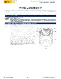
© Copyright 2026