
Cytokinin-like activity of N,N%
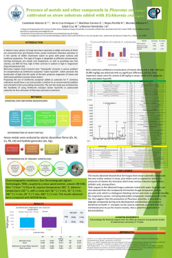
Plant Science 160 (2001) 1055– 1065 www.elsevier.com/locate/plantsci Cytokinin-like activity of N,N%-diphenylureas. N,N%-bis-(2,3-methylenedioxyphenyl)urea and N,N%-bis-(3,4-methylenedioxyphenyl)urea enhance adventitious root formation in apple rootstock M26 (Malus pumila Mill.) Ada Ricci a,*, Angela Carra a, Anna Torelli a, Cesare Augusto Maggiali b, Giovanni Morini b, Camillo Branca a a Dipartimento di Biologia E6oluti6a e Funzionale, Via delle Scienze 11 /A, I-43100 Parma, Italy b Dipartimento Farmaceutico, Via delle Scienze 7, I-43100 Parma, Italy Received 18 July 2000; received in revised form 23 January 2001; accepted 29 January 2001 Abstract Vegetative propagation of cuttings is a widespread method to multiplicate plants. Adventitious root formation is a key step in vegetative propagation and considerable progress has recently been made in understanding root formation. But, in spite of the efforts made, no new rooting treatments have been developed. Here, we report for the first time, that N,N%-bis-(2,3-methylenedioxyphenyl)urea and N,N%-bis-(3,4-methylenedioxyphenyl)urea enhance adventitious root formation in microcuttings of Malus pumila Mill. rootstock M26. Roots emerge without auxin supplementation in the darkness, transfer in hormone free medium, or callus formation. With the use of different bioassays, we also demonstrate that these two diphenylurea derivatives do not show cytokinin- or auxin-like activity. © 2001 Elsevier Science Ireland Ltd. All rights reserved. Keywords: Adventitious rooting; Auxin-like activity; Cytokinin-like activity; Diphenylurea derivatives; Malus pumila Mill. 1. Introduction Several billion rooted plantlets transferred to the soil per year are obtained by vegetative propagation of cuttings, whether by classical multiplication methods or, in many cases, by in vitro micropropagation (MP). This equals a commercial value of many billion dollars, 35–75% of which is spent for adventitious root formation [1,2]. These high costs are due to the steps necessary to induce adventitious root formation, such as a 3–10 day period of darkness in the presence of a hormonal supplementation and a transfer to a hormone free (HF) medium in the light [2,3], and to the losses that can occur during the acclimatization phase. * Corresponding author. Fax: +39-0521-905403. E-mail address: [email protected] (A. Ricci). Owing to the economic importance, many authors have tried to improve rooting and have investigated the effects of any kind of plant growth regulators [4] and the influence of non-hormonal compounds in vitro. Putrescine seems to be involved in the inductive phase of the adventitious rooting process, but it is unable per se to induce adventitious root formation when supplemented at physiological doses [5]. Phenolic compounds (i.e. pyrogallol, catechol, phloroglucinol, ferulic acid) are reported to act synergistically with auxin on adventitious rhizogenesis [6,7]. Auderset et al. [8,9] have shown that root formation of soybean and apple cuttings is stimulated by dithiothreitol and reduced glutathione in presence of auxin shock. Despite the great amount of research, no rooting substance leads to a practical application without some kind of auxin supplementation. 0168-9452/01/$ - see front matter © 2001 Elsevier Science Ireland Ltd. All rights reserved. PII: S 0 1 6 8 - 9 4 5 2 ( 0 1 ) 0 0 3 5 9 - 4 1056 A. Ricci et al. / Plant Science 160 (2001) 1055–1065 It is a well-known fact that some phenyl- and diphenylurea (DPU) derivatives show cytokininlike activity, such as a strong positive regulation of cell division and shoot regeneration [10]. Among these kinds of compounds, the Nphenyl-N%-1,2,3-thiadiazol-5-ylurea (thidiazuron, TDZ) which was developed for mechanized harvesting of cotton bolls, has proved to be a highly efficacious bioregulant of morphogenesis in the tissue culture of many plant species. In our work on synthetic auxin- and cytokinin-like compounds [11 – 14], we synthesized some DPU derivatives (Fig. 1). They were tested as cytokinins on tomato regeneration test, Malus pumila Mill. M26 rootstock cutting MP, chlorophyll level determination in cucumber cotyledons, Pg5-GUS gene expression in tobacco protoplasts in the presence of auxin. Owing to the unexpected root formation in M26 cutting MP, a suitable root inducing test on M26 microcuttings was performed. The same compounds were tested on a root inducing assay designed to confirm their root inducing capacity using Lycopersicon aesculentum var. Alice seedlings. The possible auxin-like activity was also tested on pea stem elongation test, tomato cotyledon rooting test, Pg5-GUS gene expression in tobacco protoplasts in the presence of cytokinin and apple stem slice rooting test. 2. Materials and methods 2.1. Chemicals 2.1.1. General notes The compounds tested, all neosynthesized but compound 4 [15], were recrystallized from EtOH, EtOH – H2O, or 1,4-dioxane. The various reactions were checked by TLC on silica gel (Kieselgel 60, F 254; Merck), with CHCl3 –EtOH (95:5) or (98:2), as eluents. For Soxhlet extractions, calcium-type silica gel (NM Kieselgel 60, 0.06 – 0.2 mm) was used. Mps: uncorrected. Only the most significant peaks of IR (KBr) are reported. 1H-NMR spectra (DMSO-d6) agree with the structural formulae. The results of elemental analysis (C, H, N) were within 90.3% of the theoretical values. 2.1.2. Compound 4, N,N%-bis-(3,4 -methylenedioxyphenyl)urea, and 3,4 -(methylenedioxy)aniline According to the method of Neville et al. [16], diphenylphosphoryl azide (17.4 g, 0.06 mol) and triethylamine (6.50 g, 0.06 mol) were added in one batch to a boiling solution of 3,4-methylenedioxybenzoic acid (piperonylic acid) (10 g, 0.06 mol) in 1,4-dioxane (150 ml) and ter-butyl alcohol (150 ml); the mixture were stirred under reflux for a further 10 h, then concentrated in vacuo. The residue was taken up in CHCl3 –CCl4 (1:1; 330 ml): an insoluble bright crystalline white solid was isolated by filtration and identified as N,N%-bis-(3,4-methylenedioxyphenyl)urea, compound 4 (3.40 g). The remaining filtrate was washed with successive portions of 5% aqueous citric acid (3×50 ml), water, saturated aqueous sodium hydrogen carbonate, and finally with saturated brine; a further 1.3 g of compound 2 were recovered from the ‘‘interphase’’. The organic layer was dried (Na2SO4), silica gel (10 g) was added and the solvents evaporated. The residual powder was placed in a Soxhlet apparatus and exhaustively extracted (24 h) with etherlight petroleum (30:70) solvent mixture (500 ml). Evaporation of the solvents gave a product (4 g; crude t-butyl-3,4-methylenedioxyphenylcarbamate) which was directly hydrolyzed (EtOH 90 ml, 5 N HCl 65 ml; 24 h stirring at room temperature). After evaporation of the ethanol solvent, the aqueous layer was washed with ether (3×45 ml) and to the resulting acid solution, cooled in an ice-bath, solid potassium hydroxide was added, to pH 7; the filtrate extracted with ether (3×70 ml) and the organic layers dried. The solvent was evaporated to leave 3,4(methylenedioxy)aniline, a yellow viscous oil (1.8 g), which was used as such in the next reaction (compound 1). Likewise, compounds 2, 6, 8 and the respective amines were prepared. 2.1.3. Compounds 1,3,5 and 7 The appropriate amine (0.0085 mol) was dissolved in benzene (30 ml) and to the boiling solution phenylisocianate (1.12 g, 0.0095 mol) in one batch was added. The mixture was stirred under reflux for 1 h, then filtrated and the whitish solid recrystallized. Yield 60%. A. Ricci et al. / Plant Science 160 (2001) 1055–1065 Fig. 1. Scheme of synthesis of the DPU derivatives. 1057 1058 A. Ricci et al. / Plant Science 160 (2001) 1055–1065 2.1.4. Analytical data Compound 1: N-(2,3-methylenedioxyphenyl)-N%phenylurea, m.p. 195 –196°C; IR (cm − 1) 3300, 1630; 1H-NMR(d) 8.94 (1H, s, NH); 8.32 (1H, s, NH); 7.54 (1H, d, Ph); 7.43 (2H, dd, Ph); 7.26 (2H, t, Ph); 6.98-6.93 (1H, m, Ph); 6.77 (1H, t, Ph); 6.59 (1H, dd, Ph); 6.04 (2H, s, OCH2O). Compound 2: N,N%-bis-(2,3-methylenedioxyphenyl)urea, m.p. 293 –294°C; IR (cm − 1) 3280, 1650; 1 H-NMR(d) 8.74 (2H, s, NH); 7.54 (2H, d, Ph); 6.76 (2H, t, Ph); 6.04 (4H, s, OCH2O). Compound 3: N-(3,4-methylenedioxyphenyl)-N%phenylurea, m.p. 204 –205°C; IR (cm − 1) 3280, 1640; 1H-NMR(d) 8.54 (1H, s, NH); 8.50 (1H, s, NH); 7.42 (2H, dd, Ph); 7.28-7.19 (3H, m, Ph); 6.95 (1H, t, Ph); 6.83-6.73 (2H, m, Ph); 5.96 (2H, s, OCH2O). Compound 4: N,N%-bis-(3,4-methylenedioxyphenyl)urea, m.p. 275 –277°C; IR (cm − 1) 3280, 1620; 1 H-NMR(d) 8.43 (2H, s, NH); 7.16 (2H, d, Ph); 6.82-6.71 (4H, m, Ph); 5.95 (4H, s, OCH2O). Compound 5: N-(2,3-dimethoxyphenyl)-N%-phenylurea, m.p. 142 –143°C; IR (cm − 1) 3320, 1650; 1 H-NMR(d) 9.29 (1H, s, NH); 8.33 (1H, s, NH); 7.81 (1H, dd, Ph); 7.45 (2H, d, Ph); 7.28 (2H, t, Ph); 6.97 (2H, m, Ph); 6.67 (1H, dd, Ph); 3.80 (3H, s, OCH3); 3.76 (3H, s, OCH3). Compound 6: N,N%-bis-(2,3-dimethoxyphenyl)urea, m.p. 178 – 179°C; IR (cm − 1) 3280, 1640; 1 H-NMR(d) 9.05 (2H, s, NH); 7.82 (2H, d, Ph); 6.97 (2H, t, Ph); 6.67 (2H, d, Ph); 3.79 (6H, s, OCH3); 3.75 (6H, s, OCH3). Compound 7: N-(3,4-dimethoxyphenyl)-N%-phenylurea, m.p. 180 –182°C; IR (cm − 1) 3280, 1620; 1 H-NMR(d) 8.53 (1H, s, NH); 8.45 (1H, s, NH); 7.43 (2H, dd, Ph); 7.28-7.23 (2H, m, Ph); 7.19 (1H, s, Ph); 6.97-6.86 (3H, m, Ph); 3.73 (3H, s, OCH3); 3.70 (3H, s, OCH3). Compound 8: N,N%-bis-(3,4-dimethoxyphenyl)urea, m.p. 209 – 210°C; IR (cm − 1) 3300, 1630; 1 H-NMR(d) 8.36 (2H, s, NH); 7.17 (2H, s, Ph); 6.85 (4H, m, Ph); 3.73 (6H, s, OCH3); 3.70 (6H, s, OCH3). All the DPU derivatives were dissolved in dimethylsulfoxide (DMSO) and the final concentration of DMSO in the medium did not exceed 0.2% [16]. 2.2. Tomato regeneration test Twelve cotyledon explants, obtained from seedlings cultivated in vitro, were plated on MS medium [17] plus the DPU derivatives, as putative cytokinins at 1, 5 and 10 mM, both alone and in the presence of 20 mM 1,2-benzisoxazole-3-acetic acid (BOAA), as auxin [18]. The results were compared with that TDZ in the same culture conditions. After 2 week incubation, the cotyledon explants were transferred to a hormone free medium (HF) and the percentage of regenerated shoots was checked 2 weeks later. The experiments were carried out in triplicate, and repeated three times. 2.3. Micropropagation of apple cuttings Microcuttings of M. pumila Mill. rootstock M26 were obtained from Dr. E. Caboni (Istituto Sperimentale per la Frutticoltura, Roma, Italy). The cuttings deprived of apices were propagated on a MP medium (MS salts plus 0.4 mg l − 1 thiamine HCl, 0.5 mg l − 1 nicotinic acid, 0.5 mg l − 1 pyridoxine HCl, 100 mg l − 1 myo-inositol, 10 g l − 1 sucrose, 20 g l − 1 sorbitol, 0.65% agar B&V, 5.8 pH). The MP medium was supplemented with 1.3 mM benzylaminopurine (BAP), as control, or 1.3 mM DPU derivatives, as cytokinins, and 0.25 mM indole-3-butyric acid (IBA). Incubation was at 26°C at a light intensity of 27 mmol m − 2 s − 1 under 16 h photoperiod. After 3 week culture the microcuttings were checked. 2.4. Chlorophyll le6el determination Chlorophyll levels were determined as previously described [19] with minor modifications. Cucumber seeds (Cucumis sati6us L.) were soaked for 4 h in running tap water and germinated on moist filter paper in the darkness at 26°C for 1 week. Expanded cotyledons were excised in dim green light and floated in 5 cm Petri dishes containing 5 ml of DPU derivative solutions at three different concentrations: 0.1, 1 and 10 mM. Each plate contained 10 cotyledons with the adaxial face down. Control was performed with water and the same concentrations of TDZ. After 17 h incubation in the darkness at 26°C, the dishes were moved to a light intensity of 27 mmol m − 2 s − 1 for 3 h at 26°C. Chlorophyll was then extracted from weighed cotyledons with 80% acetone, and the chlorophyll concentration was calculated as described [20]. Experiments were carried out in triplicate, and repeated three times. A. Ricci et al. / Plant Science 160 (2001) 1055–1065 2.5. Bioassay for Pg5 -GUS gene expression The bioassay for Pg5-GUS gene expression is based on the expression of the chimeric Pg5-GUS gene in protoplasts of Nicotiana tabacum transformed plants [21]. This chimeric gene, consisting of A. tumefaciens T-DNA gene 5 promoter (Pg5) fused to the b-glucuronidase coding sequence (GUS), is slightly induced by cytokinin or auxin alone, but is strongly activated when both growth regulators are added to the culture medium. We exploited this fact and tested compounds 2 and 4 separately either in the presence of cytokinin, to check their auxin nature, or in the presence of auxin, to check their cytokinin nature. Seeds of Pg5-GUS transformed N. tabacum cv. Petit Havana (SR1) were a gift from W. Boerjan (Universiteit Gent, Belgium). Seed sterilization, in vitro plant growth and tobacco protoplast preparation were carried out as previously described [22]. An average of 5× 10⁶ protoplasts were resuspended in 2 ml of K3 medium [22] containing 0.4 M glucose. Culture conditions were as follows: hormone free medium; 1 mM naphthalene-acetic acid (NAA) or 1 mM zeatin riboside (ZR); 1 mM NAA plus the following concentrations of the DPU derivatives: 0.02, 0.2, 2 and 20 nM, to test their cytokinin-like activity; 1 mM NAA plus the same concentrations of TDZ (0.02, 0.2, 2 and 20 nM) used as control; 1 mM ZR plus the following concentrations of the DPU derivatives: 1, 10 and 100 nM, to test their auxin-like activity; and 1 mM ZR plus the same concentrations of NAA (1, 10 and 100 nM) used as control. The rest of the experimental procedure was performed according to Ricci et al. [22]. Experiments were carried out in duplicate, and repeated twice. 2.6. Rooting of apple cuttings M. pumila Mill. rootstock M26 microcuttings, 2 – 2.5 cm in length excised from 3-week old cultures, were incubated in MP medium supplemented with 1, 5, 10 and 15 mM of N,N%-bis(2,3-methylenedioxyphenyl)urea (compound 2) or N,N%-bis-(3,4-methylenedioxyphenyl)urea (compound 4) under the following culture conditions: 1059 at light intensity of 27 mmol m − 2 s − 1 under 16 h photoperiod; for 6 days in the darkness, then at light intensity of 27 mmol m − 2 s − 1 under 16 h photoperiod; and for 6 days in the darkness, then transferred in HF medium at light intensity of 27 mmol m − 2 s − 1 under 16 h photoperiod. For each culture condition, controls were performed with microcuttings cultured in HF medium and in the presence of the same concentrations of IBA. In all the experiments the roots were counted after 2 weeks. For each experiment 50 microcuttings were used. 2.7. Rooting of tomato seedlings Tomato seeds (L. aesculentum var. Alice) were sown and germinated under sterile conditions in half strength MS medium without sucrose, 0.8% agar, 5.8 pH. Incubation was at 26°C and 27 mmol m − 2 s − 1 light intensity under 16 h photoperiod. One-week old seedlings were divided into two groups: in one, the seedlings were deprived of their roots; and in the other, the seedlings were deprived of their cotyledons too. The two groups were then treated in the same way: the seedlings were plated in MS medium containing 30 g l − 1 sucrose and 0.8% agar, 5.8 pH. Four different concentrations of compounds 2 or 4 were used: 0.1, 0.5, 1 and 5 mM. HF medium and the same concentrations of IBA were used as controls. The number of roots was counted after 10 days. For each experiment 50 seedlings were used. 2.8. Pea stem elongation test The activity of the compounds 2 and 4 on cell elongation was tested on etiolated pea stem segments (Pisum sati6um var. Alaska) measuring their increase in fresh weight [24]. The compounds were tested at four different concentrations: 0.1, 1, 10 and 100 mM. Water and the same concentrations of indole-3-acetic acid (IAA) were used as controls. The experiments were carried out in triplicate, and repeated twice. 2.9. Rooting of tomato cotyledons Tomato cotyledons, var. Alice, were obtained and cultivated in vitro as previously described [18]. A. Ricci et al. / Plant Science 160 (2001) 1055–1065 1060 Ten cotyledon explants were plated on MS medium plus compounds 2 and 4 separately, as auxins, at 0.5, 1 and 5 mM. HF medium and the same concentrations of IBA were used as controls. The number of roots was counted after 10 days. For each experiment 50 samples were used. 2.10. Rooting of apple stem slices The experiment was performed according to Van der Krieken et al. [23]. M. pumila Mill. rootstock M26 stem slices having :1 mm thickness were cut with a razor blade from shoots of 1.5 –2 cm obtained with the MP method described above. Groups of 25 slices were cultured in Petri Table 1 Effect of DPU derivatives on shoot regeneration at three different concentrations (1, 5 and 10 mM) in the tomato cotyledon bioassay, in the absence (−) and presence (+) of 20 mM BOAA as auxina Concentration (mM) Explants forming shoots (%) 1 Compound Compound Compound Compound Compound Compound Compound Compound TDZ a 1 2 3 4 5 6 7 8 5 − + − 0 0 0 0 0 0 0 0 0 0 0 0 0 0 0 0 0 0 0 4.3 0 0 0 0 0 43 59 10 + − 8.5 0 0 0 0 0 0 0 5.6 0 4.5 0 0 0 0 0 83 68 The cytokinin-like activity of all the DPU derivatives was tested on the following assays. + 2.6 0 0 0 4.8 0 0 0 92 Table 2 Effect of DPU derivatives on apple cutting MP at 1.3 mMa All the DPU derivatives are unable to induce shoot regeneration at all the concentrations tested, both alone and in the presence of 20 mM BOAA, as auxin (Table 1). 3.2. Micropropagation of apple cuttings All the DPU derivatives are unable to induce the formation of new microcuttings in the MP of M. pumila Mill. rootstock M26 (Table 2) but, surprisingly, compounds 2 and 4 induced adventitious roots. 3.3. Chlorophyll le6el determination Mean shoot number Concentration (mM) 1.3 Compound Compound Compound Compound Compound Compound Compound Compound BAP 0 0 0 0 0 0 0 0 12 a 3. Results 3.1. Tomato regeneration test The same concentrations of TDZ were used as control. 1 2 3 4 5 6 7 8 dishes with the apical side on a nylon mesh put on MP medium supplemented with compounds 2 and 4 separately, as auxin at 1, 3.2, 5 and 10 mM. The dishes were incubated upside down at light intensity of 27 mmol m − 2 s − 1 at 26°C under 16 h photoperiod. Control was performed with apple stem slices cultured in HF medium and the same concentrations of IBA. In another set of experiments, apple stem slices were incubated in the darkness under the same conditions described above. After 6 days of darkness the slices were transferred to HF medium and incubated at light intensity of 27 mmol m − 2 s − 1 at 26°C under 16 h photoperiod [23]. The number of roots was counted after 14 days. The experiments were carried out in triplicate, and repeated twice. The same concentration of BAP was used as control. All the DPU derivatives are unable to increase chlorophyll level at all the concentrations tested (Table 3). 3.4. Bioassay for Pg5 -GUS gene expression (as cytokinins) All the DPU derivatives are unable to induce Pg5-GUS activity, when tested as cytokinins in the presence of 1 mM NAA. Fig. 2(A) shows only the relative graphs for compounds 2 and 4. A. Ricci et al. / Plant Science 160 (2001) 1055–1065 Table 3 Effect of DPU derivatives on chlorophyll level at three different concentrations (0.1, 1 and 10 mM) using the cucumber cotyledon bioassaya Concentration (mM) Compound Compound Compound Compound Compound Compound Compound Compound TDZ 1 2 3 4 5 6 7 8 Chlorophyll level of the control (%; H2O) 0.1 1 10 −34.35 3.68 −23.92 −50.31 −18.41 −46.01 −34.35 7.97 6.13 −25.76 −3.06 −21.47 −49.07 −17.79 −29.44 −19.63 −34.96 8.58 −24.54 −17.17 −17.79 −37.42 −7.36 −23.31 −3.06 −29.44 11.65 1061 were observed when the microcuttings cultured in the continuous presence of compounds 2 and 4 separately, were transferred to the light after 6 days in the darkness. When compounds 2 and 4 were separately tested at 5, 10 and 15 mM, the a The results are expressed as a percentage of the control (H2O alone) by the formula (T−C/C)×100. The same concentrations of TDZ were used as control. The standard error is less than 10% for each measurement (n= 9). According to these results, no DPU derivative shows cytokinin-like activity. However, during the apple MP, N,N%-bis-(2,3-methylenedioxyphenyl)urea (compound 2) and N,N%-bis-(3,4methylenedioxyphenyl)urea (compound 4) induced adventitious roots. Therefore, these two compounds were tested in the rooting bioassays described below. 3.5. Rooting of apple cuttings Compounds 2 and 4 enhanced root formation in microcuttings of M. pumila Mill. rootstock M26. The best rooting stimulation was obtained when the microcuttings were cultured in the light, in the constant presence of 1 mM of compound either 2 or 4. In 70% and 91% of the microcuttings respectively (Table 4), the roots emerged directly from the base of the microcuttings without callus formation. Under the same culture conditions, in the presence of 1 mM IBA, the percentage of rooted microcuttings was 75%, and only few roots emerged from a large-sized callus. This percentage went up to 86% when the microcuttings were transferred to HF medium in the light, after a 6 day treatment with 1 mM IBA in the dark (best auxinic rooting treatment). In this condition, several roots emerged from each microcutting, but a callus formation was always present. No significant differences in rooting percentages Fig. 2. (A) Effect of TDZ, compounds 2 and 4 (as cytokinins) on Pg5-GUS expression in the tobacco bioassay at four different concentrations (0.01, 0.02, 0.2 and 2 nM) in the presence of 1 mM NAA. The results express GUS activity as pmol of 4-methyl-umbelliferone (MU)/mg protein/min. The standard error is less than 10% for each measurement (n=4). (B) Effect of NAA, compounds 2 and 4 (as auxins) on Pg5-GUS expression in the tobacco bioassay at three different concentrations (1, 10 and 100 nM) in the presence of 1 mM ZR. The results express GUS activity as pmol of 4-methylumbelliferone (MU)/mg protein/min. The standard error is less than 10% for each measurement (n= 4). A. Ricci et al. / Plant Science 160 (2001) 1055–1065 1062 Table 4 Effect of compounds 2 and 4 on adventitious rooting of apple microcuttings at 1 mMa Concentration (mM) Rooted cuttings (%) 0 Compound 2 Compound 4 IBA HF 1 0 70.4 91.1 75.2 28.5 1 0 1 68.3 87.5 79.3 43.7 50.1 86.5 18.7 18.7 a L – the microcuttings, cultured in the continuous presence of the compounds, in the light; D–L – the microcuttings are cultured in the continuous presence of the compounds, are transferred to the light after 6 days in the darkness; D–L (HF) – after 6 day culture in the darkness in the presence of the compounds, the microcuttings are transferred to HF medium in the light. HF and the same concentration of IBA were used as control. The results express the percentage of rooted microcuttings. The standard error is less than 10% for each measurement (n= 50). rooting percentage was not significantly different from that of the HF condition. With the increasing concentrations of IBA the number of rooted microcuttings diminished in all the culture conditions tested. 3.6. Rooting of tomato seedlings When the tomato seedlings were only deprived of their root system, compounds 2 and 4 were able to increase the number of adventitious roots. At 5 mM the mean root number was similar to that obtained with the same concentration of IBA (Table 5). When the seedlings were deprived both of their root system and their cotyledons, only IBA at 5 mM was able to increase root formation (Table 6). According to the results obtained with the rooting bioassays, the possible auxin-like activity of compounds 2 and 4 was tested. 3.7. Bioassay for Pg5 -GUS gene expression (as auxins) Compounds 2 and 4 are unable to induce Pg5GUS activity, when tested as auxins in the presence of 1 mM ZR (Fig. 2(B)). stem at all the concentrations tested (Table 8). The increase in fresh weight is not significantly different from that obtained with H2O, used as control. Table 5 Effect of compounds 2 and 4 on adventitious rooting of tomato seedlings at 0.1, 0.5, 1 and 5 mMa,* Concentration (mM) 0 Compound 2 Compound 4 IBA HF Compounds 2 and 4 are unable to induce cell elongation on the third internode of etiolated pea 0.5 1 5 7.6 9 7.7 8.5 10.4 7.3 9 9 7.3 10.3* 11.7* 10.8* 6.6 HF and the same concentrations of IBA were used as control. The results express the mean root number obtained per seedling. The standard error is less than 10% for each measurement (n= 50). * Significantly different at 95% (F test). Table 6 Effect of compounds 2 and 4 on adventitious rooting of tomato seedlings deprived of cotyledons at 0.1, 0.5, 1 and 5 mMa,* Concentration (mM) Mean root number 0 Compound 2 Compound 4 IBA HF a 3.9. Pea stem elongation test 0.1 a 3.8. Rooting of apple stem slices Compounds 2 and 4 are unable to induce roots in apple stem slices at all the concentrations tested (Table 7). Mean root number 0.1 0.5 1 5 4.1 3.8 3.9 4.1 4 3.4 4 3.7 4.2 3.7 3.2 6* 3.9 HF and the same concentrations of IBA were used as control. The results express the mean root number obtained per seedling. The standard error is less than 10% for each measurement (n= 50). * Significantly different at 95% (F test). A. Ricci et al. / Plant Science 160 (2001) 1055–1065 1063 Table 7 Effect of compounds 2 and 4 on root formation using the apple stem slice bioassay at four different concentrations (1, 3.2, 5 and 10 mM)a Concentration (mM) Mean root number per slice 1 L Compound 2 Compound 4 IBA HF 0 D 3.2 5 10 L D L D L D L D 0 0 0.5 0 0 0.7 0 0 1 0 0 2.3 0 0 1 0 0 0.7 0 0 0.7 0 0 0.9 0 a Control was performed with the same concentrations of IBA. L: the apple stem slices are cultured in continuous light; D: after 6 days in the darkness, the apple stem slices are transferred to HF medium in the light. The standard error is less than 10% for each measurement (n=6). 3.10. Rooting of tomato cotyledons Compounds 2 and 4 are unable to affect root formation in tomato cotyledon explants at all the concentrations tested (Table 9). The number of roots is not significantly different from that obtained in HF condition. 4. Discussion None of our neosynthesized N,N%-diphenylurea derivatives showed cytokinin-like activity. This is consistent with the knowledge that substituents of N,N%-diphenylurea phenyl rings with great steric hindrance lower the cytokinin-like activity of the compounds [10]. When we tried to micropropagate microcuttings of M. pumila Mill. rootstock M26, surprisingly compound 2 (N,N%-bis-(2,3-methylenedioxyphenyl)urea) and compound 4 (N,N%-bis-(3,4-methylenedioxyphenyl)urea) enhanced adventitious root formation. Even this particular activity is related to specific molecular structures. Our asymmetrical compounds are actually unable to enhance adventitious root formation. But symmetrical molecular structure is not the only requirement to be met in order to obtain a greater number of adventitious roots. In fact N,N%-bis-(2,3-dimethoxyphenyl)urea and N,N%-bis-(3,4-dimethoxyphenyl)urea, i.e. compounds 6 and 8, respectively, are unable to enhance adventitious root formation. Besides, neither N-(2,3-methylenedioxyphenyl)-N%-phenylurea nor N-(3,4-methylenedioxyphenyl)-N%-phenylurea, i.e. compound 1 and 3 respectively, are able to increase the number of adventitious roots, as only one methylenedioxy benzo-condensed group is linked to urea matrix. To verify if compounds 2 and 4 could be responsible per se for this activity, apple cuttings were cultured with different doses of these two DPU derivatives alone in the light. The results were definitely surprising. When tested at 1 mM, compounds 2 and 4 allowed root formation in were separately tested at 5, 10 and 15 mM, the 70% and 91% of the cuttings, respectively, as shown in Table 4. Roots emerged after 15 days with no auxin supplementation, no darkness treatment, no transfer to HF medium in the light and no callus formation (Fig. 3). Under the same cultureconditions, 1 mM IBA allowed root formation in 75% of the cuttings, in which only few roots emerged from a large-sized callus. This is Table 8 Effect of compounds 2 and 4 on cell elongation measured as the increase in fresh weight of the third internode of etiolated pea stem, at four different concentrations (0.1, 1, 10 and 100 mM)a Concentration (mM) Compound 2 Compound 4 IAA H2O a Increase of fresh weight (%) 0.1 1 10 100 15 11 39* 19 14 48* 15 14 54* 18 18 54* 11 Water and the same concentrations of IAA were used as control. The standard error is less than 10% for each measurement (n=6). * Significantly different at 95% (F test). A. Ricci et al. / Plant Science 160 (2001) 1055–1065 1064 Table 9 Effect of compounds 2 and 4 on root induction at three different concentrations (0.5, 1 and 5 mM) using the tomato cotyledon explant bioassaya,* Concentration (mM) Mean root number 0.5 Compound 2 Compound 4 IBA HF 2.2 6.1 7.1 5.1 1 90.6 91.2 90.9 91.2 4.1 6.9 6.7 5.1 5 91.5 90.9 91.3 91.2 2.2 6.3 14.9* 5.1 90.6 90.9 91.3 91.2 a HF and the same concentrations of IBA were used as control. The standard error is less than 10% for each measurement (n=50). * Significantly different at 95% (F test). Fig. 3. Example of rooted microcuttings obtained under the best culture conditions. 1 mM IBA – after 6 days of IBA treatment in the darkness, the microcuttings were transferred to HF medium in the light (27 mmol m − 2 s − 1 under 16 h photoperiod); 1 mM compound 2 – the microcuttings are cultured in the continuous presence of 1 mM compound 2 in the light (27 mmol m − 2 s − 1 under 16 h photoperiod); 1 mM compound 4 – the microcuttings are cultured in the continuous presence of 1 mM compound 4 in the light (27 mmol m − 2 s − 1 under 16 h photoperiod). Control was performed with microcuttings cultured in HF medium. Picture was taken after 15 days of culture. consistent with the knowledge that after 96 h of treatment, auxin is no longer required and becomes inhibitory during the rooting differentiation phase [2]. In fact, the rooting percentage increases (86%) when the IBA period treatment was reduced. As compounds 2 and 4 were able to enhance root formation also in the tomato seedlings deprived of their own roots, we performed different bioassays to verify their possible auxin-like activity. The results show that the N,N%-bis-(2,3methylenedioxyphenyl)urea and the N,N%-bis-(3,4-methylenedioxyphenyl)urea do not behave as auxins. In conclusion, our data demonstrate that 1. the rooting activity of N,N%-bis-(2,3-methylenedioxyphenyl)urea and N,N%-bis-(3,4-methylenedioxyphenyl)urea is strictly related to the symmetrical presence of methylenedioxy benzo-condensed group on urea matrix; 2. they might be able to cooperate with molecules originating from other parts of the plants, as, when the two compounds were tested on apple stem slices or tomato seedlings deprived of roots and cotyledons, root induction was absent; 3. the N,N%-bis-(2,3-methylenedioxyphenyl)urea and the N,N%-bis-(3,4-methylenedioxyphenyl) urea do not show inhibitory effect on root formation, as the best rooting condition is A. Ricci et al. / Plant Science 160 (2001) 1055–1065 obtained when the apple microcuttings are left in continuous contact with them (Table 4); and 4. they are the first non-hormonal compounds showing a direct rhizogenetic activity. They could be the starting point of a new category of specific compounds favouring root formation, that could cut down the adventitious rooting costs in horticultural, agricultural and forestry vegetative multiplication. Work is in progress to elucidate the mechanism by which the N,N%-bis-(methylenedioxyphenyl) ureas might enhance rooting. References [1] P.C. Debergh, L.J. Maene, A scheme for commercial propagation of ornamental plants by tissue culture, Scientia. Hort. 14 (1981) 335–345. [2] G.J. De Klerk, W. Van der Krieken, J.C. De Jong, The formation of adventitious roots: new concepts, new possibilities, In Vitro Cell Dev. Biol. Plant 35 (1999) 189– 199. [3] C. Moncousin, Rooting of microcuttings: general aspects, Acta. Hort. 289 (1991) 301–310. [4] B.E. Haissig, T.D. Davis, A historical evaluation of adventitious rooting research in 1993, in: T.D. Davis, B.E. Haissig (Eds.), Biology of adventitious root formation, Plenum Press, New York, 1994, pp. 275–331. [5] C. Kevers, C. Bringaud, J.F. Hausman, T. Gaspar, Putrescine involvement in the inductive phase of walnut shoots rooting in vitro, Saussurea 28 (1997) 47–57. [6] C.E. Hess, Internal and external factors regulating root initiation, in: W.J. Whittington (Ed.), Root growth, Butterworths, London, 1969, pp. 42–64. [7] D.J. James, I.J. Thurbon, Shoot and root initiation in vitro in the apple rootstock M9 and the promotive effects of phloroglucinol, J. Hort. Sci. 56 (1981) 15 – 20. [8] G. Auderset, C. Moncousin, J. O’Rourke, D.J. Morre´, Stimulation of root formation by thiol compounds, Hort. Sci. 31 (1996) 240–242. [9] G. Auderset, C. Moncousin, J. O’Rourke, D.J. Morre´, Stimulation of root formation in difficult-to-root woody cuttings by dithiothreitol, Int. J. Plant Sci. 158 (1997) 132–135. [10] K. Shudo, Chemistry of phenylurea cytokinins, in: D.W.S. Mok, M.C. Mok (Eds.), Cytokinins Chemistry, Activity and Function, CRC Press, Boca Raton, 1994, pp. 35–42. . 1065 [11] C. Branca, V. Plazzi, M. Vitto, F. Bordi, D. SerafiniFracassini, N. Bagni, Auxin-like activity of 1,2-benzisothiazole derivatives, Phytochemistry 14 (1975) 2545– 2550. [12] C. Branca, C.A. Maggiali, G. Morini, F. Ronchini, D. Ricci, V. Scoccianti, Synthesis and cytokinin-like activity of 7-chloro-imidazo[1,2-c]pyrimidines, Phytochemistry 26 (1987) 25 – 29. [13] C. Branca, G. Bucci, P. Domiano, A. Ricci, A. Torelli, M. Bassi, Auxin structure and activity on tomato morphogenesis in vitro and pea stem elongation, Plant Cell Tiss. Org. Cult. 24 (1991) 105– 114. [14] A. Ricci. A. Carra, A. Torelli, C.A. Maggiali, P. Vicini, F. Zani, C. Branca, Cytokinin-like activity of N%-substituted N-phenylureas, Plant Growth Reg. (2000). [15] C.F. Neville, M.F. Grundon, V.N. Ramachandran, J. Reisch, Quinoline alkaloids. Part 27. Synthesis of the Ptelea alkaloids pteleflorine, neohydroxylunine, Omethylhydroxyluninium salt and hydroxylunine, J. Chem. Soc. Perkin. Trans. 1 (1991) 259– 262. [16] R.Y. Schmitz, F. Skoog, The use of dimethylsulfoxide as a solvent in the tobacco bioassay for cytokinins, Plant Physiol. 45 (1970) 537– 538. [17] T. Murashige, F. Skoog, A revised medium for rapid growth and bioassay with tobacco tissue cultures, Physiol. Plant 15 (1962) 473– 497. [18] C. Branca, A. Torelli, M. Bassi, Effects of benzisothiazole on tomato plant regeneration in vitro, Plant Cell Tiss. Org. Cult. 21 (1990) 17 – 19. [19] R.A. Fletcher, D. McCullagh, Cytokinin-induced chlorophyll formation in cucumber cotyledons, Planta 101 (1971) 88 – 90. [20] C.O. Miller, F. Skoog, M.H. von Saltza, F.M. Strong, Kinetin: a cell division factor from deoxyribonucleic acid, J. Am. Chem. Soc. 77 (1955) 1392. [21] W. Boerjan, C. Genetello, M. Van Montagu, D. Inze´, A new bioassay for auxin and cytokinins, Plant Physiol. 99 (1992) 1090– 1098. [22] A. Ricci, C.A. Maggiali, A. Torelli, S. Amorosi, F. Ronchini, C. Branca, Methoxylation modifies the activity of 1,2-benzisoxazole-3-acetic acid: 6,7-dimethoxy-1,2benzisoxazole-3-acetic acid is an auxin antagonist in cytokinin mediated processes, Plant Sci. 117 (1996) 151– 158. [23] W.M. Van der Krieken, H. Breteler, M.H.M. Visser, D. Mavridou, The role of the conversion of IBA into IAA on root regeneration in apple: introduction of a test system, Plant Cell Rep. 12 (1993) 203– 206. [24] C. Branca, G. De Lorenzo, F. Cervone, Competitive inhibition of the auxin-induced elongation by 2-D-oligogalacturonides in pea stem segments, Physiol. Plant 72 (1988) 499– 504.


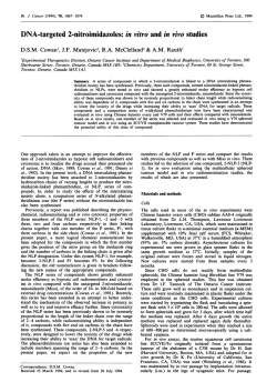

© Copyright 2026