
Fall/Winter 2014 - Pediatrics Nationwide
Fall/Winter 2014 Pediatrics NATIONWIDE Building the Modern-Day Vaccine Advancing the Conversation on Child Health 20 BUILDING THE MODERN-DAY VACCINE Table of Contents Vaccine development used to be straightforward. Now, the challenges are many and the successes are few. What will it take to overcome the obstacles presented by both immunology and society? 2 PediatricsNationwide.org | Fall/Winter 2014 10 D E PA R T M E N T S 4 34 36 In Practice: News from the Field In Sight: Working Up the Nerve Second Opinions: Adult Congenital Heart Disease: Let’s Finish What We Started 38 Connections: Continuing the Conversation F E AT U R E S 10 16 Enteral Therapy on Trial 24 Thinking Outside the (Tool) Box 28 Body, Heal Thyself 24 Building the Modern Day Vaccine The ‘Eureka!’ moment for me was accepting that if preeclampsia is a disease of improper protein folding, there should be connections with other [such] diseases. – Irina Buhimschi, MD, Nationwide Children’s Hospital (page 24) It’s an absolutely fundamental discovery. They’ve shown that in a state of health, the kidney is protected from microbial invasion by its ability to produce very high concentrations of some very powerful antimicrobial agents. – Michael Zasloff, PhD, Georgetown University School of Medicine (page 28) Fall/Winter 2014 | PediatricsNationwide.org 3 In Practice An Unwelcome Blast from the Past Physicians in Tennessee reported a rash of cases of VKDB last year. Now, the CDC wants to know if it’s a sign of a national trend. M ost pediatric specialists who began practicing medicine in the mid-1960s have probably never seen a case of vitamin K deficiency bleeding. Robert Sidonio, MD, had seen just one, and that was during his fellowship. And then, in February 2013, an infant came in to the emergency room with bleeding in the brain, one of the most lethal problems of VKDB. “One case was surprising, but nothing to get alarmed about,” says Dr. Sidonio, a pediatric neurologist at Vanderbilt University Medical Center. “But then a few months later, we saw another one, then another one, and I knew something wasn’t right.” Reports of VKDB in the United States have been rare since the American Academy of Pediatrics issued a recommendation in 1961 that newborns receive vitamin K shots shortly after birth to protect against bleeding disorders. Studies had shown that the body doesn’t manufacture enough of the vital clotting agent until around 6 months of age, leaving infants vulnerable to VKDB, which can cause bleeding in the brain and other organs. Vitamin K isn’t transferred easily from mother to child in breast milk or from the placenta, so supplementing at birth or using infant formula — which is fortified with the nutrient — are the only ways to lower the bleeding risk. Each of the families of infants with VKDB diagnosed by Dr. Sidonio last year had declined the vitamin K booster after birth and exclusively breastfed. Concerned that their refusal may signal a growing trend, he asked the Centers for Disease Control and Prevention for guidance. Working with the Tennessee Health Department and Dr. Sidonio, the CDC launched an investigation that uncovered a total of seven cases of VKDB, five of which included brain bleeds. Four of those children demonstrated cognitive and developmental delays and at least one will have lifelong disabilities as a result. 4 PediatricsNationwide.org | Fall/Winter 2014 This image depicts intracranial hemorrhages in four children with vitamin K deficiency bleeding. Reprinted from Pediatric Neurology, Schulte R et al., Rise in late onset vitamin K deficiency bleeding in young infants because of omission or refusal of prophylaxis at birth, 50(6):564-8, 2014 Jun, with permission from Elsevier and Dr. Sidonio. Because the condition is rare, VKDB could be easily misdiagnosed, Dr. Sidonio says. So to spread the word, Dr. Sidonio published an article in February in Pediatric Neurology, detailing the cases at Vanderbilt and urging clinicians to advocate for newborn vitamin K supplementation. Later this year, the CDC and the American Society of Pediatric Hematology/Oncology will conduct a nationwide survey to see if the increase in VKDB is confined to Tennessee or is more widespread. — Kelli Whitlock Burton The End of “Under 12” UNOS board eliminates age classification for child lung transplant candidates and alters rules for pediatric heart transplants. M any in the pediatric field were surprised last year when a federal judge ordered that a 10-year-old Pennsylvania girl with cystic fibrosis be added to the national adult waiting list for a double-lung transplant. She had spent 18 months on the pediatric list to no avail and without new lungs, her doctors said, she would die within weeks. The lawsuit, filed by her parents, asked the court to overrule a policy known as the “under-12 rule,” which separated lung transplant waiting lists between children and adults based solely on their age, instead of on medical need. In response to the judge’s order, leaders of the Organ Procurement and Transplant Network, established by the U.S. Congress in 1984, and the United Network for Organ Sharing, which operates the OPTN, temporarily suspended the age classification. Now, the governing board of the two networks has permanently done away with the under-12 rule. Under the new policy, if a patient age 11 years or younger meets certain criteria, a hospital’s transplant team can ask OPTN/UNOS to list the patient on both the child and adult waiting lists for donor lungs. If adult lungs become available and the child’s case is more dire than that of the patient at the top of the adult list, the adult lungs will go to the pediatric patient. “These are never easy decisions because every time you prioritize one group of patients, you deprioritize another,” says William T. Mahle, MD, incoming vice chair of the Pediatric Transplantation Committee for OPTN/ UNOS and medical director for the heart transplant program at Children’s Healthcare of Atlanta. Dr. Mahle was on the committee that recommended the temporary suspension of the under-12 rule. Changes to the network’s organ procurement and transplant policies aren’t unusual, but officials with OPTN/UNOS say this was the first time policy revisions stemmed from a lawsuit. “I think UNOS felt like they couldn’t be on the wrong side of this issue when there was some positive public sentiment supporting it and the courts supporting it,” Dr. Mahle says. Less controversial but equally important are two changes the OPTN/UNOS board made to policies governing pediatric heart transplants. Child heart transplant candidates are rated as either status 1A, 1B or 2, with 1A getting first priority when a donor organ becomes available. In the past, the 1A ranking was based solely on how long a child had been on the waiting list. Now, 1A status is reserved for the sickest patients. Children whose condition is stabilized with medication, such as those with certain types of cardiomyopathy that can be managed with the drug inotrope, will be classified as 1B. The board also voted to allow children under the age of 2 years to receive hearts from donors with a different blood type. The change follows new research that shows young patients often do well in these procedures, known as ABO-incompatible transplants. — Kelli Whitlock Burton Fall/Winter 2014 | PediatricsNationwide.org 5 Mystery Rising Data shows that the global incidence of type 1 diabetes is increasing. What the data doesn’t show is why. T ype 1 diabetes used to be rare. Late 19th-century estimates put its incidence at about 0.004 percent of the world’s population. But by the end of the 20th century, most nations reported a number 350 times that rate. With many countries continuing to experience a steady rise in new cases of up to 4 percent annually, the word “epidemic” has crept into the scientific literature. the increase. Although they vary widely, most of these hypotheses aim to explain, in part, why the incidence in type 1 diabetes spiked so dramatically in the second half of the 1900s — a period underscored by improved medicine and cleaner surroundings, but also a rise in artificial diets and more time spent indoors. Although some of these theories offer compelling evidence and strong associations with the increase in type 1 incidence, Dr. Mayer-Davis, who also has served as vice president of health care and education for the American Diabetes Association, suggests it is too early to pinpoint the reason behind the rise. “This is indeed an alarming trend because of the impact that type 1 diabetes has on the daily life and the long-term health of those affected,” says Elizabeth Mayer-Davis, PhD, national chair of the SEARCH for Diabetes in Youth Study, a multi-center effort funded by the National Institute of Diabetes and Digestive and Kidney Diseases and the Centers for Disease Control and Prevention. “The trend of increasing incidence is particularly worrisome because we do not know its cause.” “Type 1 diabetes is caused by a combination of genetic and environmental or behavioral effects. I would not speculate on the precise cause,” she says. “International studies are ongoing that will be critical in identifying the environmental or behavioral factors that are acting on genetic risk. From those studies, we hope to identify approaches to prevention.” Speculators list theories such as the hygiene hypothesis, intestinal parasite eradication, rotavirus infection, vitamin D deficiency, early introduction of cow’s milk and a combination of such factors as possible causes of — Katie Brind’Amour AVERAGE ANNUAL CHANGES IN TYPE 1 DIABETES INCIDENCE AFRICA 3.0% ASIA 4.0% NORTH AMERICA 5.3% SOUTH AMERICA 5.3% EUROPE Global average annual increase: 2.8% Increases are among children ages 0-14. The highest continental average annual increase is in ages 0-4, with rates topping 11% per year. Source: DIAMOND Project Group. Incidence and trends of childhood Type 1 diabetes worldwide 1990-1999. Diabetic Medicine. 2006 Aug, 23(8):857-66. 6 PediatricsNationwide.org | Fall/Winter 2014 3.2% OCEANIA 3.2% CENTRAL AMERICA AND WEST INDIES -3.6% Getting to the Point Most hospitals have replaced sharp needles with plastic, blunt tips to reduce the risk of needlestick injury. Are these “safety” needles actually safe? S harp hypodermic needles are, as the name implies, sharp. Just ask the nearly 400,000 U.S. health care workers who are accidentally pricked each year. Most needlesticks aren’t serious, but the potential for exposure to bloodborne diseases has led many hospitals to discontinue the use of sharp needles in certain areas, including the operating room. When administering perioperative anesthesia, most specialists now use plastic “safety” needles too blunt to easily puncture skin. But a study published in a recent issue of the Journal of Clinical Anesthesia suggests these needles may have safety issues of their own. While sharp needles glide easily through rubber stoppers atop medication vials, plastic needles require more force to pierce the vial tops. Some anesthesiologists reported seeing tiny pieces of rubber floating inside vials from a stopper punctured by a plastic needle. “There were reports of this all the time, so we wanted to see if there was anything to it,” says Tariq Wani, MD, an anesthesiologist at Nationwide Children’s Hospital and lead author of the study. Collaborating with The Ohio State University and the University of Louisville in Kentucky, Dr. Wani studied this phenomenon, called “coring,” in 465 empty medication vials collected from operating rooms. The vials were topped with new rubber caps that certified anesthesiology specialists punctured with either an 18-gauge sharp hypodermic needle or a blunt plastic safety needle. They then used a filter to catch any rubber shavings. Coring occurred in 40.8 percent of the vials pierced with safety needles, compared to just 4.2 percent of those punctured with a sharp needle. “The concern is that these pieces could be drawn into the syringe and injected into a patient,” Dr. Wani says, noting that the majority of shavings were smaller than 1 mm. Although it’s unknown whether they would be Blunt steel needles with a built-in filter (left) prevent certain risks associated with sharp (middle) and unfiltered blunt needles (right). harmful, Dr. Wani says it’s possible that patients with latex allergies could suffer a reaction or that the pieces could cause lung clots in people repeatedly exposed to anesthesia. Some hospitals, including Nationwide Children’s, have switched to blunted steel needle tips, which puncture stoppers without coring but are still less likely to pierce skin accidentally. Another option, Dr. Wani says, is a filter needle, which is currently used on vials that have glass stoppers. — Kelli Whitlock Burton Fall/Winter 2014 | PediatricsNationwide.org 7 IN NUMBERS Pediatric Food Allergies 50 — percent increase in food allergies among U.S. children from 1997 to 2011 6 million — estimated number of food-allergic children in the United States 1 12 $25 billion — annual economic cost of children’s food allergies 90 — percent of food-allergic reactions in — proportion of U.S. kids with food allergies caused by the top eight allergens (milk, eggs, soy, peanuts, tree nuts, wheat, fish and shellfish) 300,000 9,500 — number of hospital discharges related to food allergies per year 480 — number of emergency room visits per day due to food allergy reactions 240 — number of those daily visits due to anaphylaxis 38.7 — percent of food-allergic children with a history of severe reactions 30 — percent of food-allergic children with multiple food allergies 50 — estimated percent of reactions triggered by food consumed away from home 15+ — percent of school-aged, food— number of ambulatory care visits related to food allergies per year allergic children who have had a reaction at school SOURCE: Food Allergy Research & Education and the American Academy of Allergy, Asthma & Immunology. Stressed Out Abuse, neglect and family dysfunction can lead to “toxic stress,” a growing public health concern that can disrupt a child’s brain development. S tress, in small doses, can be good for children. When they argue with another child over a toy or attend a new school or daycare, the experience can teach them valuable coping skills. Even a more intense event, such as the death of a loved one or a frightening accident, can be beneficial developmentally with the support of caring adults. But what happens if the stress exposure lasts longer — days, weeks, months or even years? Research shows that this so-called “toxic stress” can lead to permanent neurological damage in children. “The brain architecture actually changes because of these exposures,” says Diane Abatemarco, PhD, MSW, associate professor of pediatrics at Thomas Jefferson University in Philadelphia. The phenomenon — often the byproduct of abuse, neglect and parental dysfunction — is becoming a growing public health issue. In June, the American Academy of Pediatrics hosted a Symposium on Child Health, Resilience & Toxic Stress in Washington, D.C., to discuss the lasting health impact of childhood adversity. Emerging science is driving the interest. Toxic stress, studies show, can lead to all kinds of long-term health problems, from diabetes and heart disease to depression and substance abuse. Preventing toxic stress isn’t easy. Complex socioeconomic problems surround the issue. “Really, when you talk about toxic stress, you’re talking about poverty, you’re talking about parents who might have disabilities, who might not have economic resources,” says Dr. Abatemarco, who also is the director of pediatric population health research at Nemours Children’s Health System in Wilmington, Del. But that doesn’t mean pediatricians are powerless. Dr. Abatemarco stresses the importance of universal screening tools such as SEEK, Bright Futures and Practicing Safety, which can help identify signs of toxic stress before too much damage is caused. She also advocates for including social workers and case managers in pediatric practices to better meet the needs of children and their parents. Another effective measure can be mindfulness, says Dr. Abatemarco, whose research focuses on practical ways physicians can identify and prevent toxic stress. If pediatricians give their patients their full attention by listening with compassion and an open mind, they may notice things that can make a big difference. “When you’re more open, you’re going to see more of the real-life issues in that well-child visit,” she says. — Dave Ghose 8 PediatricsNationwide.org | Fall/Winter 2014 Delays and Diff iculties Use of fecal microbiota transplantation to treat a wide range of disorders is in limbo while the FDA decides how to regulate the therapy. B etween 1997 and 2007, Clostridium difficile bacterial infections among U.S. children more than doubled and its mortality rate among all U.S. cases more than quadrupled. Clinicians are turning to a number of treatment options, including the use of fecal microbiota transplants, or FMT. The therapy reportedly has a 90 percent cure rate against C. diff, but some physicians claim that repeated regulatory changes by the U.S. Food and Drug Administration are hindering its use. FMT is the transfer of fecal matter from one person to the gastrointestinal tract of another to repopulate the recipient’s gut with healthy bacteria, helping the patient defeat C. diff, which causes debilitating diarrhea and recurs in up to 30 percent of patients. In 2013, the FDA announced that an investigational new drug application would be necessary for any FMT, including in the treatment of recurrent C. diff. However, the FDA revised its decision following complaints that the rule was onerous and counterproductive. Although physicians can now use FMT to treat C. diff without the application, IND applications are still required before FMT can be used for other illnesses. “I understand why they’re wary,” says Jonathan M. Gisser, MD, a gastroenterologist at Nationwide Children’s Hospital. “Fecal transplant is like a bottle of pills with each capsule or dose being different — it’s a scenario the FDA has never had to deal with before.” Dr. Gisser’s team, along with more than a dozen others nationwide, has performed multiple colonoscopic fecal transplants for children with recurrent C. diff infections and is collecting data to measure the therapy’s effectiveness. The FDA is now accepting public input on its guidelines for the therapy against C. diff, which currently requires fecal matter donors to be known to the patient or the physician. The FDA will likely need to develop a more comprehensive policy, Dr. Gisser says, as use of the therapy is explored for conditions such as irritable bowel syndrome and Crohn’s disease. — Katie Brind’Amour Join the conversation: Find out more about the FDA’s regulation of FMT online at PediatricsNationwide.org/FMT-Difficulties. C. diff image courtesy of the Centers for Disease Control and Prevention. Fall/Winter 2014 | PediatricsNationwide.org 9 ENTERAL THERAPY ON TRIAL M icah Cohen sat down at the dining room table in his family’s Columbus, Ohio, home and took the first sip of a new therapy he hoped would relieve the symptoms of his Crohn’s disease. The thick, sweet chocolate shake, rich with nutrients, felt heavy in the 14-year-old’s stomach. Can I really drink six of these a day for the next 12 weeks? he wondered silently. Will my friends tease me every time I gulp down a bottle of Ensure® in the school cafeteria? More importantly, will this make me better? When Micah’s family first read about the therapy, called enteral nutrition, used to induce remission for Crohn’s disease, it was a rarely prescribed treatment plan in the United States, despite its widespread use in Europe, Great Britain, Japan and elsewhere. Now, following a published statement by the European Society for Pediatric Gastroenterology, Hepatology and Nutrition endorsing the therapy, more U.S. doctors are acquainting themselves with enteral therapy as a first-line option for their pediatric Crohn’s patients. For Micah, it offered a welcome alternative to the intense series of hours-long drug injections he would otherwise face for acute relapses. First diagnosed with Crohn’s at age 11, Micah had been taking a chemotherapy medication 10 PediatricsNationwide.org | Fall/Winter 2014 MAKING THE CASE FOR ENTERAL THERAPY IN PEDIATRIC CROHN'S DISEASE. by Katie Brind’Amour called 6-mercaptopurine (6-MP) to manage his disease. It kept him in remission for two years, until he experienced a flare-up that even increased dosages were unable to control. When his growth and weight started faltering as well, an MRI confirmed his disease was active and severe. The prospect of a lifetime of immunosuppressant and steroid treatments left Micah and his parents ready to try something else. Studies suggest that enteral nutrition by mouth or nasogastric tube is as effective for pediatric Crohn’s as steroids — without the same side effects. But the majority of U.S. physicians still rely on drugs to treat children’s flare-ups. As Micah drained that first can of Ensure®, he didn’t know what to think about the therapy. He thought back to his last episode of Crohn’s inflammation — the cramps in his gut, the mood swings, the feelings of futility. Could an over-thecounter meal replacement drink be the wonder drug he’d been hoping for? A REVIEW OF THE EVIDENCE Pitting nutritional shakes or at-home tube feeding against potent pharmaceuticals may seem like a naïve approach to therapy for a chronic inflammatory condition of the gastrointestinal tract. With Crohn’s, the entire thickness of the intestinal wall can be involved and any part of the GI tract — from mouth to anus — can be affected. As many as 70,000 children in the United States have the disease, with symptoms ranging from intestinal bleeding and persistent diarrhea to malnutrition and fevers. Traditionally, patients with moderate to severe cases receive short-term corticosteroids during a Crohn’s flare to reduce inflammation and calm the immune system. With extended use, however, the drugs can stunt growth and cause hair loss, insomnia and a number of other undesirable side effects. Furthermore, they do not heal the mucosal lining of the gastrointestinal tract — a key disease severity indicator in Crohn’s disease. Immunomodulators are typically prescribed to maintain disease remission in children. These drugs help reduce dependency on steroids but carry their own Fall/Winter 2014 | PediatricsNationwide.org 11 risks, including increased susceptibility to infections, nausea, inflammation of the pancreas or liver and even a heightened risk of certain cancers. The condition is monitored by regular blood tests that look for increased levels of inflammatory markers that signal a flare up. In many cases, the drugs are effective at inducing or maintaining remission. But the severity of the side effects and the appeal of treating a GI condition with nutrition have led many researchers to investigate enteral therapy’s potential. Studies published in the Cochrane Database System Review, the Journal of Pediatric Gastroenterology and Nutrition, Inflammatory Bowel Diseases, Gut and elsewhere have concluded that near-exclusive or exclusive enteral nutrition (EEN) can induce remission of Crohn’s in pediatric patients. Researchers have found that the therapy and steroids are about equivalent in their effectiveness, although children respond to enteral nutrition better than adults with Crohn’s. The exclusively liquid diet requires that 90 to 100 percent of the child’s calories come from either a standard meal-replacement shake, such as Ensure® or Boost®, or a prescription formula containing broken-down proteins. Studies suggest it is effective in up to 90 percent of pediatric patients, particularly if the therapy begins shortly after diagnosis. The mechanisms behind the effectiveness of enteral therapy are not completely understood, says Sandra Kim, MD, medical director of the Inflammatory Bowel Disease Center at Nationwide Children’s Hospital. However, she and other proponents of enteral therapy are less concerned about the precise biochemical mechanism than the safety profile of the treatment and the clinical outcomes their patients achieve. EEN may be offered for 8 to 12 weeks to reduce inflammation, initiate mucosal healing and induce “ remission. Its effectiveness in partial use as a maintenance therapy is less established, though early research is promising. Side effects are limited to nausea or an uncomfortable feeling of fullness, which most patients can overcome by adjusting how quickly or at what intervals they drink the shakes, and it has the considerable benefit of reversing malnutrition and growth delays due to Crohn’s patients’ inability to absorb nutrients normally. Despite these findings, fewer than 12 percent of U.S. pediatric gastroenterologists currently recommend enteral therapy as a treatment option to patients experiencing Crohn’s flare-ups, compared to nearly two-thirds of European specialists. The success of the treatment in other countries has led some U.S. families, including Micah’s, to request the alternative treatment even before doctors offer it. “When we first asked about enteral nutrition, Micah’s doctors didn’t feel comfortable recommending it as a treatment,” says Donna Cohen, Micah’s mother. The family decided to try enteral therapy anyway, while following the drug regimen recommended by Micah’s physician, but couldn’t maintain an effective EEN protocol on their own. “When they did start offering it as a prescribed therapy, it was so much easier — their support was essential to giving Micah a real chance to heal using nutrition.” After just one week on 90 percent enteral nutrition, Micah’s symptoms had subsided significantly, and blood tests showed that two of the three chief Crohn’s disease markers were back in the normal range. “When I started on the shakes, I felt like I had nothing to lose,” Micah says. “Then I got better so quickly. And when I got used to making the most out of my 250 calories of food per day, I was much happier to be able to use the formula than a serious medication.” That’s When I started on the shakes, I felt like I had nothing to lose...I was much happier to be able to use the formula than a serious medication. 12 PediatricsNationwide.org | Fall/Winter 2014 – Micah Cohen, 14, Crohn’s patient Crohn’s patient Micah Cohen, 14, shares dinner with his brother and parents. By eating mostly vegetables and lean protein, he maximized his 250-calorie allowance for solid food during his 90 percent enteral nutrition, remission-induction treatment plan. Micah remains healthy and has maintained a 60 percent liquid diet as part of his maintenance therapy since early 2014. how he knew he could manage another 11 weeks of chocolate shakes. ESTABLISHING PRECEDENT Although Micah’s determination to make it through his initial enteral therapy regimen seems impressive, his response is not unique. “The medical community has long appreciated the importance of proper nutrition in maintaining health,” says Dr. Kim, who chairs pediatric committees for the Crohn’s and Colitis Foundation of America and the multi-institutional inflammatory bowel disease collaborative called ImproveCareNow. “What is emerging more recently is the key role of specific nutrition-based therapies in targeted disease outcomes.” Dr. Kim is one of the physicians leading the enteral therapy movement in the United States, speaking nationally about her center’s experiences with EEN. Led by her close collaborator Jennifer Smith, RD, their team of pediatric dieticians and gastroenterologists has developed and shared a protocol for initiating and maintaining the therapy. “We’ve had considerable success helping our patient population adopt oral enteral therapy as their primary treatment during flare-ups,” Dr. Kim says. “Many families appreciate the non-steroid option and, contrary to popular expectations, have very good compliance with the program.” Their success is not an isolated occurrence. Children’s Hospital of Philadelphia has also spearheaded the United States’ use of enteral therapy for pediatric Crohn’s, largely through the work of Robert Baldassano, MD, director of their Center for Pediatric Inflammatory Bowel Disease. He has prescribed EEN for dozens of young patients, but his program has achieved its principle successes with an entirely different approach from Dr. Kim’s: nighttime nasogastric tube feeding. Fall/Winter 2014 | PediatricsNationwide.org 13 “ This option isn’t a drug. It’s just nutrition with the power of a medicine and virtually none of the negatives. – Sandra Kim, MD, Nationwide Children’s Hospital ” “Long-term compliance is difficult if you expect a child to drink a substantial portion of their caloric needs each day, even if it tastes good,” argues Dr. Baldassano, who has published a number of studies on EEN use for pediatric Crohn’s disease. “We have found that long-term compliance, tension in the household regarding enteral nutrition and the overall success of therapy is better by nasogastric tube.” Enteral therapy didn’t become part of Nationwide Children’s GI team’s arsenal overnight. Despite Dr. Kim’s vocal advocacy for the therapy, there was some initial hesitation on the part of her department’s physicians. Dr. Baldassano uses enteral therapy to control his own severe Crohn’s disease. His personal experience engenders confidence among his colleagues and his patients. “Even though everyone understood its efficacy, I think people mainly were uncomfortable recommending it because it was new to them and wasn’t as simple as prescribing a pill,” she says. “But I think they also were hesitant because they didn’t think patients would be able to comply with the diet.” “When I have problems, I use the therapy,” he says. “It has made much more of a difference in my condition than any medication I’ve ever been on — and I’ve been on them all.” When Dr. Kim first started promoting EEN in her center a year ago, there were only a handful of patients following the protocol. Now they prescribe it to two patients per week, and their numbers are increasing. Regardless of whether patients use nasogastric tubes or drink their formulas, actively supporting the families is essential, says Smith, who implemented her enteral therapy protocol at Nationwide Children’s in June of 2013. “We are constantly available for them if they have questions or need more suggestions about managing the diet, but we’re also learning a lot from them,” she adds. “They’re sharing great tips and strategies for making oral EEN work.” “Many times, families who were considered noncompliant with their prescriptions in the past do much better with the enteral nutrition,” Smith says, “since their prior noncompliance was due to the fact that they just didn’t like their medications.” Together with the dietitians, patient families at Nationwide Children’s have built 200-calorie lists for common foods and restaurants and are even creating an Enteral Therapy Cookbook. Tools like these help families adjust to changes in diet and make the most of the limited calories that can come from food each day, Smith says. Experimentation with flavors and texture by adding food-grade essential oils and freezing or blending the formula help add variety, she says. Options for delivery mode, such as through popsicles or poured into a coffee-house tumbler, may also help bring a sense of normalcy to even this unusual diet. 14 OVERCOMING THE OPPOSITION PediatricsNationwide.org | Fall/Winter 2014 Other chief barriers to the uptake of EEN as a first-line therapy include lack of physician training on and exposure to the practice, Dr. Baldassano suggests, and the increased time commitment it requires from a doctor if dietitians or other support staff are not available as patient resources. Furthermore, he says, getting insurance companies to cover it can be a long and arduous process, although Dr. Kim has found a way around that. “Medical necessity letters to insurers often help,” she says. “But even when they don’t, the over-the-counter cost of the formula doesn’t often exceed the cost of regular meals per month.” The mystique behind the therapy for physicians who have not yet guided a patient through it is slightly more complicated, however. Although reaching out to large centers with experience in enteral therapy for assistance may help increase comfort levels for physicians before they begin treating patients on their own, Dr. Kim suggests it may also be helpful to try something slightly less conventional. One such idea is Enteral Therapy Thursday, a program Smith and another dietitian initiated at Nationwide Children’s to introduce the treatment option to medical staff. To understand what Crohn’s patients experience on EEN, every member of the GI team — dietitians, nurses and clinicians — spent an entire day on 90 percent enteral nutrition, trying a variety of formula flavors and coming up with their own choices for their solid food calorie allotment. The insight the staff gained was tremendous, says Dr. Kim. “It’s pretty rare that physicians can try out a therapy just to see how it feels, without having to worry about adverse effects,” Dr. Kim explains. “But this option isn’t a drug. It’s just nutrition with the power of a medicine and virtually none of the negatives.” • Begin exclusive enteral nutrition (see table) • Establish plan for maintenance therapy • Schedule practitioner visit at 4-6 weeks to determine response • Note–Dietition visit at 1-2 weeks (if available) • Determine 90% vs. 100% EN Significant response or remission at 4-6 weeks? YES Continue EN for a total of 12 weeks NO Consider additional or alternate therapy EN for maintenance therapy? For a complete oral EEN induction and maintenance protocol, visit the website below. This type of hands-on experience may help doctors better assist patients, she says, since physician commitment and willingness appears almost as crucial for patient success as family motivation. “It’s hard to tell a patient that 90 percent of their calories need to come from these shakes instead of from food,” Dr. Kim says. “But if the physician believes in maximizing the outcome while minimizing side effects, enteral therapy is a way to do it.” Dr. Kim also encourages practitioners new to EEN to follow protocols developed by CHOP and Nationwide Children’s and draw on the resources of organizations such as the North American Society of Pediatric Gastroenterology, Hepatology and Nutrition and ImproveCareNow. Doing so may help bring enteral therapy into the realm of common first-line care for pediatric Crohn’s, and eventually, Dr. Kim forecasts, for maintenance therapy as well. “I’m looking forward to the day that enteral therapy is the norm,” she says. “The reduction in side effects and the avoidance of pharmaceuticals may be enough to interest many families in attempting it for inducing remission or for long-term use.” For patients with a lifelong condition such as Crohn’s, enteral therapy may never appear “normal” to the rest of the world, and Micah, for one, knows that. He just doesn’t care. Having maintained a 60 percent enteral nutrition diet for over six months, Micah does not plan to go back to a regular diet. Ever. Although his experience may not be typical, Micah has already been weaned off his maintenance medication and his disease has been in clinical remission since he initiated EEN. He simply increases the percentage of his calories from formula temporarily if his regular lab testing indicates a rise in disease markers. “I can imagine that, eventually, the shakes or just a careful diet will be my only therapy for Crohn’s,” Micah says. Dr. Baldassano shares his optimism. “In the future, enteral therapy will be used as the primary treatment for IBD flares,” he predicts. “I would even go so far as to say that it will be used to prevent IBD problems.” Lend us your voice: What would persuade you to prescribe enteral therapy for a Crohn’s patient? Read your colleagues’ comments and contribute your own at PediatricsNationwide.org/Enteral-Therapy-On-Trial. Fall/Winter 2014 | PediatricsNationwide.org 15 Building the Modern-Day Vaccine Vaccine development used to be straightforward. Now, the challenges are many and the successes are few. What will it take to overcome the obstacles presented by both immunology and society? by Katie Brind’Amour F or 160 years, vaccine after vaccine succeeded at safely and effectively preventing its targeted illness using a set of standard strategies. Scientists knew they simply had to weaken or kill a virus or toxin and it was “mission accomplished.” But one fatal mistake in the 1960s rocked the field’s foundation, calling into question the future of vaccinology. On paper, the vaccine had looked perfect. Its concept was in line with the strategy that had brought well over a dozen other vaccines to the realm of medical miracles. As the deadliest infectious diseases approached global eradication, vaccine scientists turned their sights toward respiratory syncytial virus (RSV), another threat to human health, first recognized in the 1950s. RSV 16 PediatricsNationwide.org | Fall/Winter 2014 continues to hospitalize millions of infants each year and causes up to 200,000 childhood deaths annually around the world. Following the process of their predecessors, RSV vaccinologists designed a vaccine containing the killed virus and formalin, a water-based solution of formaldehyde. Initial research suggested the vaccine stimulated strong immunization against RSV, and clinical studies in human infants began in Washington, D.C., in 1966. The vaccine failed with tragic consequences, leaving vaccinologists shocked, confused and saddened. Among the study’s control children, who were vaccinated against parainfluenza, 5 percent of those who naturally developed an RSV infection ended up in the hospital, all of whom survived. By comparison, 80 percent of the RSV-vaccinated infants who developed the illness were hospitalized, and two died. Instead of receiving protection from the vaccine, the immunized children had developed a more serious form of the disease. In the aftermath of the RSV vaccine of 1966, immunology specialists had to confront a new world of challenges in their field. Public sentiment and changing ideas about medical ethics reshaped the landscape of research on human subjects, money became a serious player in the decision-making process about vaccine research and development, and perhaps most importantly, vaccines for the “easy” diseases were already developed. In short, the RSV vaccine disaster was a dramatic wake-up call signaling a new era for vaccinology. ONE SIZE DOES NOT FIT ALL After nearly 1,000 years of crude inoculation strategies against smallpox, the first formal vaccination from the cowpox virus was developed by Edward Jenner in 1796. His technique gave birth to the concept of stimulating the body’s immune response with a weakened or altered virus so that, when threatened by the actual infectious disease, the body would already have the cells in place to recognize the invader and mount a rapid, effective response. This vaccination and similar vaccines for conditions such as yellow fever, influenza and polio have since saved the lives of hundreds of millions of people throughout the world. Fall/Winter 2014 | PediatricsNationwide.org 17 Yet in a way, Edward Jenner’s smallpox vaccine targeted a type of disease that scientists now consider low-hanging fruit. That’s because modern-day targets are harder to locate and refine, requiring a much deeper understanding of the organisms involved. In some cases, multiple pathogens can be responsible for a single disease. In others, the virus mutates quickly, creating a moving target for a vaccination. Researchers now must also frequently consider using adjuvants, additives that enable a vaccine to hit its antibody and cellular targets more effectively or stimulate the immune response in a specific way. These adjuvants bridge the gap between what should work and what the body actually requires for the expected immunologic response. The advancement of the field of vaccinology from the days of single-organism, clear-target vaccines to the challenges of today have required a host of new technologies. Microbiology and pathology are at least as important to vaccine creation as an understanding of immunology, and the many unknowns about the immune system make progress slow and difficult. According to John Clements, PhD, professor and chair of Microbiology and Immunology at Tulane University School of Medicine, the pioneer vaccinologists were fortunate in one respect. “Early vaccines for people were against diseases like diphtheria and tetanus, for which an extracellular toxin is the primary manifestation. If you could neutralize the toxin, then you could prevent the disease,” he says. “Whole-killed or live attenuated organisms were also early targets, as with typhoid fever and polio. Most of these efforts were directed at making antibodies against the bacterium or virus.” development to improve techniques for existing or promising vaccines for diseases such as tuberculosis, rotavirus, polio and diarrheal illness caused by Escherichia coli. And once a safe and effective vaccine is created, he says, the question of delivery comes into play. Not every vaccine works best as a shot in the arm, and most vaccines require a “cold chain,” or refrigeration from creation to injection. Nontraditional delivery routes — such as intradermal, oral, sublingual and transcutaneous administration — thus offer promise for improving outcomes in resource-poor areas where people are still suffering from vaccine-preventable diseases. Dr. Clements and his research team, funded by the National Institutes of Health and the Bill & Melinda Gates Foundation, investigate ways to reduce the costs of existing vaccines, augment their function, increase their shelf life or improve their accessibility. By developing alternatives for delivery method and storage, they could reduce or eliminate the need for a cold chain or for administration by trained health care workers. “Imagine a transcutaneous vaccine on an adhesive bandage that would allow you to skip the needle and syringe while inducing a mucosal immune response,” Dr. Clements says. “Theoretically it could be a huge advantage, since most of the pathogens we encounter first infect mucosal surfaces. If we were able to immunize through the skin and induce a response at the level of the mucosal surface, then that could stop an infection before it begins.” Effective transcutaneous immunization may be closer than many vaccine scientists think. Now, anticipating and controlling the antibody response is only half the battle, Dr. Clements says. Scientists need to learn how to manage the cellular immune response, understanding the immune system and the exact mechanisms by which organisms cause disease. Much of Dr. Clements’ work involves adjuvant “Imagine a transcutaneous vaccine on an adhesive bandage that would allow you to skip the needle and syringe while inducing a mucosal immune response. Theoretically it could be a huge advantage.” – John Clements, PhD, Tulane University School of Medicine 18 PediatricsNationwide.org | Fall/Winter 2014 Photo credit: Paula Burch-Celentano “There is a tremendous amount of responsibility associated with developing something that’s going to be injected into humans. You have to do the work very diligently.” – Lauren Bakaletz, PhD, Nationwide Children’s Hospital LENDING AN EAR Lauren Bakaletz, PhD, director of the Center for Microbial Pathogenesis in The Research Institute at Nationwide Children’s Hospital, thinks transcutaneous delivery of a vaccine could be just the ticket to preventing — and even curing — chronic ear infections in children around the world. Her own approach to vaccination for otitis media, or middle-ear inflammation, is a novel one. It hinges on finding the right target and helping the body mount an effective immune response to particular bacterium depending on its location in the body. The bacteria known to be involved in ear infections naturally inhabit a specific region within the respiratory tract. But when an upper respiratory tract viral infection disrupts the homeostasis of the bacterium’s normal environment, it travels and begins to cause trouble. “If you put a vaccine on top of that skin, cells can send dendrites up to grab that vaccine antigen and take them to the regional lymph nodes for processing,” Dr. Bakaletz explains. “This is an ideal site to immunize for a disease that’s right there — in the middle ear.” Her research, funded by the National Institutes of Health and the National Institute on Deafness and Other Communication Disorders (NIDCD), focuses on otitis media for both a skin patch and an injectable vaccine. Dr. Bakaletz and her team hope the two strategies can yield a way to thwart and treat chronic ear infections in diverse populations. “We don’t want to get rid of them all, because they do have some beneficial qualities,” Dr. Bakaletz explains. “So we are aiming at a way to titrate these vaccines to keep the number of bacteria from getting to the disease-causing level, because that’s when they go where they don’t belong — up into the ears, into the sinuses or down into the lungs.” “We’re trying to prevent ear infections from ever occurring. In this scenario, the bacteria would never get into your ear and you’d never develop chronic or recurrent disease,” Dr. Bakaletz predicts. “But if you could develop a vaccine that could cure someone with existing otitis media and also confer some sort of preventive benefits so that they don’t get otitis media again, then that would be ideal.” A transcutaneous vaccine that could target an immune response just where the bacteria are causing inflammation and infection could offer a solution to an undesirable system-wide attack on our natural community of bacteria. The outer layer of the skin in all mammals is arranged in a staggered pattern, like brick and mortar, forming an impermeable barrier that complicates transcutaneous vaccine delivery. But the skin behind the ear, called the post-auricular skin, is the only place in mammals, including humans, where the cells of the outer layer of skin are stacked in linear layers that are more amenable to vaccination. Estimates put the prevalence of chronic secretory otitis media between 65 and 330 million children worldwide. These children have long-term infections causing pus to drain from their ears through perforated ear drums, which can impact their ability to hear and, in turn, limit their ability to learn language. Even curative treatments are difficult to distribute in developing countries, where the problem is most widespread. But in most cases, antibiotics are not useful for treating recurrent ear infections because of biofilms, sticky scaffolds formed by groups of bacteria that prevent the drugs from reaching the target organisms. Fall/Winter 2014 | PediatricsNationwide.org 19 In the process of examining this barrier to effective antibiotic delivery, Dr. Bakaletz and her colleagues discovered that the nonmotile bacterium Haemophilus influenzae — one of the bacteria commonly involved in chronic ear and respiratory tract infections — was, in fact, motile. It twitched its way into groups that grew into the sticky biofilm scaffolds. “That discovery changed the way the whole world thought about this microorganism,” says Dr. Bakaletz of that research, published in 2005 in the journal Infection and Immunity. “To see this big protein sticking out of the bacterium that allowed it to latch onto surfaces and other bacteria in the area and then learn how important that protein was to the production of biofilms — that screamed vaccine candidate to me.” to figure out the mechanisms at play and whether this approach could work for other diseases, too.” Theoretically, if she could prevent the bacterium from sticking to a cell or from latching onto other bacteria to form the biofilm, it could be managed by drugs or the body’s immune system. The target was clear. By aiming to disrupt the function of that protruding protein in the bacteria, Dr. Bakaletz and her team showed that the bacteria were unable to effectively build the biofilms. Even better, inhibiting the protein caused the collapse of bacterial biofilms, and the bacteria then became susceptible to antibodies and antibiotics. For the first time in history, a therapeutic vaccine for this microbe was in reach. Four decades after its tragic start, Fernando Polack, MD, the Cesar Milstein Professor of Pediatrics specializing in pediatric infectious disease research at Vanderbilt University School of Medicine, and his team set out to solve the mystery of why an effective vaccine for the deadly respiratory condition is still out of reach. In 2008, Dr. Polack and his team demonstrated that the 1966 RSV vaccine failure likely resulted from not properly priming the immune system. And much like work with the otitis media vaccine, a new adjuvant may play a critical role in overcoming the RSV vaccine’s backfire. IT TAKES A VILLAGE…AND THEN SOME If any vaccine saga affirms that adage, it is perhaps that of RSV — the story with a rocky beginning and an uncertain end. “He said, ‘Why don’t you take your vaccine and rub it on the ear and see what happens?’” she recollects. “Sure enough, it worked beautifully, so we decided to put it on an adhesive bandage and place it on the skin just behind the ear in our animal models, and it worked. Again and again.” The study, published in Nature Medicine, investigated the impact of a very similar RSV vaccine in mice with and without the addition of an adjuvant. The new adjuvant was targeted at boosting the body’s natural affinity maturation — a process by which repeated exposure to the same antigen stimulates the body’s white blood cells to respond with increased numbers of antibodies. Dr. Polack’s research showed that by activating a key pathway in the immune system’s response to the virus, the body may develop the proper protective antibodies against RSV. When a variation of the killed vaccine was administered with the new adjuvant, the mice did not suffer from the heightened disease symptoms caused by the 1966 human vaccine. The current version of her team’s vaccine relies on Dr. Clements’ adjuvant to improve the mucosal and systemic response to the vaccine. “Now, the onus is on us to prove how it works,” Dr. Bakaletz says of the work ahead of her team. “People are skeptics. We are skeptics. It’s one thing to see the end result but another Unfortunately, the solution to the decades-long mystery may not be as simple as adding a single adjuvant that enables a better immune response. Scientists and regulators are reluctant to try a killed virus again. Instead, many researchers believe that live attenuated viruses and purified viral protein vaccines may be the best approach. “The fact that Dr. Bakaletz’s approach is both therapeutic and prophylactic is significant,” says Dr. Clements of her research. He learned of Dr. Bakaletz’s vaccine candidate and suggested the behind-the-ear idea that she is now developing with funding from the NIH, the NIDCD and the National Center for Advancing Translational Sciences. 20 The patch application of the vaccine is now in pre-clinical studies. If Dr. Bakaletz and her team succeed in bringing the vaccine to the clinical realm any time soon, it will be one of the few contemporary examples of such a feat being accomplished during the career of a single scientist. But as her collaboration with experts in adjuvant and biofilm research demonstrates, no researcher works in isolation when it comes to building the modern-day vaccine. PediatricsNationwide.org | Fall/Winter 2014 The second problem, says Dr. Peeples, is related to what his own team studies. The monkey cell line that is used to grow the experimental live attenuated RSV vaccines actually weakens the virus, limiting the production of a live RSV vaccine and making it expensive to manufacture. His team has decided to work around this barrier. “When we grow the RSV virus in monkey cells, its attachment protein, called G glycoprotein, is clipped and no longer functions, so that the virus cannot attach to its receptor on human airway cells,” Dr. Peeples says of the discovery he published in the Journal of Virology in 2009. “That means that these cell lines, which the World Health Organization approved and which most RSV vaccine scientists use, don’t work well for RSV.” Rather than studying the virus in those established cell lines, Dr. Peeples’ team isolates cells directly from the airways of organ donors, because they have discovered that RSV uses a different receptor in these cells and, most likely, in living people. Mark Peeples, PhD, showcases a 3-D model of the RSV fusion protein that helps him study how the protein’s transformation is triggered, enabling the virus to enter healthy cells. He hopes that work to untangle the activities and mechanisms of the viral proteins will soon lead to a life-saving vaccine and effective antiviral drugs. About 30 variations of a new RSV vaccine are currently in development, and although some show considerable promise, many attempts at a vaccine have already been proven ineffective. “There’s not going to be a one-size-fits-all approach,” Dr. Clements suggests. Because of this likelihood, scientists are taking a step back and surveying the landscape of RSV vaccine creation with a systematic approach. “There are three technical problems that are central to making a live attenuated RSV vaccine work,” says Mark Peeples, PhD, principal investigator in the Center for Vaccines and Immunity at Nationwide Children’s. “The first one is the virus has developed a very potent mechanism for preventing cells from producing interferon, the main actor in the initial innate immune response, and that likely in turn dampens the adaptive immune response, antibodies and T cells.” According to Dr. Peeples, the third problem is finding the best method for attenuating RSV, ideally by methodically searching for the strategy that produces viruses that are progressively weaker, then selecting the most effective ones for trial vaccines. Once these three problems are solved, Dr. Peeples believes a successful RSV vaccine will follow. To that end, he and collaborators at The Ohio State University and the University of South Florida are using NIH funding to attack different angles of the RSV vaccine mystery. Dr. Peeples believes that their investigations and those performed by RSV vaccinologists around the world will solve the enigma during his lifetime. But the collective efforts are crucial, he says. “The days of Jonas Salk, of inventing a vaccine by yourself, are probably behind us,” Dr. Peeples says. “RSV, HIV, hepatitis C — these are viruses that have much more complex problems than the diseases that succumbed to standard vaccine techniques for many decades. They must be attacked in a different way.” But Dr. Peeples and his colleagues are up for the challenge. “The good news is that we’ve learned a lot about these organisms and developed a lot of tools that we didn’t have back in those days,” he says. “I” IS FOR IMMUNOME The widespread focus on various strategies and the tools Fall/Winter 2014 | PediatricsNationwide.org 21 “We need to step back to ask more fundamental questions about what components the immune system is comprised of — we need a ‘parts’ list.” – James Crowe, Jr., MD, Vanderbilt Vaccine Center developed over the last 50 years may help vaccinologists address a range of challenges confronting the field. For instance, what is the best age for administering a vaccine? How long will immunity last? How much risk is acceptable? James Crowe, Jr., MD, an immunologist, board-certified pediatric infectious disease specialist and director of the Vanderbilt Vaccine Center, wants to find answers to those and other questions. He is a vocal advocate for a Human Vaccines Project, modeled after the Human Genome Project, with the goal of pushing vaccine science forward in part by mapping and analyzing the human “immunome.” To Dr. Crowe, this effort could hold the key to propelling vaccinology toward an effective vaccine for RSV, HIV and other complex diseases. “We need to step back to ask more fundamental questions about what components the immune system is comprised of — we need a ‘parts’ list,” says Dr. Crowe, who believes that researchers may be able to harness the appropriate information to create the next generation of vaccines. “Tools are emerging that will allow us to understand the underlying mechanisms of the immune system, which could be applied to all manner of disease targets.” The human immunome has a number of gene segments that are put together in combinations to make antibodies or T cell receptors, Dr. Crowe explains. The number of potential combinations of these segments is estimated to be 1013, boiling the science of the immune system down to a problem of data. “It’s an ambitious 10-year goal,” Dr. Crowe says. “Once we have a list of all the antibodies and T cell receptors that the human population can make, we’d also have to understand how they relate. We need to connect the scientific community with the big data community and use those types of tools for large-scale storage and data analysis that are currently being used for commercial purposes.” According to Dr. Crowe, when these substantial accomplishments are achieved, scientists can move toward rationally developing vaccines that are tailored to fit typical human immune responses. In the case of 22 PediatricsNationwide.org | Fall/Winter 2014 Photo credit: Susan Urmy RSV, Dr. Crowe and his collaborators used an approach called reverse vaccinology to identify the antibody they wanted the immune system produce and then worked backward to figure out where the antibody would fit onto the virus. In collaboration with colleagues who designed a small protein on the computer that mimicked the antibody-binding site of RSV, Dr. Crowe’s team was then able to induce immunity and stimulate antibody production in animal models that were nearly identical to the one they had expected. “We manipulated the immune system of animals to make the antibody we had created on the computer,” says Dr. Crowe, who believes this application of vaccine study is just scraping the surface of what will be possible with computer design in the future. WHERE SCIENCE AND SOCIETY MEET In the modern-day development and delivery of vaccines, creating an effective vaccine and knowing how best to administer it are only part of the picture. Standards for medical ethics and safety, as well as the social, political, ethical and financial climates surrounding vaccine delivery, present challenges for every scientist bringing a new vaccine to clinical trials. “There is a tremendous safety barrier — society has an exceptionally low tolerance for adverse events,” Dr. Crowe says. This is even the case for vaccines targeting otherwise pervasive, deadly diseases. “Any vaccine has to pass a very high bar,” Dr. Peeples agrees. Vaccine scientists understand the need to not only minimize actual risk, but also to assuage societal concerns, which can make or break a vaccine’s success once brought to market. “There is a tremendous amount of responsibility associated with developing something that’s going to be injected into humans, and I think that responsibility is even greater when the population you’re targeting is newborn babies,” Dr. Bakaletz says. “You have to do the work very diligently.” But even a high safety profile and demonstrations of success may not be enough to influence popular opinion in some cases, these researchers suggest. “People get resistant or lackadaisical about vaccinating and don’t realize that effective, robust immunization programs are what keep diseases away,” Dr. Bakaletz says. “There has to be constant vigilance, people have to become more aware. And now that many vaccine-preventable diseases are recurring, it is sparking a new societal conversation on the importance of vaccinations.” Although broad social support may begin to swell in the aftermath of vaccine-preventable disease outbreaks, these researchers say that the path to vaccine distribution depends most heavily on one thing: money. Funding determines which research advances, priorities at pharmaceutical companies dictate which vaccines will be advantageous to pursue, and countries must decide which vaccines make the best use of limited health care dollars. “You’re typically looking at over $1 billion in research and development to get a reproducible, affordable vaccine to market,” says Dr. Crowe, whose work on the reverse-engineered RSV vaccine is currently in preclinical studies. “The vaccine business is not a high-margin industry. But if you’ve been a medical provider and held a baby who died in your arms, it changes your view on life. You want to do something about it. Money and technical obstacles pale in comparison to that.” His dedication to the end goal of vaccinology — saving human lives — is not unique. The passion and determination to protect human life, not make money, is a common motivator among vaccine scientists. “We’re all on the same page: We want these vaccines developed and we don’t want cost to be the reason they’re not,” Dr. Clements says. His own work with adjuvants is licensed by PATH and Tulane, who will license them to anyone, royalty free, for use in developing countries. Understandably, vaccinologists don’t want their efforts to be in vain. “If you make a good vaccine, it should be used,” Dr. Peeples says. “But if nobody’s going to produce it because of money, what’s the point?” “Political, financial, societal and cultural issues have always presented challenges to vaccine development and acceptance programs,” Dr. Bakaletz concedes. “But there is so much proof that they are the most cost-effective way to manage infectious diseases, they must have a place in the future of medicine. Access to a vaccine shouldn’t be defined by money or politics.” Her work with otitis media and biofilms may also hold clues impacting other diseases of the airway, including chronic obstructive pulmonary disease and even cystic fibrosis. Once scientists understand the biological mechanisms at work in the human immune system and their target diseases, they can apply the latest technology toward interrupting or preventing the infection process with their vaccines. At that point, some believe the vaccine floodgates will open. “When we overcome these scientific challenges, the field of opportunity for effective vaccines won’t be limited to only infectious diseases,” Dr. Crowe says. “Think of cancer vaccines, therapeutic vaccines for chronic illness — we should take a longer-term view of what’s going to come out of these basic science projects.” Despite all the barriers, vaccinologists aren’t discouraged, and many teams work at the cusp of critical discoveries that can advance the science of immunization. They are tackling the difficulties presented by their target diseases from every conceivable angle, and laboratory techniques are keeping pace with advances in their understanding of disease mechanisms. They are up for the challenge, and the future of vaccinology, though complicated, looks bright. Join the conversation: How do you think scientists might overcome the economic and logistic barriers to vaccine development and distribution? Lend us your voice at PediatricsNationwide.org/Building-Vaccines. Fall/Winter 2014 | PediatricsNationwide.org 23 Thinking Outside the (Tool) Box HOW TECHNIQUES FROM ALZHEIMER’S RESEARCH ARE ILLUMINATING THE PATHOPHYSIOLOGY OF PREECLAMPSIA. by Katie Brind’Amour 24 PediatricsNationwide.org | Fall/Winter 2014 I rina Buhimschi, MD, has a habit of getting lost. In July 2007, that poor sense of direction proved to be a fortunate flaw. Having wandered into the wrong presentation at the Protein Society’s national conference, she hovered in the back to get her bearings. Up front, a speaker discussed how misfolded proteins accumulate in the brains of Alzheimer’s patients, clogging up cellular pathways and wreaking havoc on the organ’s function. As she listened to the findings, a light went off in her head. Two years earlier, in Dr. Buhimschi’s lab at Yale University, a frustrated laboratory researcher had shown her a smeared mass in a gel test of a urine sample from a woman with preeclampsia. The clumps of misassembled proteins were unlike anything the team or any other preeclampsia researcher had observed before — except when experiments erred. And this result was not a mistake. As Dr. Buhimschi listened to Charles Glabe, PhD, a molecular biochemist at the University of California, Irvine, talk about the misfolded proteins he’d identified in Alzheimer’s disease, she realized that the jumbled proteins of preeclampsia shared features with that seemingly unrelated condition. Protein clumps themselves were worthy of research, Dr. Glabe explained, and could actually be a cause of disease. Then he told the audience what tools he used to study them. Dr. Buhimschi may have been lost when she entered that lecture hall, but when she left, she had found a new direction for her research on preeclampsia — and a whole new set of tools to understand a pregnancyrelated condition that kills up to 75,000 women worldwide each year. A DISEASE TO DYE FOR Preeclampsia has long baffled researchers searching for clues to its cause. Before 2007, Dr. Buhimschi was Fall/Winter 2014 | PediatricsNationwide.org 25 “Congo Red dye correctly identified 100 percent of the women with preeclampsia just from their urine.” – Irina Buhimschi, MD, Nationwide Children’s Hospital among that puzzled lot. Then a serendipitous wrong turn led her to Dr. Glabe’s talk and for the first time, that mystery started to unravel. Alzheimer’s is one of a group of conditions known as protein conformational disorders. To function normally, proteins must take a specific shape. When they fold incorrectly, as is the case with Alzheimer’s, they accumulate, causing lesions or blocking cell pathways. When Dr. Buhimschi realized that the misfolded proteins in her preeclamptic samples were so similar to those in Dr. Glabe’s Alzheimer’s samples, she began to wonder. What if preeclampsia caused a logjam of misfolded proteins that resulted in the condition’s symptoms? Or what if women with an existing protein folding problem were the ones to develop the condition? “The ‘Eureka!’ moment for me was accepting that if preeclampsia is a disease of improper protein folding, there should be connections with other diseases, such as Alzheimer’s and mad cow disease,” says Dr. Buhimschi, now the director of the Center for Perinatal Research at The Research Institute at Nationwide Children’s Hospital. That also meant that she could take the tools used to study those conditions and apply them to her work. Irina Buhimschi, MD, director of the Center for Perinatal Research at The Research Institute at Nationwide Children’s Hospital. 26 PediatricsNationwide.org | Fall/Winter 2014 One of those tools is Congo Red. Developed in Germany in the late 1800s as a textile dye for cotton and paper, Congo Red is renowned among biochemists for its attraction to misconfigured proteins. The dye binds to such proteins on contact, unveiling its characteristic crimson color. Dr. Glabe and others use it to identify the misfolded amyloid precursor protein (APP) that goes on to form the characteristic plaques found in the brains of Alzheimer’s patients. Dr. Buhimschi says. “And beyond that, the test also accurately predicted how severe a woman’s preeclampsia would be. We stumbled upon one heck of a test.” ONE DOT AT A TIME Word spread quickly, and funding agencies took notice. The team received a Grand Challenges for Development award from the U.S. Agency for International Development and similar initiatives around the world, funds that allowed them to design a point-of-care diagnostic test using plain paper and inexpensive plastic pipettes pre-filled with a tiny drop of Congo Red. Created specifically for resource-poor settings, the Congo Red Dot test is now being piloted in Ohio, Bangladesh, Mexico, South Africa and elsewhere. The alpha prototype of Dr. Buhimschi’s point-of-care test will enable low-cost, convenient preeclampsia screening. The handheld kit represents the latest improvement on early iterations of the multistage laboratory testing process, which was described in Science Translational Medicine earlier this year. Complications from preeclampsia usually occur when the condition goes undetected, a common occurrence in regions where prenatal care is scarce or nonexistent. A simple diagnostic test could save thousands of lives, but before the perplexing gel plate, scientists had made little headway in developing precise and affordable screens. Now, Dr. Buhimschi at least knew what to look for — misfolded proteins in urine. Although the test appears to be easily understood and readable by even unskilled health workers, a smartphone app can also be used to diagnose samples from a picture of the Congo Red dot test results — eliminating the need for a high-tech laboratory. Early diagnosis allows physicians to monitor preeclamptic patients for spikes in blood pressure, put at-risk women on bed rest and prepare for induced early delivery if necessary. Although childbirth is currently the only known remedy for preeclampsia, Dr. Buhimschi believes effective therapies may not be far off. “Some of the drugs used for treatment of other protein conformational disorders could be great candidates for pregnant women at risk of preeclampsia,” Dr. Buhimschi says. “Drugs targeting protein aggregates that don’t pass the blood-brain barrier may not cross the placenta to the fetus, so the ideal preventive treatment or cure for preeclampsia might come from what they thought was a failed drug for Alzheimer’s.” If Congo Red stained the abnormally shaped proteins from other conditions, Dr. Buhimschi thought it might do the same for preeclampsia samples. And it did. This open-minded approach is what has helped her interdisciplinary team progress so rapidly, Dr. Buhimschi claims, and it’s what she is hoping will help them overcome the many challenges ahead. “Congo Red dye correctly identified 100 percent of the women with preeclampsia just from their urine,” “None of this would have been possible without thinking outside the box,” she says. And a little getting lost. Join the conversation: Should preeclampsia be formally redefined as a protein conformational disorder? Lend us your voice at PediatricsNationwide.org/Preeclampsia-Toolbox. Fall/Winter 2014 | PediatricsNationwide.org 27 WHAT THE URINARY TRACT’S FRONT-LINE DEFENSES CAN TEACH US ABOUT OUR INNATE ABILITY TO SELF-HEAL…AND THWART ANTIBIOTIC RESISTANCE. by Katie Brind’Amour 28 PediatricsNationwide.org | Fall/Winter 2014 A ntibiotic resistance is on the rise, health care-acquired infections are becoming harder to treat and even simple infectious illnesses account for billions of dollars per year in spending in the United States alone. As with most health science challenges, there is no magic bullet, no one-size-fitsall solution. But there is an increasingly attractive opportunity for a solution to these problems: the body’s ability to self-heal. Imagine a therapy that boosts our own natural abilities to combat bacteria and defeat infection. That’s the hope of many researchers studying human antimicrobial peptides, a group of cell types with potent antibiotic activity that naturally occur in the skin, colon and a number of other organs. These killer cells shield the body from daily assaults by infectioncausing pathogens, helping the body heal itself or even prevent illness altogether. Antimicrobial peptides are champions of innate immune function. Although they often exist only in small concentrations, they can be lethal to a wide range of enemies. They are potent and effective and, even when they succumb to invaders, they go down fighting, ensuring a coordinated response from the acquired immune system. Innate immunity’s role as a first-line defense for the body is well known, but some of its many mechanisms still evade scientific understanding. Antimicrobial peptides are increasingly being studied for their ability to teach us two things: how the body protects itself and how we can design therapies with the power to thwart antibiotic resistance. More than 23,000 people in the United States die each year from infections caused by antibiotic-resistant bacteria. An endogenous defender that kills even drug-resistant bacteria at concentrations far smaller than those of typical antibiotic medications would seem too good to be true. And yet, researchers across the country are studying just that. Uncovering Potential Present in everything from algae and agriculture to rabbits and human beings, antimicrobial peptides — AMPs for short — are a highly preserved defense mechanism. The first animal AMPs were discovered in silk moths and rabbits. But our current understanding of AMPs owes its origin to novel work with an equally unexpected creature: the African clawed frog, Xenopus laevis. In the 1980s, Michael Zasloff, PhD, then a scientist at the National Institutes of Health, was studying eukaryote RNA expression in the ovaries of frogs. He noticed that the surgical sites of the amphibians rarely got infected, despite non-sterile surgical procedures and the less-than-sanitary tank water the animals inhabited. The AMP group he discovered, which he named magainins, offered researchers a glimpse into the arsenal of the animal kingdom’s defenses against microorganisms. The field exploded. Human counterparts were identified almost immediately and the ranks of known AMPs in all species has since swelled to over 2,000. Less than 15 years after his landmark discovery of magainins, Dr. Zasloff, now a professor of surgery at Georgetown University School of Medicine, began another milestone in AMP history: he is turning a frog-derived AMP into a therapeutic for human use. The antimicrobial skin cream, called Pexiganan, targets infected foot ulcers in diabetics and is on track to become one of the first medicinal applications of AMP research approved by the Food and Drug Administration. The therapeutic potential of AMPs may be broadly recognized, but scientists’ understanding of the peptides’ various functions remains limited. For instance, researchers don’t know why, if killer AMPs are truly as potent as they suspect, people still fall prey to infection. They also don’t know the best way to harness AMPs for medicines that boost the body’s self-immunity or if over-expressing AMPs to fight an infection could be harmful. In short, scientists have a lot to learn. Fall/Winter 2014 | PediatricsNationwide.org 29 A Rainbow of Protection The greenish arc in the lower right is the urothelium of an infected mouse kidney at 40x magnification. Red and pink monocytes and macrophages — white blood cells that engulf pathogens and recruit other immune system defenses — were positive for the glycoprotein CD68, while the bright green and yellow cells were positive for Ribonuclease 6, an AMP first identified in the kidneys by Drs. Spencer and Becknell. The inflammatory cells in the upper left also mobilize to protect the urinary tract. Research Downstream According to research on the human urinary tract by John David Spencer, MD, and Brian Becknell, MD, PhD, nephrologists at Nationwide Children’s Hospital, a bit of urine may help resolve some of AMPs’ most fundamental mysteries. The human kidneys and urinary tract are bombarded with nasty bacteria, but only about 3 percent of all children develop urinary tract infections each year. Although that accounts for more than 1 million visits to the pediatrician annually, Dr. Zasloff theorizes the number would be much higher without the urinary tract’s family of front-line defenders. “I think AMPs are pretty much the reason why most of us — men, women and children — do not have daily infections of the urinary tract,” says Dr. Zasloff, who also is scientific director of Medstar Georgetown Transplant Institute. He credits much of that knowledge to the work of Drs. Spencer and Becknell, who have spent the past five years characterizing the innate immune defense of the urinary tract. They started with only the basic knowledge that the kidneys housed an AMP known as beta-defensin, identified from an isolated project from another team about a decade ago. Now, their studies of the human and mouse urinary tracts have revealed a new family of ribonucleases. Called the RNase A Superfamily, these 30 PediatricsNationwide.org | Fall/Winter 2014 enzymes have a number of appealing microbicidal functions that could be exploited for drug development. “It’s an absolutely fundamental discovery,” says Dr. Zasloff of their research, begun under the guidance of Andrew Schwaderer, MD, research director of Nephrology at Nationwide Children’s, and former colleague David S. Hains, MD, now program director of nephrology fellowship training at Le Bonheur Children’s Hospital. “They’ve shown that in a state of health, the kidney is protected from microbial invasion by its ability to produce very high concentrations of some very powerful antimicrobial agents.” Clearly, many people still suffer from UTIs, some of whom contract recurrent, often-serious infections. Drs. Spencer and Becknell, both principal investigators in the Center for Clinical and Translational Research at The Research Institute, believe that AMP production — or lack thereof — may be at the root of that problem, too. “There is incredible genetic diversity in AMP production among humans,” Dr. Becknell explains. “Sometimes the differences can be so great that certain individuals might not express a certain AMP at all. And if that AMP is required by the body to shield against infection, that might be the individual who’s getting UTIs.” “ IT’S A SIMPLE BUT COMPELLING VISION TO TRY TO INDUCE YOUR OWN ANTIMICROBIAL FORCES AGAINST INFECTION. – Brian Becknell, MD, PhD, Nationwide Children’s Hospital ” Nephrologists may have an advantage in studying the range of human AMP diversity and production. Patients with chronic kidney infections often require hospitalization and catheterization, so urine samples are easy to collect. Scientists could study AMP activity during and after infection as well as variation among patients or from day to day — even hour to hour — within individuals. Catheterization for these patients also presents a potential fix for another AMP challenge: mode of administration. own AMP defenses, we need to find the best way to deliver it,” Dr. Spencer explains. “Whether we end up administering a drug that contains synthesized AMPs or something to stimulate the body’s “It’s a simple but compelling vision to try to induce your own antimicrobial forces against infection,” Dr. Becknell As with any pharmaceutical, the mode of administration matters, but localized or targeted delivery may be especially important for these antimicrobial forces. Upregulating them throughout the body may not be ideal, Drs. Spencer and Becknell theorize, so direct administration of AMPs or an upregulator — on a catheter already being placed for individuals with kidney infections, for example — could offer a way to avoid systemic effects. The image on the left displays cells of non-infected human kidney tissue at 20x magnification. Nuclei of the kidney cells appear in blue and aquaporin 2, which identifies the kidney’s collecting ducts and is involved in water reabsorption, appears in red. Green denotes RNase 7 expression in the intercalated cells of the renal collecting duct. On the right, cultured human kidney cells are shown at 100x magnification. Green staining identifies RNase 7 — the main AMP of interest in the team’s innate immunity research. Fall/Winter 2014 | PediatricsNationwide.org 31 “ IT’S AN ABSOLUTELY FUNDAMENTAL DISCOVERY. THEY’VE SHOWN THAT IN A STATE OF HEALTH, THE KIDNEY IS PROTECTED FROM MICROBIAL INVASION BY ITS ABILITY TO PRODUCE VERY HIGH CONCENTRATIONS OF SOME VERY POWERFUL ANTIMICROBIAL AGENTS. – Michael Zasloff, PhD, Georgetown University School of Medicine says. “But it’s also predicated on this assumption that more AMPs are actually good for us.” Not ones to rely on assumptions, Drs. Becknell and Spencer are currently studying AMP upregulation in mice to determine whether high concentrations are toxic to the host. Certain diseases, such as psoriasis, have been tentatively linked to overexpression of these innate immunity defenders. If AMPs do have negative impacts when over-activated, local upregulation may become a crucial requirement of turning these defense mechanisms into therapeutics. “Could AMP upregulation alter the natural bacterial flora of certain organs? Could it harm our healthy, native tissue?” Dr. Spencer asks. “We don’t know yet.” Learning how AMPs truly function in vivo is a critical first step in developing the concept of these defenders as self-healing medicine, the team says. Thankfully, the challenge of targeted administration has been overcome before. Drugs to treat UTIs, for example, tend to work best when they are metabolized by the kidneys instead of the liver, and their effects are concentrated in the urinary tract. “The one advantage AMPs have over all conventional antibiotics is that we make them,” Dr. Zasloff says, “And in principle, we could turn them on or up to higher levels should we need to do so.” Even in very specific areas, he suggests. If researchers can design a pill that upregulates or synthesizes AMPs and primarily affects the kidneys and bladder, it could minimize systemic effects and target the urinary tract for receipt of its medicinal payload. 32 PediatricsNationwide.org | Fall/Winter 2014 ” Once such barriers are overcome, AMP researchers believe the potential of these peptides to evade the modern problem of antibiotic resistance is immense. Cautiously Optimistic Unlike most antibiotic drugs, AMPs target and defeat invading bacteria by attacking parts of the pathogens’ membranes that cannot be easily altered. That means that AMPs may not lose potency due to antibiotic resistance. “If these things have been there since Genesis Chapter One and they’re still having efficacy,” Dr. Spencer says, “the chances of pathogens developing resistance is probably lower.” These killer cells are also active against a broad spectrum of bacteria, which could make upregulation of even a single AMP an effective treatment for multiple pathogens, he says. But before the nation defects from antibiotics and starts clamoring for synthesized AMP pills, researchers need to determine whether it’s best to maintain a higher level of defenses or to simply boost our response during infection. “Your innate immune system is there at baseline, keeping things at bay,” Dr. Spencer says. “But it’s like any machine — it’s probably not good to leave it revved up all the time. Maybe it could burn out, resistance could develop to it or being so fired up all the time could harm the body or the tissue it’s trying to protect.” For these reasons, the researchers believe that AMP therapeutics will likely take the shape of short-course John David Spencer, MD, and Brian Becknell, MD, (right) have published their research on antimicrobial peptides in the mouse and human urinary tract in Kidney International, PLoS One and Pediatric Nephrology. “This is just another reason why kidney patients are great candidates for helping us open the door to AMPbased therapeutics,” says Dr. Becknell. “They would be some of the patients most likely to benefit from something that could replace preventive antibiotic drugs.” The team is currently working to connect the dots of the AMP profile of the urinary tract and individual AMPs’ effectiveness at preventing urinary tract infections. They are also beginning to explore mechanisms and tools for upregulating AMP production and whether high-risk patient populations for these infections have a decreased production of any particular AMPs. And although the search for AMP therapeutics is just getting started, Dr. Becknell is hopeful that the journey won’t take too long. “There may be an existing FDAapproved drug that upregulates AMPs and we just don’t know it yet — it could be a drug for blood pressure, diabetes, an existing antibiotic or even a vitamin,” he says. Whatever the challenges, these AMP researchers are optimistic. “I think traditional antibiotics will always have a place in medicine,” Dr. Spencer says, “But I don’t think it is farfetched to imagine a future where AMPs have a significant role in the treatment of a wide range of infections.” treatments rather than a long-term drug, although they are unwilling to rule that option out. Join the conversation: What would you still want to know about AMPs before prescribing an upregulator to your patients? Lend your voice at PediatricsNationwide.org/AMP-Potential. Fall/Winter 2014 | PediatricsNationwide.org 33 In Sight Working Up the Nerve Using regional anesthesia to numb nerves reduces pain and speeds recovery in pediatric orthopedic surgery. Ultrasound-guided regional anesthesia has been used in adult patients for more than a decade but is now being used more regularly in pediatric patients, especially for orthopedic procedures. Femoral nerve block, in which the femoral nerve is numbed, is among the techniques gaining in popularity among anesthesiology and orthopedic specialists. A recent study by clinician-scientists at Nationwide Children’s Hospital found that FNB reduces the need for opioids, leads to fewer inpatient days for those who do require hospitalization and allows the majority of patients to go home within hours of surgery. In fact, as many as 98 percent of all pediatric knee surgeries performed at Nationwide Children’s are now done in an outpatient setting, say study co-authors Tarun Bhalla, MD, director of Acute Pain and Regional Anesthesia, and Kevin E. Klingele, MD, chief of Orthopedics. Nationwide Children’s is among 13 medical centers participating in the Pediatric Regional Anesthesia Network, a collaboration designed to support the collection of highly audited data on practice patterns and complications and to facilitate collaborative research in regional anesthetic techniques in infants and children. The technique There are different kinds of regional anesthesia, depending on the anatomy to be numbed. But in all cases, an ultrasound is used to guide a needle to the nerves near the targeted surgical site and deliver a local anesthetic. The type of anesthetic used varies, but the most common are Bupivacaine and Ropivacaine. A catheter may also be used to give continuous numbing medication next to the nerve. The continuous femoral nerve block technique is similar to the single-injection procedure; however, insertion of the needle at a different angle may be necessary to facilitate threading of the catheter. For a single injection the block can last from 12-24 hours, while a continuous catheter can last for approximately three days. Inguinal ligament Femoral nerve Femoral triangle Femoral artery Femoral vein Blocking nerves The femoral nerve runs close to the femoral artery and conducts signals running along the front of the thigh, the inner leg and the foot. By numbing this nerve, all feeling to those regions is blocked. Peripheral nerve catheter Deep artery of the thigh Femoral nerve block placement Ultrasound transducer Saphenous nerve Needle insertion for femoral nerve block Needle insertion location Sources: Tarun Bhalla, MD, Nationwide Children’s Hospital, Churchill Livingstone Medical Library, UT Southwestern Acute Pain Medicine & Regional Anesthesia Graphic by: Christina Ullman, Ullman Design 34 PediatricsNationwide.org | Fall/Winter 2014 Knee Ultrasound-guided regional anesthesia process Insertion location The ultrasound transducer is placed over the femoral triangle site to locate the femoral nerve. 1 Needle Needle with catheter Ultrasound waves Femoral artery Femoral nerve 2 3 Once the correct area is located, anesthetic is injected with a needle around the nerve area near the femoral artery and vein. Catheter The needle may be re-inserted a second time to numb tissue around the nerve or to insert a catheter to provide longer-term anesthetic. Finding the right location Ultrasound application allows the practitioner to monitor the spread of the anesthetic and needle placement and make appropriate adjustments to adequately numb the nerve. The femoral nerve is easily visualized near the femoral artery in most patients. Femoral nerve Femoral artery Femoral vein Needle ULTRASOUND VIEW Fall/Winter 2014 | PediatricsNationwide.org 35 Second Opinions Adult Congenital Heart Disease: Let’s Finish What We Started by Curt Daniels, MD C ongenital heart disease is the most common birth defect, diagnosed in nearly 1 percent of all births in the United States. Traditionally, life expectancy in many infants with severe CHD was limited to months. However, advances in medical and surgical care have led to remarkable improvements in survival: the median age of those living with severe disease has increased from 11 years in 1985 to 17 years in 2000. Indeed, there are now more than 1 million people over the age of 18 living with congenital heart disease (ACHD) in the United States, which means that two-thirds of the entire CHD population in this country are adults. As we celebrate these tremendous accomplishments, we must also recognize, however reluctantly, that this success can be short-lived for many patients. Increased morbidity and early mortality appear significant from available data registries. Arrhythmias and heart failure are common. Many patients require cardiac re-operation and carry a risk for silent — and often fatal — vascular complications. Hospitalization rates are two to three times higher for certain age groups, and the mean age of death from sudden cardiac arrest among patients with moderate and severe CHD diagnoses is younger than 40 years. Therefore, it is time to finish what we started. The attention, organization, collaboration, training, program building and research funding for ACHD must match the magnitude of the population and the overwhelming need to improve outcomes. We can get there within our own institutions by improving the care of our adult patients through better coordination between pediatric and adult hospitals. But to achieve true success in improving outcomes for all ACHD patients, we must change or create sustainable models in the field. First, we must look at who cares for the ACHD patient. Traditional cardiology training consists of three years 36 PediatricsNationwide.org | Fall/Winter 2014 of either pediatric or internal medicine training followed by another three years of pediatric or internal medicine cardiology. While that instruction offers knowledge in the required curriculum, rotations, procedures and documented competencies, there is no training in the actual care of the ACHD patient — and all that this care involves. The lack of specialized programs means that most cardiologists trained via traditional pathways are not adequately skilled or experienced in managing this complex population. The intricacies of anatomy, physiology, surgery and post-repair changes in these patients warrant a separate knowledge base and hands-on experience in caring for the specific needs of ACHD patients. To address this problem, a petition to develop ACHD board certification was initiated in 2007 with support from both the American Board of Pediatrics and the American Board of Internal Medicine. In 2012, the petition was approved and in 2015, the first set of training guidelines will be offered and the first board exam in ACHD will be administered. Second, we must look at how we care for ACHD patients. Currently, these patients are treated in either a pediatric or adult hospital, or, in some cases, both. The staff and personnel availability may vary from city to city and those performing high-risk procedures may or may not have experience in treating ACHD patients. The care models across the United States are inconsistent. Recent data suggests that the lack of a dedicated team or program for ACHD patients may affect overall outcome and survival. For this to change, these programs must be held to a higher standard, with a minimum set of criteria based upon improving the quality of care delivered. Physicians and nurses must be trained in the field of ACHD and be dedicated to this patient population. Those performing procedures, both catheter-based and surgical, must be either certified or have specific skills and knowledge in ACHD, and the environment must be appropriate for adult patients, including the availability of technical staff and equipment. Last, to realize improved outcomes we must develop and complete robust, meaningful research to guide therapies. This patient population is complex, often presenting with variations in anatomy and physiology within the same diagnosis. This requires large data sets to accurately study the disease and provide results that will shape future practice. Toward that end, a multi-center research consortium, the Alliance for Adult Research in Congenital Cardiology, was created to develop and implement large multi-center trials for ACHD. Several important studies have already emerged from this group, with many more in progress. This is all a part of recognizing that CHD patients require specialized treatment by cardiologists trained to care for them — from infancy through adulthood — and that providing this care requires models focused on the ACHD patient, supported through rigorous research into the mechanisms of disease. This is how we’ll finish what we started. Curt Daniels, MD, is director of the Adolescent and Adult Congenital Heart Disease Program at The Heart Center at Nationwide Children’s Hospital and the Dottie Dohan Shepard Endowed Chair Professor in Internal Medicine and Pediatrics at The Ohio State University College of Medicine. Join the conversation about the future of ACHD care. Lend us your voice at PediatricsNationwide.org/ACHD-Finish-Line. Fall/Winter 2014 | PediatricsNationwide.org 37 Connections Visit PediatricsNationwide.org for online-exclusive content dedicated to advancing the conversation on child health. Out of Drugs: Planning for the Unthinkable by Dave Ghose Chemotherapy shortages are an unfortunate reality of pediatric cancer treatment. Is your hospital prepared for the repercussions? To read the full story, visit PediatricsNationwide.org/Chemo-Shortages. Using Zinc for Growth Delays in NICU Babies by Katie Brind’Amour Extremely low birth-weight infants suffering from chronic lung disease often face another uphill battle: poor growth trajectories. Now, preliminary research suggests that a simple supplement may help these vulnerable neonates gain weight. To read the full story, visit PediatricsNationwide.org/Zinc-Growth. A Better Strategy for Suicide Prevention by Dave Ghose Earlier this year, the National Action Alliance for Suicide Prevention set its most ambitious suicide prevention goal ever: decrease the number of deaths by suicide in the United States by 20 percent over the next five years and 40 percent over the next decade. To read the full story, visit PediatricsNationwide.org/Suicide-Prevention. Reframing Hope in Pediatric End-of-Life Care by Tiasha Letostak Physicians often refrain from telling patients and families the truth about poor end-of-life expectations, fearing the news will lead to hopelessness. But studies on prognostication suggest that patients and their families prefer more honest and accurate communication. To read the full story, visit PediatricsNationwide.org/Reframing-Hope. 38 PediatricsNationwide.org | Fall/Winter 2014 The aim of argument, or of discussion, should not be victory, but progress. – Joseph Joubert C I TAT I O N S Mystery Rising 1. Egro FM. Why is type 1 diabetes increasing? Journal of Molecular Endocrinology. 2013 Jul 12, 51(1):R1-13. 2. Gale EAM. The rise of childhood type 1 diabetes in the 20th century. Diabetes. 2002 Dec, 51(12):3353-3361. 3. Tuomilehto J. The emerging global epidemic of type 1 diabetes. Current Diabetes Reports. 2013, 13:795-804. Delays and Difficulties 1. U.S. Food and Drug Administration. Draft guidance for industry: Enforcement policy regarding investigational new drug requirements for use of fecal microbiota for transplantation to treat Clostridium difficile infection not responsive to standard therapies. U.S. Department of Health and Human Services, FDA.gov. 2014 Mar. 2. Critch J, Day AS, Otley A, King-Moore C, et al. NASPGHAN IBD Committee. Use of enteral nutrition for the control of intestinal inflammation in pediatric Crohn disease. Journal of Pediatric Gastroenterology and Nutrition. 2012 Feb, 54(2):298-305. 7. Kim HW, Canchola JG, Brandt CD, Pyles G, et al. Respiratory syncytial virus disease in infants despite prior administration of antigenic inactivated vaccine. American Journal of Epidemiology. 1969 Apr, 89(4):422-34. 3. Day AS, Burgess L. Exclusive enteral nutrition and induction of remission of active Crohn’s disease in children. Expert Review of Clinical Immunology. 2013 Apr, 9:375-384. 8. Kwilas S, Liesman RM, Zhang L, Walsh E, et al. Respiratory syncytial virus grown in Vero cells contains a truncated attachment protein that alters its infectivity and dependence on glycosaminoglycans. Journal of Virology. 2009 Oct, 83(20):10710-8. 4. Gupta K, Noble A, Kachelries KE, Albenberg L, et al. A novel enteral nutrition protocol for the treatment of pediatric Crohn’s disease. Inflammatory Bowel Disease. 2013 Jun, 19(7):1374-8. 5. Johnson T, Macdonald S, Hill SM, Thomas A, Murphy MS. Treatment of active Crohn’s disease in children using partial enteral nutrition with liquid formula: a randomized controlled trial. Gut. 2006 Mar, 55:356-361. 2. Redelings MD, Sorvillo FJ, Mascola L. Increase in Clostridium difficile-related mortality rates, United States, 1999-2004. Emerging Infectious Diseases. 2007 Sep, 13(9). 6. Newby EA, Sawczenko A, Thomas AG, Wilson D. Interventions for growth failure in childhood Crohn’s disease. Cochrane Database of Systematic Reviews. 2005 Jul 20, 3:CD003873. 3. Committee on Infectious Diseases, American Academy of Pediatrics. Policy statement: Clostridium difficile infections in infants and children. Pediatrics. 2013 Jan 1, 131(1):196-200. 7. Ruemmele FM, Veres G, Kolho KL, Griffiths A, et al. Consensus guidelines of ECCO/ESPGHAN on the medical management of pediatric Crohn’s disease. Journal of Crohn’s & Colitis. 2014 Jun 5. [Epub ahead of print] 4. Youngster I, Sauk J, Pindar C, Wilson RG, et al. Fecal microbiota transplant for relapsing Clostridium difficile infection using a frozen inoculum from unrelated donors: a randomized, open-label, controlled pilot study. Clinical Infectious Diseases. 2014 Jun, 58(11):1515-22. 5. Austin M, Mellow M, Tierney WM. Fecal microbiota transplantation in the treatment of Clostridium difficile infections. The American Journal of Medicine. 2014 Jun, 127(6):479-83. To the Point 1. Rosenstock L. Statement for the Record on Needlestick Injuries by the director of the National Institute for Occupational Safety and Health, Centers for Disease Control and Prevention. House Subcommittee on Workforce Protections Committee on Education and the Workforce. Washington, D.C. 2000 Jun 22. 8. Soo J, Malik BA, Turner JM, Persad R, et al. Use of exclusive enteral nutrition is just as effective as corticosteroids in newly diagnosed pediatric Crohn’s disease. Digestive Diseases and Sciences. 2013 Dec, 58(12):3584-91 9. Stewart M, Day AS, Otley A. Physician attitudes and practices of enteral nutrition as primary treatment of paediatric Crohn disease in North America. Journal of Pediatric Gastroenterology and Nutrition. 2011 Jan, 52(1):38-42. Building the Modern-Day Vaccine 1. Bakaletz LO, Baker BD, Jurcisek JA, Harrison A, et al. Demonstration of Type IV pilus expression and a twitching phenotype by Haemophilus influenzae. Infection and Immunity. 2005 Mar, 73(3):1635-43. 2. Wani T, Wadhwa A, Tobias JD. The incidence of coring with blunt versus sharp needles. Journal of Clinical Anesthesia. 2014 Mar, 26(2):152-4. 2. Briney BS, Willis JR, Finn JA, McKinney BA, Crowe JE Jr. Tissue-specific expressed antibody variable gene repertoires. PLoS One. 2014 Jun 23, 9(6):e100839. An Unwelcome Blast from the Past 1. Schulte R, Jordan LC, Morad A, Naftel RP, Wellons JC 3rd, Sidonio R. Rise in late onset vitamin K deficiency bleeding in young infants because of omission or refusal of prophylaxis at birth. Pediatric Neurology. 2014 Jun, 50(6):564-8. 3. Brockson ME, Novotny LA, Mokrzan EM, Malhotra S, et al. Evaluation of the kinetics and mechanism of action of anti-integration host factor-mediated disruption of bacterial biofilms. Molecular Microbiology. 2014 July 29. [Epub ahead of print]. 2. Warren M, Miller A, Traylor J, Sidonio R, et al. Notes from the field: Late vitamin K deficiency bleeding in infants whose parents declined vitamin K prophylaxis — Tennessee, 2013. Morbidity and Mortality Weekly Report. 2013 Nov 15, 62(45):901-902. Enteral Therapy on Trial 1. Akobeng AK, Thomas AG. Enteral nutrition for maintenance of remission in Crohn’s disease. Cochrane Database of Systematic Reviews. 2009, 3:CD005984. 4. Correia BE, Bates JT, Loomis RJ, Baneyx G, et al. Proof of principle for epitope-focused vaccine design. Nature. 2014 Mar 13, 507(7491):201-6. 5. Delgado MF, Coviello S, Monsalvo AC, Melendi GA, et al. Lack of antibody affinity maturation due to poor Toll-like receptor stimulation leads to enhanced respiratory syncytial virus disease. Nature Medicine. 2009 Jan, 15(1):34-41. 9. McLellan JS, Ray WC, Peeples ME. Structure and function of respiratory syncytial virus surface glycoproteins. Current Topics in Microbiology and Immunology. 2013, 372:83-104. 10. Nair H, Nokes DJ, Gessner BD, Dherani M, et al. Global burden of acute lower respiratory infections due to respiratory syncytial virus in young children: a systematic review and meta-analysis. Lancet. 2010 May 1, 375(9725):1545-55. 11. Novotny LA, Clements JD, Bakaletz LO. Transcutaneous immunization as preventative and therapeutic regimens to protect against experimental otitis media due to nontypeable Haemophilus influenzae. Mucosal Immunology. 2011 Jul, 4(4):456-67. 12. Novotny LA, Clements JD, Bakaletz LO. Kinetic analysis and evaluation of the mechanisms involved in the resolution of experimental nontypeable Haemophilus influenzae-induced otitis media after transcutaneous immunization. Vaccine. 2013 Jul 25, 31(34):3417-26. Thinking Outside the (Tool) Box 1. Buhimschi IA, Nayeri UA, Zhao G, Shook LL, et al. Protein misfolding, congophilia, oligomerization, and defective amyloid processing in preeclampsia. Science Translational Medicine. 2014 Jul 16, 6(245):245-92. Body, Heal Thyself 1. Becknell B, Eichler TE, Beceiro S, Li B, et al. Ribonucleases 6 and 7 have antimicrobial function in the human and murine urinary tract. Kidney International. 2014 Jul 30. [Epub ahead of print.] 2. Becknell B, Spencer JD, Carpenter AR, Chen X, et al. Expression and antimicrobial function of beta-defensin 1 in the lower urinary tract. PLoS One. 2013 Oct 21, 8(10):e77714. 3. Spencer JD, Hains DS, Porter E, Bevins CL, et al. Human alpha defensin 5 expression in the human kidney and urinary tract. PLoS One. 2012, 7(2):e31712. 4. Spencer JD, Schwaderer AL, Becknell B, Watson J, Hains DS. The innate immune response during urinary tract infection and pyelonephritis. Pediatric Nephrology. 2014 Jul, 29(7):1139-49. 5. Zasloff M. Magainins, a class of antimicrobial peptides from Xenopus skin: Isolation, characterization of two active forms, and partial cDNA sequence of a precursor. Proceedings of the National Academy of Sciences of the United States of America. 1987 Aug, 84(15):5449-53. 6. Graham, BS. The current state of RSV vaccine development. Presentation at the 2014 Conference of the American Academy of Asthma, Allergy and Immunology. 2014 Mar 3. Pediatrics NATIONWIDE Co-Editors Jan Arthur Tonya Lawson-Howard Staff Writer Katie Brind’Amour Contributing Writers Dave Ghose Kelli Whitlock Burton Advancing the Conversation on Child Health Art Director and Designer Tanya Burgess Bender Photographers Brad Smith Dan Smith Illustrator Christina Ullman PEDIATRICS NATIONWIDE is published by Nationwide Children’s Hospital, 700 Children’s Drive, Columbus, Ohio 43205-2696. All opinions and recommendations stated in these articles are those of the authors and interviewees – not necessarily of the editors, the medical staff or the administration of Nationwide Children’s. Inclusion of products, services or medications in this publication should not be considered an endorsement by Nationwide Children’s. These articles are not intended to be medical advice and physicians should be consulted in all cases. The material contained in this journal is the property of Nationwide Children’s Hospital. No articles may be reproduced in whole or in part without the written consent of Nationwide Children’s Hospital. For permission to reprint any articles, for additional copies and for information regarding the material contained herein, please contact Nationwide Children’s Hospital at (614) 355-0485. Disclaimer: All images in PEDIATRICS NATIONWIDE are used for artistic or illustrative purposes only. The persons displayed appear voluntarily and do not necessarily reflect the subject matter in real life. Copyright 2014 by Nationwide Children’s Hospital. All rights reserved. Fall/Winter 2014 | PediatricsNationwide.org 39 NONPROFIT ORG. U.S. POSTAGE PAID COLUMBUS, OH PERMIT NO. 777 Nationwide Children’s Hospital 700 Children’s Drive Columbus, Ohio 43205-2696 NEUROBLASTOMA IN FOCUS T his trichrome-stained microscopic view of neuroblastoma comes from the lab of Timothy Cripe, MD, PhD, chief of Hematology/Oncology/BMT at Nationwide Children’s Hospital. The tumor cells (deep red) are the most common cancer of infancy and arise from immature nerve cells. The tumor’s collagen matrix (in blue) and blood vessel (the small red cells near the center) help support its growth. Dr. Cripe’s Phase I clinical trial examining the effects of a herpes simplex virus-based therapy against solid childhood tumors is currently underway at Nationwide Children’s. His and other teams are also developing new neuroblastoma therapeutics based on oncolytic viruses and small molecule inhibitors. Watch a video and learn more about Dr. Cripe’s work online at PediatricsNationwide.org/Neuroblastoma-In-Focus. 9354


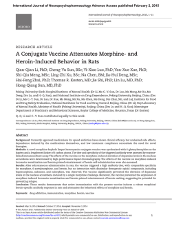
![Download [ PDF ] - journal of evolution of medical and dental sciences](http://s2.esdocs.com/store/data/000475167_1-3fd66abd823a299e32f0bb1f6c6f6a60-250x500.png)
© Copyright 2026