
Recurrent Ischemic Strokes and Headaches Originating from
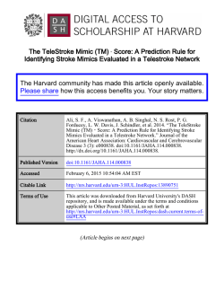
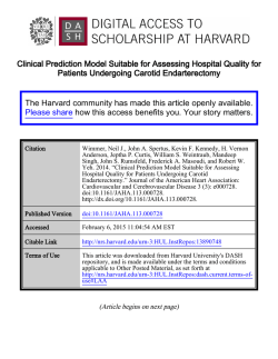
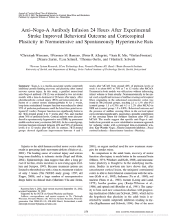
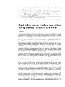
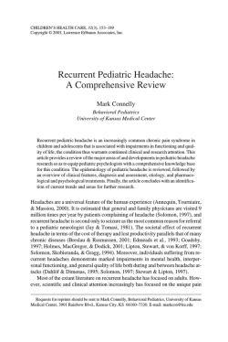
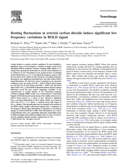
International Journal of
Dumitrascu and Tsimerinov. Int J Neurol Neurother 2015, 2:1
Neurology and Neurotherapy
Case-Report: Open Access
Recurrent Ischemic Strokes and Headaches Originating from Lambl’s
Excrescences: A Case-Report
Oana Dumitrascu* and Evgeny Tsimerinov
Department of Neurology, Cedars-Sinai Medical Center, Los Angeles, CA, USA
*Corresponding author: Oana Dumitrascu, Department of Neurology, Cedars-Sinai Medical Center, 127 S.
San Vicente Blvd, 6th floor, Los Angeles, CA 90048, USA, Tel: 310-423-6472, Fax: 310-423-0148, E-mail:
[email protected]
Abstract
Determining recurrent stroke etiology and subsequent therapeutic
approaches is an important, but not always straightforward task.
Lambl’s Excrescences (LE) are cardiac valve strands that can be
a source of recurrent cerebral ischemic events. We report the case
of a 60 year old female that was seen in neurologic consultation
for recurrent ischemic strokes. The patient developed migraine
headache with complex auras at the age of 59. She reported
three admissions for ischemic strokes in the past year. Stroke
work-ups were unrevealing. The patient remained on daily Aspirin.
Upon current admission, a brain MRI showed a combination of
sub acute and chronic infarcts in diverse brain areas bilaterally.
Further diagnostic evaluation revealed a relatively unremarkable
CT head and neck angiogram. Transcranial Doppler (TCD) showed
increased pulsatility index throughout the insonated vessels.
Transesophageal echocardiogram (TEE) showed a trileaflet aortic
valve with a 10 mm long thin fibrin strand prolapsing into the left
ventricle outflow tract in diastole, meeting echocardiographic
criteria for an LE. The patient refused anticoagulation and surgical
intervention. She opted instead for a combination of two antiplatelet
agents. Recurrent cerebral infarcts presenting concurrently with
late-age onset migraine with aura should raise a clinical suspicion
of cardiogenic valvular etiology. The use of TEE in evaluating
patients with unexplained recurrent stroke is imperative so that
LE is not missed. Anti-platelet agents, anti-coagulants, and
cardiothoracic surgical intervention are proposed in the literature
for the management of long-term complications associated with LE.
Asymptomatic patients should be monitored, counseled, and made
aware of LE complications, neurological symptoms, and alarm
signals. Migraine headaches associated with LE should raise a red
flag and prompt treatment with anti-platelet agents.
association with recurrent migraine headaches [2].Current data on
LE cerebral complications and their management is limited. The
treatment selection criteria and indications are variable and not
evidence-based [3].
Case Report
We report the case of a 60 year old right-handed female seen in
neurologic consultation for recurrent stroke management. The patient
presented with acute onset of binocular blurry vision, and an abnormal
left peri-ocular sensation that started during casual conversation with
a family member, and lasted about 2 hours. The event was associated
with slurred speech and a persistent frontal headache. Upon arrival
to our emergency department, visual and speech disturbances were
resolved but a moderate headache remained. An initial neurological
examination was unremarkable. A brain MRI reveals a subacute
infarct in the left parietal lobe with petechial hemorrhages, old infarcts
in the bilateral centrum semiovale, and chronic right occipital infarct
(Figure 1). The patient reported three prior admissions for acute stroke
Keywords
Ischemic stroke, Migraine headache with aura, Cardiac valvular
disease
Introduction
Recurrent ischemic stroke is still a challenge for the modern
neurologist. Rare cardiac culprits include Lambl’s Excrescences
(LE), which are native cardiac valve strand variants. LE is associated
with cerebral thromboembolism, recurrent cerebral infarcts, and
transitory ischemic attacks which are more common in younger
patients [1]. Microemboli coming from LE have been reported in
ClinMed
International Library
Figure 1: Brain MRI without contrast at presentation
Subacute right parietal infarct with petechial hemorrhage is illustrated on
DWI (A), SWI (B) and FLAIR (C) images. Chronic right occipital (D-FLAIR
sequence), old bilateral corona radiata (E- FLAIR sequence) and right frontal
lobe (F- T2W sequence) infarcts are identified.
Citation: Dumitrascu O, Tsimerinov E (2015) Recurrent Ischemic Strokes and Headaches
Originating from Lambl’s Excrescences: A Case-Report. Int J Neurol Neurother 2:019
Received: January 11, 2015: Accepted: January 29, 2015: Published: January 31,
2015
Copyright: © 2015 Dumitrascu O. This is an open-access article distributed under the terms
of the Creative Commons Attribution License, which permits unrestricted use, distribution,
and reproduction in any medium, provided the original author and source are credited.
outflow tract in diastole, strongly suggestive of Lambl`s excrescences,
based on echocardiographic criteria (Figure 2). No other valvular,
atrial or ventricular abnormalities were identified. There were no
thrombi and absent intracardiac shunts. A moderate (2-4mm)
atheroma was seen in the ascending aorta.
Figure 2: Transesophageal echocardiogram view of giant Lambl’s
excrescences deriving from the aortic valve (A- valve is closed; B- valve is
opened)
management. The first admission was for ataxia and visual changes,
five months prior to the current presentation. Two months later, the
patient was admitted for sudden-onset of non-fluent aphasia and
right upper extremity weakness. Finally, she was admitted for right
upper and lower extremity weakness, one month prior to the current
presentation. Stroke work-ups performed during all three admissions
did not reveal any specific causes for the stroke recurrences. She was
left with multiple residual neurological symptoms after each event,
including mild subjective gait abnormality which did not require
assistive devices, intermittent episodes of stuttering speech, transitory
visual symptoms (difficulties focusing on objects and reading because
of “jumping” letters, and intermittent left-sided hemianopsia), and
abnormal skin sensations around the left eye. In addition, the patient
reported recurrent unilateral throbbing headaches of a mild intensity
lasting hours, sometimes in association with visual and sensory
symptoms. Despite recurrent strokes, the patient was prescribed
Aspirin 162mg daily.
Upon arrival to our emergency department, a medical history
was remarkable for treated controlled hypertension, untreated
obstructive sleep apnea, dyslipidemia, left upper extremity DVT in
2005 of unknown etiology (was on warfarin for 2 years), and bilateral
knee osteoarthritis. Family history was positive for cardiac ischemic
disease and negative for migraine headaches. Besides Aspirin 162mg
daily, the patient was also taking Lipitor 10mg daily and two antihypertensive agents to maintain good blood pressure control.
Outside hospital medical records showed a normal
Transthoracic Echocardiogram (TTE) and negative work-up for
some hypercoagulable disorders. Although the majority of her comorbidities were taken into consideration as independent stroke
risk factors, we decided to conduct further investigation into her
recurrent and frequent cerebrovascular events. Serologic work-up
revealed normal cell blood count, normal complete metabolic profile,
normal TSH, HbA1c, and hypercoagulable profile (consisting of
unremarkable anti cardiolipin antibodies, prothrombin time, INR,
lupus anticoagulant, protein C and S activities, activated protein
C resistance, factor V Leiden, homocysteine, beta 2 glycoprotein,
fibrinogen), and an LDL of 112mg/dl.
A CT angiogram of the head and neck revealed minimal
atherosclerotic changes in the right greater than in the left internal
carotid artery at the high cervical level; a moderate irregularity in
the right A1 segment and mild to moderate narrowing bilaterally
in the M1-M2 junctions. No aneurysm or dissection was identified
in the anterior circulation. Evaluation of the posterior circulation
demonstrated a dominant right vertebral artery and a fetal origin
of the right posterior cerebral artery. No flow-limiting stenosis,
aneurysm, or dissection was identified in the posterior circulation.
A large mixed soft tissue and fat-containing mass was incidentally
noticed on the CT angiogram in the left neck. The mass biopsy
revealed a fibrolipoma.
As no clear causes for the recurrent strokes were currently
identified, and initial cardiac work-up was negative at the other
institutions, we elected to conduct a TEE. The TEE revealed a trileaflet
aortic valve with mildly calcified aortic cusps and a 10 mm long thin
fibrin strand on the aortic valve prolapsing into the left ventricle
Dumitrascu and Tsimerinov. Int J Neurol Neurother 2015, 2:1
A cardiothoracic team was consulted to address management
of the giant LE. The patient refused anticoagulation treatment and
surgical intervention. She opted instead for management with two
antiplatelet medications, Aspirin 81 mg and Clopidogre l75mg, daily.
The patient was discharged home in stable condition. A nine-month
follow-up confirmed that the patient remained free from headaches
and stroke recurrence.
Discussion
Recurrent cerebral infarct scan arise from endovascular plugs,
vascular stenosis and vascular malformations, coagulation path way
dysfunctions due to genetic defects, or autoimmune disorders. One of
the most common sources is cardiogenic, arising from hemodynamic
instability due to structural and/or diverse cardiac valve disorders
[4,5]. Valvular strands contribute to systemic thromboembolisms
and cerebrovascular pathology [2,3,5-9]. In daily clinical practice
the terms LE, cardiac papillary fibroelastoma, and valvular strands
are sometimes interchangeable based on echocardiographic
characterization alone; however, their management and prognosis
should be individualized [3].
LE are usually thin (1mm) and long (4-10mm), mobile, filiform
projections from native cardiac valves, more commonly seen on mitral
than on aortic valve leaflets. They consist of a collagenous structure
(elastic fibers surrounded by loose connective tissue), which gathers
subsequent fibrin deposition [1,8]. Little is known about the exact
prevalence and incidence of cerebral embolism from LE because of
their rare occurrence and possible under-recognition. Standardized
diagnostic criteria are not established due to paucity of retrospective
and prospective clinical trials. The mechanism of recurrent cerebral
infarcts in patients with LE, in absence of other risk factors, has been
explained by the formation of thrombi along the endothelial surface
[3]. The influence of LE size, morphology and location on these
thromboembolic events has not been elucidated [1].
While TTE can be used as a screening tool for cardiac sources
of cerebral embolism, TEE is necessary and remains the gold
standard in determining the etiology of multiple recurrent strokes.
Echocardiographic criteria of LE are described in the literature [3,5,9].
Multi-slide computed tomography can be used as an alternative
diagnostic tool if a TEE cannot be performed [10].
Current scientific literature does not provide strong
recommendations for stroke prevention in patients with LE [1],
emphasize that asymptomatic patients do not require treatment.
Other authors suggest that antiplatelet agents are indicated after the
first stroke, while anticoagulation or cardiothoracic intervention
should be reserved for recurrent events [3]. Controversially, Azis and
Baciewitz strongly support anticoagulation as an initial intervention,
followed by LE surgical excision if stroke prevention is not achieved
[6].
Migraine headaches are known to be associated with intracardiac
shunts and structural heart anomalies [11]. Migraine headaches with
aura and amaurosis fugax have been reported as a first presentation
of cerebral ischemia, preceding the stroke manifestations in patients
with LE [2]. No clinical or scientific studies are available to guide
headache management in these cases. Some clinicians support the
prevention of migraine headaches with antithrombotic medications
based on a presumed mechanism of micro-emboli originating from
LE [2].
Our case report suggests that the presentation of multiple
recurrent strokes in concurrence with late-age onset migraine with
aura should raise a clinical suspicion of cardiogenic valvular etiology.
Early TEE use is strongly indicated to diagnose cardiac valvular strands
such as LE in these patients. Even though our experience and data are
• Page 2 of 3 •
very limited, we feel safe managing our patient on the combination of
two antiplatelet agents for stroke and migraine prevention. Further
prospective clinical trials should be conducted to establish benefits of
different therapeutic options and better management guidance once
diagnosis of LE is made in patients with recurrent strokes.
References
1. Roberts JK, Omarali I, Di Tullio MR, Sciacca RR, Sacco RL, et al. (1997)
Valvular strands and cerebral ischemia. Effect of demographics and strand
characteristics. Stroke 28: 2185-2188.
2. Liu RZ, Yu SY, Li Y (2012) Migraine-like headache and ischemic strokes in
two patients with Lambl’s excrescences. Chin Med J (Engl) 125: 3346-3348.
3. Wolf RC, Spiess J, Vasic N, Huber R (2007) Valvular strands and ischemic
stroke. Eur Neurol 57: 227-231.
4. Wu TY, Gerber IL, Roxburgh RH (2013) Thrombo-embolic cerebral infarction
secondary to giant Lambl’s excrescence. J Clin Neurosci 20: 1632-1634.
Dumitrascu and Tsimerinov. Int J Neurol Neurother 2015, 2:1
5. Sun JP, Asher CR, Yang XS, Cheng GG, Scalia GM, et al. (2001) Clinical and
echocardiographic characteristics of papillary fibroelastomas: a retrospective
and prospective study in 162 patients. Circulation 103: 2687-2693.
6. Aziz F, Baciewicz FA Jr (2007) Lambl’s excrescences: review and
recommendations. Tex Heart Inst J 34: 366-368.
7. Kalavakunta JK, Peddi P, Bantu V, Tokala H, Kodenchery M (2010) Lambl’s
excrescences: a rare cause of stroke. J Heart Valve Dis 19: 669-670.
8. Nakahira J, Sawai T1, Minami T1 (2014) Pathologic examination of Lambl’s
excrescence. J Cardiothorac Vasc Anesth 28: e3-4.
9. Leitman M, Tyomkin V2, Peleg E2, Shmueli R2, Krakover R2, et al. (2014)
Clinical significance and prevalence of valvular strands during routine echo
examinations. Eur Heart J Cardiovasc Imaging 15: 1226-1230.
10.Rbaibi A, Bonnevie L, Guiraudet O, Godreuil C, Martin D, et al. (2002)
[Importance of transesophageal echocardiography and computed
tomography in the differential diagnosis of a case of papillary fibroelastoma
revealed by a neurologic accident]. Arch Mal Coeur Vaiss 95: 601-605.
11.Schwedt TJ (2009) The migraine association with cardiac anomalies,
cardiovascular disease, and stroke. Neurol Clin 27: 513-523.
• Page 3 of 3 •

© Copyright 2026