
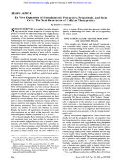
This is the author-manuscript version of this work
This is the author-manuscript version of this work - accessed from
http://eprints.qut.edu.au
Bartold, P. Mark and Xiao, Yin and Lyngstaadas, S. Petter and Paine, Michael L. and Snead,
Malcolm L. (2006) Principles and applications of cell delivery systems for periodontal regeneration.
Periodontol 2000 41(1):pp. 123-135.
Copyright 2006 Blackwell Publishing
Principles and Applications of Cell Delivery Systems for Periodontal Regeneration
PM Bartold1, Y Xiao2, SP Lyngstaadas3, ML Paine4, ML Snead4
1
Colgate Australian Clinical Dental Research Centre, University of Adelaide, Adelaide, Australia,
2
Tissue BioRegeneration and Integration, Science Research Centre, Queensland University of
Technology, Brisbane, Australia,
3
Faculty of Dentistry, University of Oslo, Oslo, Norway,
4
School of Dentistry, University of Southern California, Los Angeles, California
Short title: Cell Delivery for Regeneration
Address for Correspondence
PM Bartold
University of Adelaide
Colgate Australian Clinical Dental Research Centre,
Dental School
Frome Road
Adelaide, South Australia 5005
AUSTRALIA
1
2
Introduction
The management of periodontal defects has been an ongoing challenge in clinical
periodontics. This is mainly due to the fact that the tissues which comprise the periodontium,
the periodontal ligament, cementum and alveolar bone, represent three unique tissues in their
own right. Thus, reconstruction of the periodontium is not just a simple matter of
regenerating just one tissue but involves at least three quite diverse and unique tissues.
Resective surgical therapy with or without osseous recontouring was considered the
norm during the 1950’s and into the 1960’s in the belief that attainment of shallow pocket
depths was a worthwhile goal. More recently attention has been focussed more on
regenerative and reconstructive therapies rather than resective therapies. Currently clinical
and scientific research is focussing on a number of approaches for periodontal regeneration.
One approach requires the introduction of a “filler” material into a periodontal defect
in the hope of inducing bone regeneration. Various types of bone grafts have been
investigated to determine their ability to stimulate new bone formation. Of these, the
following have been studied in detail: (1) Alloplastic materials which are generally synthetic
filler materials; (2) Autografts which are grafted tissue from one site to another in the same
individual; (3) Allografts of tissue between individuals of the same species but with different
genetic composition; (4) Xenografts which consist of grafted materials between different
species. Although utilization of such grafting materials for periodontal defects may result in
some gain in clinical attachment levels and radiographic evidence of bone fill, careful
histologic assessment usually reveals that these materials have little osteoinductive capacity
and generally become encased in a dense fibrous connective tissue (19).
In another approach to induce periodontal regeneration, polypeptide growth factors
3
have been locally applied to the root surface in order to facilitate the cascade of wound
healing events that lead to new cementum and connective tissue formation. Among the
myriad growth factors currently characterized and available, platelet-derived growth factor
(PDGF) and insulin-like growth factor-1 (IGF-I), have been noted to enhance regeneration of
periodontal defects in beagle dogs and monkeys (42, 61). Another promising group of
polypeptide growth factors is the bone morphogenetic proteins (BMP), which offer good
potential for stimulating bone and cementum regeneration (59). An extension of growth
factor application to root surfaces is the application of cell free matrix constructs to the root
surface to aid cell repopulation and enhance regeneration. Enamel matrix proteins, are such
an example and there is some evidence that these proteins can assist in the regeneration of
periodontal tissues (24, 80). It is postulated that the enamel matrix derivative acts as a matrix
enhancement factor, creating a positive environment for cell proliferation, differentiation and
matrix synthesis (43, 23).
Yet another approach, known as guided tissue regeneration (GTR), has been
developed to achieve periodontal regeneration. This utilizes barrier membranes to guide and
instruct the specialized cellular components of the periodontium to participate in the
regenerative process. The GTR concept was founded on sound scientific research, and based
on the premise that the periodontal ligament contained all of the progenitor cells required for
the formation of bone, cementum and periodontal ligament (21, 35, 52). Through
repopulation of the wound site by the progenitor cells periodontal regeneration could be
induced. Although this procedure became widely accepted as a clinical procedure (35, 48),
recent clinical evaluation has indicated that the clinical improvements obtained by this
procedure are small and highly variable (10, 56, 78).
4
It now seems likely that a combination of several techniques may offer the most likely
chance of a beneficial outcome. Through a combination of transplanted biomaterials
containing appropriately selected and primed cells, together with an appropriate mix of
regulatory factors and extracellular matrix components to allow growth and specialization of
the cells, new therapies are emerging of significant clinical potential (4).
Tissue engineering is defined as the reconstruction of living tissues to be used for the
replacement of damaged or lost tissue / organs of living organisms and is founded on
principles of cell biology, developmental biology and biomaterials science (49, 71, 60). This
developing area of applied biomedical research is attracting considerable attention from both
the private and government sectors because of its considerable economic and therapeutic
potential (44, 54). A clear distinction should be made between tissue engineering, which is
the implantation of in vitro seeded cells and matrices, versus guided tissue regeneration which
involves the approach of using acellular matrices that are repopulated by the host after
implantation.
Successful tissue engineering requires an interplay between three components
(Figure 1) (i) the implanted and cultured cells that will create the new tissue; (ii) a biomaterial
to act as a scaffold or matrix to hold the cells and, (iii) biological signalling molecules that
instruct the cells to form the desired tissue type. This review will focus mainly on the use of
scaffold materials used to transplant cells as a means of either delivering cells or proteins to a
defect site.
Periodontal Tissue Engineering
5
The principal requirements for tissue engineering are the incorporation of appropriate
numbers of responsive progenitor cells and the presence of bioactive levels of regulatory
signals within an appropriate extracellular matrix or carrier construct. Recent advances in the
isolation of mesenchymal stem cells, growth factor biology, and biodegradable polymer
constructs have set the stage for successful tissue engineering of many tissues of which the
periodontium could be considered a prime candidate for such procedures. Preliminary studies
have indicated that periodontal ligament and bone cells can be transplanted into periodontal
sites with no adverse immunologic or inflammatory consequences (38, 46, 75, 82).
In order for successful periodontal regeneration to occur, it will be necessary to
utilize and recruit progenitor cells which can differentiate into specialized cells with a
regenerative capacity followed by proliferation of these cells, and synthesis of the specialized
connective tissues which they are attempting to repair. Clearly, a tissue engineering approach
for periodontal regeneration will need to utilize the regenerative capacity of cells residing
within the periodontium and would involve the isolation of such cells and their subsequent
within a three-dimensional framework with implantation into the defect. The use of a
prefabricated three dimensional scaffold with the appropriate cells or instructive messages
(eg, growth factors and matrix attachment factors) incorporated into it may overcome many of
the limitations associated with current regenerative technologies. With the current success
reported for other systems, a tissue engineering approach to regenerate periodontal defects
seems reasonable (4).
Despite the above positive outlook, there are still many issues that need to be dealt
with before periodontal tissue engineering becomes commonplace. There are two main
criteria for successful tissue engineering (11, 64, 5). Firstly, there are the engineering
6
principals which relate to biomechanical properties of the scaffold, architectural geometry and
space maintaining properties. The second criterion relates to the biological functions of the
engineered construct including cell recruitment, cell proliferation, cell survival in culture and
at the site of implantation, neovascularization and delivery of morphogenetic-, regulatory- and
growth-factors necessary for successful tissue regeneration. In this review we will
specifically focus on design requirements of scaffolds for cell delivery and then discuss some
of the materials and methods which have been used in recent years.
Design Requirements for Cell Seeding Scaffolds
Space maintenance within the defect site and Barrier or exclusionary functions.
The important understanding that bone will grow into an adjacent tissue space
providing that space can be maintained and soft tissue ingrowth prevented is not new (7, 30).
These early observations, which led to the principles of guided tissue regeneration, provide a
fundamental concept when considering tissue engineering and placement of bioengineered
matrices for regeneration. Thus, any engineered material should be of sufficient form and
strength to allow placement into a defect that prevents subsequent collapse of the overlying
tissues into the defect site. Indeed, the material should act in a manner consistent with the
established principles of guided tissue regeneration (62). These principles dictate that
sufficient wound space and a suitable environment for regeneration will act synergistically to
permit the uninhibited cascade of molecular and cellular events required for the regenerative
process.
The necessary design features needed to obtain adequate space maintenance will
include ease of handling and shaping, sufficient rigidity to withstand soft tissue collapse into
7
the defect and an internal structure compatible with cell attachment and colonization, as well
as permitting the in growth of tissues compatible with those to be regenerated (11, 62, 79).
Biocompatibility and design features.
An ideal material for tissue engineering scaffolds will require it to be either
biocompatible with the tissues to be regenerated or biodegradable allowing for gradual
replacement by regenerated tissue (36). Since both cell attachment and incorporation in vitro
as well as subsequent tissue maturation during in situ regeneration are crucial features of
tissue engineering, the amount of porosity and the pore size of the supporting three
dimensional structure are also important features which need to be taken into consideration
when designing tissue engineering scaffolds (79, 9). Finally, biosafety of the tissue
engineered constructs needs to be taken into consideration. Although no guidelines have yet
been established for assessment of the safety and efficacy of cell-based and tissue-based
tissue-engineered products, clearly these materials should be free from transmittable disease
and be immunologically inert while not inducing an overexuberant inflammatory response
(53). Indeed, the ability of the host to accept the implanted materials depends not only on the
material used but also the host reaction and the systemic health of the recipient (58).
Incorporation of cells with appropriate phenotype for ongoing periodontal regeneration.
Bioengineered skin substitutes with incorporated cells and extracellular matrix have
been available for some time (55). These artificial constructs can provide almost unlimited
quantities of tissue for wound management and illustrate the potential of such an approach.
With increasing knowledge of what constitutes cells with a “periodontal regenerative
8
phenotype” (32) together with the identification of adult mesenchymal stem cells within the
periodontal ligament (63) it should be possible to culture and subsequently incorporate these
cells into a suitable biodegradable scaffold for immediate introduction into a periodontal
defect.
More recently, viral vectors transformed into mesenchymal cells have been used as a
novel means of introducing specific molecules to wound sites with the intention of
stimulating tissue regeneration (51). Interestingly this technology has already been
translocated into periodontal regeneration. Using a viral vector delivery system, genetically
altered cells which express certain growth factors necessary for periodontal regeneration
(specifically PDGF) have been introduced into periodontal defects and appear to be able to
significantly enhance the regenerative response in experimental animal models (34, 1). Such
procedures introduce the problem of biosafety with regards to genetic manipulation and
control of the process, which will have to be dealt with prior to clinical acceptance.
For periodontal tissue engineering, potential sources of cells are from cementum (85),
periodontal ligament (63) and bone (81, 82). Whether the so-called progenitor cells which
reside in these tissues can be isolated and propagated in culture for future seeding remains to
be established (86).
Incorporation and bioavailability of instructive messages.
Growth and differentiation factors are essential ingredients for tissue regeneration.
Hence the synthetic scaffold used for tissue engineering should not only be bioresorbable but
also constructed from a material with a suitable affinity for the adsorption of appropriate
growth/differentiation factors as well as integrins, cell receptors and other instructive
9
molecules normally found in regenerating tissues (65, 8, 28). Notwithstanding this important
requisite, choosing the “correct” agent or agents is a formidable task. The plethora of
bioactive molecules involved in tissue regeneration will make rational selection of specific
agents very difficult. However, as our understanding of the precise signalling molecules
required for optimal growth, differentiation and gene expression become available, it is
anticipated that these agents may be incorporated into engineered matrices for regenerative
purposes based on sound biologic principles.
Although biological molecules can relatively easily be conjugated to an artificial
tissue engineered scaffold, issues relating to suitable release and delivery kinetics will become
the major focus of interest. Obstacles yet to be overcome in this regard will be controlling the
concentration, local duration and spatial distribution of these bound factors. Indeed the
control and containment of the agent is paramount for its effectiveness and safety (8).
Recently, bioengineers have devised novel methods to create a self-assembled
molecular structure that responds to ultrasonic energy by releasing a burst of entrapped drug
(37). Moreover, the self-assembling structure is a barrier to drug release in the absence of
ultrasonic energy, reducing the problem of overt leakage from an indwelling device. The
prototype for this has shown favourable release rates for insulin, as well as for an antibiotic
compound, ciprofloxacin, when triggered by ultrasonic energy. In these tests, essentially no
drug leakage occurred in the absence of an appropriate energy signature. This work also
suggests that self-assembling structures could be devised to serve as a barrier while
simultaneously serving as an ultrasonic responsive drug release device to promote tissue
regeneration strategies. Such molecularly triggered devices might also permit the placement
10
of appropriate therapeutic regimen that would be released only by the practitioner when
required to match a clinical scenario.
Regulating Cell Activity through Scaffold Design
It has been recognized for many years that the microenvironment in which a cell
resides dictates many functions and phenotypes (27). Thus it seems logical that the
construction and design of a cell seeding scaffold must take into account microenvironment
design features to induce the appropriate gene expression in cells forming new tissues. The
control of gene expression by cells within a scaffold can be regulated via interactions with the
adhesion surface, with other cells in the vicinity or, as described above, incorporated growth
and differentiation factors in the scaffold. Accordingly cell seeding scaffolds must provide
the correct combination of these factors according to the tissues to be regenerated if one is to
achieve successful gene expression and tissue regeneration. To date little work has been done
in this complex area although early studies have begun to utilize specific cell attachment
peptide sequences (“RGD” sequence for integrins), pore size, and surface texture in attempts
to improve tissue integration and regeneration.
When considering scaffold design many tissues depend upon mechanical stimuli in
order regulate gene expression and thus tissue composition. The most obvious example of
this is bone and tendon although it is likely that the periodontal ligament should also be
considered in this context. In order to engineer such functional tissues the correct mechanical
stimuli will need to be conveyed to the developing tissues within the cell/scaffold construct.
To date, because of the complexities of such systems very few studies have addressed these
issues (41).
11
Types of Cell Delivery Devices and Scaffolds
A common approach to tissue engineering is to use an exogenous three-dimensional
extracellular matrix to engineer new tissues using isolated cells. The exogenous matrix
constructs are designed to encourage cells to come into contact with it in a suitable three
dimensional environment and provide structural support for the newly forming tissues. More
recently a variant of this approach has been to isolate cells from biopsy specimens and expand
them in vitro prior to seeding onto a suitable three dimensional matrix. In doing so the cells
are allowed to either develop into a new tissue in vitro or immediately transplanted to a
particular site to create new functional tissue which is integrated within the recipient site.
Most cell seeding scaffolds are fabricated from two classes of biomaterials derived
from either synthetic or natural products. In addition they may be constructed from either a
resorbable or nonresorbable materials (Table 1). Natural products such as collagen are known
to have specific desirable biologic properties such as permitting cell interactions but have the
disadvantages of being derived from animal or human tissue leading to questions over
availability, safety and batch-to-batch variations. In contrast, synthetic materials can be
produced on a large scale to specific design criteria from generally inert, biocompatible and
biodegradable materials. Not surprisingly, there has been a plethora of materials developed
and studied over the years, each claiming specific and unique advantages over “competitor”
products. Therefore the following discussion is restricted to examples of cell carrier and
delivery devices currently under investigation and of relevance to periodontal regeneration.
Non-Resorbable Materials
12
Expanded poly tetrafluoroethylene (ePTFE, Goretex™)
Membranes made from ePTFE have traditionally been used as guided tissue barrier
membranes. However, it is possible that these membranes could also be used to nurture
specific cells that are expanded ex vivo and then delivered to a defect site. In the same context
almost any GTR membrane could be utilized in such a manner utilizing either non-resorbable
or resorbable materials (Figure 2).
Porous Ceramic Scaffolds
Several porous ceramic scaffolds have been examined for their utilization as cell
delivery materials. In general, many of these materials have been developed and investigated
with regard to bone tissue engineering (69). For these purposes the ideal scaffold should be a
porous material with good biocompatibility and possess osseointegrative capabilities, high
mechanical strength and biodegradability. Some ceramic materials have the former two
properties but to date no porous scaffolds satisfy both of the latter two properties.
Hydroxyapatite is an example of a material with good mechanical properties but due
to its porosity this materials has poor strength. Another problem with porous hydroxyapatite
is the lack of interconnectivity of the pores making neovascularization of any implant almost
impossible. Many studies have shown that hydroxyapatite scaffolds cultured with bone cells
have good osteogenic potential (16).
Biodegradable porous ceramic materials have also been developed and investigated.
Of these, the most popular material possessing high biocompatibility and biodegradability is
beta-tricalcium phosphate (TCP). When implanted alone at extraskeletal sites, TCP undergoes
rapid degradation with little bone formation. Due to this rapid degradation of TCP and its
13
associated poor mechanical properties, research has focussed on mixed calcium phosphates,
such as mixtures of beta-TCP and hydroxyapatite or beta-TCP and polymers. These hybrid
materials appear to be good vehicles for cell delivery with studies showing good tissue
formation associated with the implanted cells (22, 16).
Titanium Mesh
Another nonresorbable scaffold that has received considerable attention in recent years
is titanium mesh (33). This material has good mechanical properties with regards to stiffness
and elasticity and is relatively easy to handle during surgical placement. The lack of
bioresorbabilty of this material can be advantageous for the management of large osseous
defects whereby the mesh retains sufficient rigidity to avoid collapse which would be
expected of teflon membranes or biodegradable scaffolds. Various studies have indicated that
this material is suitable to support the growth and osteogenic expression of bone marrow cells
(74, 72, 77). Through various surface treatments, including addition of fibronectin, collagen
or calcium phosphate, the rate and amount of bone formation by implanted cells into titanium
mesh scaffolds can be regulated (76, 73).
Resorbable materials
Resorbable materials offer the significant advantage they do not need to be retrieved at
a later date from the site of implantation. These materials include materials such as polyesters
of naturally occurring alpha-hydroxy acids, amino-acid based polymers, alginate and natural
materials such as collagen and reconstituted extracellular matrix proteins.
14
Alpha Hydroxy Acids
The alpha-hydroxy acid polymers include polyglycolic acid (PGA), poly (L-lactic
acid) PLLA and copolymers of poly (lactic-co-glycolic acid) PLGA). These materials have
been used extensively for cell seeding in tissue engineering (66, 83). Their ester bonds are
quite susceptible to hydrolysis and thus degrade by nonenzymatic means. Accordingly these
natural breakdown products are removed from the site of implantation by normal tissue
respiratory routes and do not generally elicit a foreign body response resulting in massive
macrophage infiltration and chronic inflammation. Through specific chemical manipulation
these materials can be fabricated to degrade over long or short periods of time depending on
the need. These materials can also be easily manufactured into preformed sizes and shapes as
dictated by the site of the defect and its anatomy.
However, these materials are hydrophobic and are processed under quite stringent
(biologically adverse) conditions which usually makes factor incorporation and attachment or
entrapment of cells difficult. Recently a biodegradable copolymer of L-lactic acid, D-lactic
acid, glycolic acid and trimethylene carbonate have been developed (Inion, Ltd, Finland).
Although originally developed for use in dentistry as exclusionary barrier membranes, these
biodegradable membranes offer good potential as cell delivery devices with the advantage
that they seem to allow cell attachment more readily than other inert materials such as ePTFE
(Figure 2).
Alginate
An alternative to alginate gels as a carrier of cells is the incorporation of cells into
beads of alginate (Figure 3) (70). The technique is based on entrapment of individual cells
15
and tissues into an alginate droplet that is transformed into a rigid bead by gelation in a
divalent cation rich solution. The cells are surrounded by a nondegradable, selectively
permeable barrier which isolates the transplanted cells from host tissue and larger molecular
weight solutes. Such implants are considered immunoprotective as they prevent immune cells
and soluble complexes from killing the transplanted cells and this property negates the need
for use of immunosuppressants (68). While these systems may be used to deliver cells to a
specific site, because of the entrapment of the cells within an encapsulated environment, there
is little opportunity for direct and immediate cell/matrix interaction at the site of implantation.
Moreover, as a result of the semipermeable nature of the beads, the soluble factors made by
the entrapped cells can be released at the implantation site to guide regenerated tissues. In
recent years, these devices have been more appropriately developed as drug delivery devices
than cell delivery devices for tissue engineering (68).
Amino Acid Polymers
Amino acid based polymers have also been used as scaffolds for cell seeding. These
scaffolds can be synthesized using fermentation and gene transfer technology to produce
molecules which resemble natural amino acid containing matrix molecules such as collagens,
and elastin (29). While these materials have the advantage of being able to interact well with
cells, issues of biosafety (immunogenicity), large scale production and purification from
unwanted contaminants remain a problem (36)
16
Scaffolds Derived from Natural Products
A variety of materials derived from natural products have been investigated as cell
seeding scaffold materials. Cross-links between polymer chains and various chemical bonds
are often used to confer structural integrity to these products. Such materials are produced
under relatively mild conditions, possess structural and mechanical properties reminiscent of
the extracellular matrix which can act as space fillers, bioactive molecule delivery devices or
cell scaffolds. Examples of such materials are given in Table 1 and include both synthetic and
naturally derived polymers. Of these the naturally derived polymers such as alginate,
collagen chitosan and hyaluronate have been extensively studied as cell delivery vehicles.
These materials provide an excellent means to transplant cells and form three-dimensional
cell-filled matrices. Nonetheless, such materials have several problems including variability
in composition, poor mechanical properties and degradation rates which are time-limited and
difficult to control.
Hyaluronate has considerable potential as an optimal biomaterial for tissue
engineering given the significant role it plays during organogenesis, cell migration and
development in general (67). Modifications to hyaluronan include esterification and crosslinking to provide some structure and rigidity to the gel for cell seeding purposes. These
biopolymers are immunologically inert and completely biodegradable (14, 6) and support the
growth of fibroblasts, chondrocytes and mesenchymal stem cells (57, 84, 12)
Chitosan, a biopolymer that is structurally very similar to naturally occurring
glycosaminoglycans and is biodegradable in mammals, has been used quite extensively as a
tissue engineering scaffold. While chitosan can support cell attachment for cell delivery
purposes (18, 3), it is not strongly supportive of cell growth (50). Accordingly chitosan needs
17
to be either modified chemically or conjugated with other molecules or peptides to enhance its
biocompatibility for cell attachment (40, 45)
Collagen scaffolds have been investigated as a means of cell delivery device for many
years (39). Collagen is regarded as one of the most useful biomaterials due to its excellent
biocompatibility and safety due associated with its biological characteristics, such as
biodegradability and weak antigenicity (Figure 4). In this regard, collagen has been used for
tissue engineering including skin replacement, bone substitutes, artificial blood vessels and
valves. In the context of this review collagen sponges and membranes offer particular
features for cell integration and tissue engineering (Figure 5). Cells can readily be seeded into
collagen sponges or membranes, cultured and then introduced into a tissue defect site where
they can effect tissue repair and regeneration (81).
Synthetic Hydrogels
Synthetic hydrogels such as poly(ethylene glycol) (PEG) and poly(ethylene oxide) (PEO) are
also showing considerable promise for use as a three-dimensional scaffold for cell delivery.
By varying the initial cross linking density the degradation profiles of the gel can be
controlled (13). In addition it is possible to construct thermally reversible hydrogels as well
as gels which can be degraded by either hydrolytic or enzymatic means (2, 47). PEO is
currently FDA approved for several applications in medicine and together with PGA is one of
the most commonly utilized synthetic materials used for tissue engineering (17).
18
Extracellular Matrix Scaffolds
Extracellular matrix extracts or derivatives have been developed as commercial
products for cell delivery. In particular many skin and extracellular matrix substitutes such as
Matrigel™ (BD Biosciences, USA) Dermagraft™ (Advanced Tissue Sciences Inc, La Jolla,
CA, USA), Apligraf™ (Organogenesis Inc, Canton, Massachusetts) and Epidex™ (Modex
Therapeutiques SA, Lausanne, Switzerland) have been developed to allow the incorporation
of ex vivo expanded cells. Nonetheless, these products, as well as other acellular therapies
such as PV702 (GroPep Pty, Ltd, Adelaide, South Australia) and Allograft™ (Life Cell Corp,
Branchburg, New Jersey, USA), incorporate animal derived products and/or allogenic tissues
and thus constitute a potential source of pathogens; consequently they are unlikely to be
routinely used a cell delivery devices in the longer term.
In vitro produced extracellular matrix also offers potential as a biodegradable scaffold
for cell delivery. The use of extracellular matrix materials as scaffolds for the repair and
regeneration of tissues is receiving increased attention. In a recent study we have shown that
extracellular matrix formed by osteoblasts in vitro can be used as a scaffold for osteoblast
transplantation and induce new bone formation in a critical size osseous defects in vivo (82).
Human osteoblasts were cultured for 3 weeks to produce their own structured extracellular
matrix (Figure 6). The cells and self-produced matrix was then implanted into critical size
osseous defects. The cells inside the matrix could survive and proliferate at the recipient sites.
It was found that bone-forming cells differentiated from both transplanted human osteoblasts
and activated endogenous mesenchymal cells.
New Directions
19
The field of tissue engineering constructs and scaffolds is expanding at a very rapid
rate. It would, within the confines of this review, be impossible to detail all of the most recent
developments. However, there are two particular new directions that the authors are
specifically interested in which involve the co-culture of cells and nanotechnology.
In attempts to deliver cells to a complex environment such as the periodontium it is
possible that delivery of cells of multiple phenotypes may be required. For example, if one
were to want to regenerate both periodontal ligament and alveolar bone, the possibility exists
to bilaterally seed PDL cells on one side of a bioscaffold and osteoblasts on the opposite side.
While preliminary studies have begun to address such an approach little definitive data are yet
available. In a similar vein, it is not that difficult to envisage the engineering of a PDL-like
matrix from PDL fibroblasts to which one side would then be seeded with cementoblasts and
the opposite side seeded with osteoblasts. Using such an approach it may be possible to fully
reconstitute various compartments of the periodontium in vitro and then implant such
constructs into periodontal defects.
Advances in nanotechnology will also undoubtedly allow the synthesis of materials
with desirable nanoscale structures. Nanotechnology is the science of engineering at the
individual molecular level to produce materials of hitherto unthought of properties. Already,
self -assembly systems have been described and fabricated which mimic many features of the
extracellular matrix. For example, nanostructured fibrous scaffold reminiscent of extracellular
matrix can be constructed using the pH-induced self-assembly of a peptide-amphiphile. After
cross-linking, the fibres are able to direct mineralization of hydroxyapatite to form a
composite material in which the crystallographic c-axes of hydroxyapatite crystals are aligned
with the long axes of the collagen fibrils. This alignment is the same as that observed between
20
collagen fibrils and hydroxyapatite crystals in bone (25). Similarly, self-assembling
biomaterials with molecular features designed to interact with cells and scaffolds for tissue
regeneration have been reported (31). These nanofibres display attachment domains in the
form of RGD motifs that are incorporated into the amphiphiles that self-assemble into
nanofibres. The density of the RGD motif, and perhaps soon, alternative cell signalling
motifs, can be incorporated into the amphiphile for creation of a nanofibre with unique
surface properties. Cells can be embedded in the nanofibre to resemble a “native”
extracellular matrix (26). Since these materials are chemically synthesized, they present no
risk from viral contaminants as might occur for natural compounds recovered from biological
sources, such as pigs or human.
Concluding Comments
The study of scaffold materials for use in tissue engineering should lead to improved
predictability of this new technology based on cell and molecular biology. In the future it will
become increasingly important to consider the concepts of scaffolds which are not only space
making and exclusionary, but also biocompatible and able to elicit appropriate gene
expression by the cells for which it is providing the carrier capacity. Understanding the
complex design features necessary for successful tissue engineering, will help this technique
to become an accepted biomedical procedure.
21
References
1. Anusaksathien O, Jin Q, Zhao M, Somerman MJ, Giannobile WV. Effect of sustained
gene delivery of platelet-derived growth factor or its antagonist (PDGF-1308) on tissueengineered cementum. J Periodontol 2004: 75: 429-440.
2. Bae YH, Huh KM, Kim Y, Park K. Biodegradable amphiphilic multiblock copolymers
and their implications for biomedical applications. J Control Release 2000: 64: 3-13.
3. Bagnaninchi PO, Dikeako M, Veras T, Tabrizian M. Towards on-line monitoring of cell
growth in microporous scaffolds: Utilization and interpretation of complex permittivity
measurements. Biotechnol Bioeng 2003: 84: 343-350.
4. Bartold PM, McCulloch CAG, Narayanan AS, Pitaru S. Tissue engineering: A new
paradigm for periodontal regeneration based on molecular and cell biology. Periodontol
2000 2000: 24: 253-269.
5. Baum B, Mooney DJ. The impact of tissue engineering on dentistry. JADA 2000: 131:
309-318.
6. Benedetti L, Cortivo R, Berti T, Pea F, Mazzo M, Moras M, Abatangelo G.
Biocompatbility and biodegradation of different hyaluronan derivatives (HYAFF)
implanted in rats. Biomaterials 1993: 14: 1154-1160.
7. Berg A. Contributions to the technique of fusion operation on the spine. Acta Orthop
Scand 1947: 18: 1-30.
8. Boontheekul T, Mooney DJ. Protein based signalling systems in tissue engineering. Curr
Op Biotechnol 2003: 14: 559-565.
9. Boyan BD, Hummert TW, Dean DD, Schwartz Z. The role of material surfaces in
regulating bone and cartilage cell response. Biomaterials. 1996: 17: 137-146.
10. Bratthall G, Soderholm G, Neiderud AM, Kullendorff B, Edwardsson S, Attstrom R.
Guided tissue regeneration in the treatment of human intrabony defects, clinical,
radiographical and microbiological results, a pilot study. J Clin Periodontol 1998: 25:
908-914.
11. Brekke JH, Toth JM. Principles of tissue engineering applied to programmable
osteogenesis. J Biomed Mater Res 1998: 43: 380-398.
12. Brun P, Abatangelo G, Radice M, Zacchi V, Guidolin D, Daga Gordini D, Cortivo R.
Chondrocyte aggregation and reorganization into three-dimensional scaffolds. J Biomed
Mater Res 1999: 46: 337-346.
22
13. Bryant SJ, Bender RJ, Durand KL, Anseth KS. Encapsulating chondrocytes in degrading
PEG hydrogels with high modulus: Engineering gel structural changes to facilitate
cartilaginous tissue production. Biotechnol Bioeng 2004: 86: 747-755.
14. Cortivo R, Brun P, Rastrelli A, Abatangelo G. In vitro studies on biocompatability of
hyaluronic acid esters. Biomaterials 1991: 12: 727-730.
15. Dong J, Kojima H, Uemura T, Kikuchi M, Tateishi T, Taneka J. In vivo evaluation of a
novel porous hydroxyapatite to sustain osteogenesis of transplanted bone marrow derived
osteoblastic cells. J Biomed Mater Res 2001: 57: 208-216.
16. Dong J, Uemura T, Shiraski Y Tateishi T. Promotion of bone formation using highly pure
porous beta-TCP combined with mesenchymal stem cells. Biomaterials 2002: 23: 44934502.
17. Drury JL, Mooney DJ. Hydrogels for tissue engineering: scaffold design variables and
applications. Biomaterials 2003: 24: 4337-4351.
18. Fakhry A., Schneider GB, Zaharias R, Senel S. Chitosan supports the initial attachment
and spreading of osteoblasts preferentially over fibroblasts. Biomaterials 2004: 25:
2075-2079.
19. Garraway R, Young WG, Daley T, Harbrow D, Bartold PM. An assessment of the
osteoinductive potential of commercial demineralized freeze-dried bone in the murine
thigh muscle implantation model. J Periodontol 1998: 69: 1325-1336.
20. Gentile FT. Voluntary guidelines for the development of tissue-engineered products.
Tissue Eng 1998: 4: 239-266.
21. Gottlow J, Nyman S, Lindhe J, Karring T, Wennstrom J. New attachment formation in
human periodontium by guided tissue regeneration. J Clin Periodontol 1986: 13: 604616.
22. Gronthos S, Chen S, Wang CY, Robey PG, Shi S. Telomerase accelerates osteogenesis of
bone marrow stromal stem cells by upregulation of CBFA1, osterix, and osteocalcin. J
Bone Miner Res 2003: 18: 716-722.
23. Haase HR, Bartold PM. Enamel matrix derivative induces matrix synthesis by cultured
human periodontal fibroblast cells. J Periodontol 2001: 72: 341-348.
24. Hammarström L, Heijl L, Gestrelius S. Periodontal regeneration in a buccal dehiscence
model in monkeys after application of enamel matrix proteins. J Clin Periodontol 1997:
24: 669-677.
23
25. Hartgerink JD, Beniash E, Stupp SI. Peptide-amphiphile nanofibers: a versatile scaffold
for the preparation of self-assembling materials. Proc Natl Acad Sci U S A 2002: 99:
5133-5138.
26. Hartgerink JD, Beniash E, Stupp SI. Self-assembly and mineralization of peptideamphiphile nanofibers. Science 2001: 294: 1684-1688.
27. Hay ED. Role of cell-matrix contacts in cell migration and epithelialmesenchymaltransformation. Cell Differ Dev 1990: 32: 367-375.
28. Hersel U, Dahmen C, Kessler H. RGD modified polymers: biomaterials for stimulated
cell adhesion and beyond. Biomaterials 2003: 24: 4385-4415.
29. Hubbell JA. Biomaterials in tissue engineering. Biotechnology 1995: 13: 565-576.
30. Hurley LA, Stinchfield FE, Bassett AL, Lyon WH. The role of soft tissues in
osteogenesis. J Bone Joint Surg 1959: 41A: 1243-1254.
31. Hwang JJ, Iyer SN, Li LS, Claussen R, Harrington DA, Stupp SI. Self-assembling
biomaterials: liquid crystal phases of cholesteryl oligo(L-lactic acid) and their interactions
with cells. Proc Natl Acad Sci U S A 2002: 99: 9662-9667.
32. Ivanovski S, Haase HR, and Bartold PM. Characterization of regenerative phenotypes of
primary and cloned cultures of human periodontal fibroblasts. J Dent Res 2001: 80:
1665-1671.
33. Jansen JA, von Recum AF, van der Waerden JP, de Groot K. Soft tissue response to
different types of sintered metal fibre-web materials. Biomaterials 1992: 13: 959-968.
34. Jin Q, Anusaksathien O, Webb SA, Printz MA, Giannobile WV. Engineering of toothsupporting structures by delivery of PDGF gene therapyvectors. Mol Ther 2004: 9: 519526.
35. Karring T, Nyman S, Gottlow J, Laurell L. Development of the biological concept of
guided tissue regeneration-animal and human studies. Periodontol 2000 1993: 1: 26-35.
36. Kim B-S, Mooney DJ. Development of biocompatable synthetic extracellular matrices for
tissue engineering. Trends Biotechnol 1998: 16: 224-230.
37. Kwok CS, Mourad PD, Crum LA, Ratner BD. Self-assembled molecular structures as
ultrasonically-responsive barrier membranes for pulsatile drug delivery. J Biomed Mater
Res 2001: 57: 151-164.
38. Lang H, Schüler N, Nolden R. Attachment formation following replantation of cultured
cells into periodontal defects. J Dent Res 1998: 77: 393-405.
24
39. Lee CH, Singla A, Lee Y. Biomedical applications of collagen. Int J Pharm 2001: 221:
1-22.
40. Li J, Pan J, Zhang L, Yu Y. Culture of hepatocytes on fructose-modified chitosan
scaffolds. Biomaterials 2003: 24: 2317-2322.
41. Lin CY, Kikuchi N, Hollister SJ. A novel method for biomaterial scaffold internal
architecture design to match bone elastic properties with desired porosity. J Biomech
2004: 37: 623-636.
42. Lynch SE, de Castilla GR, Williams RC, Kiritsy CP, Howell TH, Reddy MS, Antoniades
HN.The effects of short-term application of a combination of platelet-derived and insulinlike growth factors on periodontal wound healing. J Periodontol 1991: 62: 458-467.
43. Lyngstadaas SP, Lundberg E, Ekdahl H, Andersson C, Gestrelius S. Autocrine growth
factors in human periodontal ligament cells cultured on enamel matrix derivative. J Clin
Periodontol. 2001: 28:181-188.
44. Lysaght MJ, Nguy NA, Sullivan K. An economic survey of the emerging tissue
engineering industry. Tissue Eng 1998: 4: 231-238.
45. Ma L, Gao C, Mao Z, Zhou J, Shen J, Hu X, Han C Collagen/chitosan porous scaffolds
with improved biostability for skin tissue engineering. Biomaterials 2003 24: 4833-4841.
46. Malekzeh R, Hollinger JO, Buck D, Adams DF, McAllister BS. Isolation of human
osteoblast-like cells and in vitro amplification for tissue engineering. J Periodontol 1998:
69: 1256-1262.
47. Metters AT, Anseth KS, Bowman CN. Fundamental studies of biodegradable hydrogels
as cartilage replacement materials. Biomed Sci Instrum 1999: 35: 33-38.
48. Nakae H, Narayanan AS, Raines EW, page RC. Isolation and characterization of
mitogenic factors from cementum. Biochemistry 1991: 30: 7047-7052.
49. Narem R, Sambanis A. Tissue engineering: From biology to biological structures. Tissue
Eng 1995: 1: 3-13.
50. Ng KW, Khor HL, Hutmacher DW In vitro characterization of natural and synthetic
dermal matrices cultured with human dermal fibroblasts. Biomaterials 2004: 25: 28072818.
51. Nussenbaum B, Rutherford RB, Teknos TN, Dornfeld KJ, Krebsbach PH. Ex vivo gene
therapy for skeletal regeneration in cranial defects compromised by postoperative
radiotherapy. Hum Gene Ther 2003: 14: 1107-1115.
25
52. Nyman S, Gottlow J, Karring T, Lindhe J. The regenerative potential of the periodontal
ligament. An experimental study in the monkey. J Clin Periodontol 1982: 9: 257-265.
53. Omstead DR, Baird LG, Christenson L, Du moulin G, Tubo R, Maxted DD, Davis J,
54. Persidis A. Tissue engineering. Nature Biotechnol 1999: 17: 508-509.
55. Pomahac B, Svensjö T, Yao F, Brown H, Eriksson E. Tissue engineering of skin. Crit
Rev Oral Biol Med 1998: 9: 333-344.
56. Pontoriero R, Lindhe J. Guided tissue regeneration in the treatment of degree III furcation
defects in maxillary molars. Clin Periodontol 1995: 22: 810-812.
57. Radice M, Brun P, Cortivo R, Scapinelli R, Battaliard C, Abatangelo G. Hyaluronanbased biopolymers as delivery vehicles for bone-marrow-derived mesenchymal
progenitors. J Biomed Mater Res 2000: 50: 101-109.
58. Rihova B. Immunocompatability and biocompatability of cell delivery systems. Adv
Drug Deliv Rev 2000: 42: 65-80.
59. Ripamonti U, Reddi AH. Periodontal regeneration: potential role of bone morphogenetic
proteins. J Periodont Res 1994: 29: 225-235.
60. Rosso F, Giordano A, Barbarisi M, Barbarisi A. From cell-ECM interactions to tissue
engineering. J Cell Physiol 2004: 199: 174-180.
61. Rutherford RB, Ryan ME, Kennedy JE, Tucker MM, Charette MF. Platelet-derived
growth factor and dexamethasone combined with a collagen matrix induce regeneration of
the periodontium in monkeys. J Clin Periodontol 1993: 20: 537-544.
62. Scantlebury TV. 1982-1992: a decade of technology development for guided tissue
regeneration. J Periodontol 1993: 64: 1129-1137.
63. Seo B-M, Miura M, Gronthos S, Bartold PM, Batouli S, Brahim J, Young M, Gehron
Robey P, Wang C-Y, Shi S. Multipotent stem cells from human periodontal ligament.
(Lancet 2004. – In Press).
64. Stock UA, Vascanti JP. Tissue engineering: Current state and prospects. Ann Rev Med
2001: 52: 443-451
65. Tabata Y. Tissue regeneration based on growth factor release. Tissue Eng 2003: 9(Supp
1): S1-S15.
66. Thomson RC, Wake MC, Yaszemski MJ, Mikos AG. Biodegradable polymer scaffolds to
regenerate organs. Adv Polym Sci 1995: 122: 245-274.
26
67. Toole BP. Hyaluronan in morphogenesis. J Intern Med 1997: 242: 35-40.
68. Tresco PA, Biran R, Noble MD. Cellular transplants as sources for therapeutic agents.
Adv Drug Deliv Rev 2000: 42: 3027.
69. Uemura T, Dong J, Wang Y, Kojima H, Saito T, Iejima D, Kikuchi M, Tanaka J, Tateishi
T. Transplantation of cultured bone cells using combinations of scaffolds and culture
techniques. Biomaterials 2003: 24: 2277-2286
70. Uludag H, DeVos P, Tresco PA. Technology of mammalian cell encapsulation. Adv
Drug Deliv Rev 2000: 42: 29-64.
71. Vacanti CA, Langer R, Schloo B, Vacanti JP. Synthetic polymers seeded with
chondrocytes provide a template for new cartilage formation. Plastic Reconstr Surg
1991: 88: 753-759.
72. van den Dolder J, Bancroft GN, Sikavitsas VI, Spauwen PH, Mikos AG, Jansen JA. Effect
of fibronectin- and collagen I-coated titanium fiber mesh on proliferation and
differentiation of osteogenic cells. Tissue Eng 2003b: 9: 505-515.
73. van den Dolder J, Spauwen PH, Jansen JA. Evaluation of various seeding techniques for
culturing osteogenic cells on titanium fiber mesh. Tissue Eng 2003a: 9: 315-325.
74. van den Dolder J, Vehof JW, Spauwen PH, Jansen JA. Bone formation by rat bone
marrow cells cultured on titanium fiber mesh: effect of in vitro culture time. J Biomed
Mater Res 2002: 62: 350-358.
75. van Dijk LJ, Schakenraad JM, van der Voort HM, Busscher HJ. Cell seeding of
periodontal ligament fibroblasts. A pilot study. J Clin Periodontol 1991: 18: 196-199.
76. Vehof JW, Spauwen PH, Jansen JA. Bone formation in calcium-phosphate-coated
titanium mesh. Biomaterials 2000: 21: 2003-2009.
77. Walboomers XF, Habraken WJ, Feddes B, Winter LC, Bumgardner JD, Jansen JA.
Stretch-mediated responses of osteoblast-like cells cultured on titanium-coated substrates
in vitro. J Biomed Mater Res 2004: 69A: 131-139.
78. Wallace SC, Gellin RG, Miller MC, Mishkin DJ. Guided tissue regeneration with and
without decalcified freeze-dried bone allografts for the regeneration of interproximal
intraosseous defects. J Periodontol 1994: 65: 244-254.
79. Whang K, Healy KE, Elenz DR, Nam EK, Tsai DC, Thomas CH, Nuber GW, Glorieux
FH, Travers R, Sprague SM. Engineering bone regeneration with bioresorbable scaffolds
with novel microarchitecture. Tissue Eng 1999: 5: 35-51.
27
80. Wilson TG. Periodontal regeneration enhanced. Clinical applications of enamel matrix
proteins. Quintessence Books, Chicago; 1999.
81. Xiao T, Qian H, Young WG, Bartold PM. Tissue engineering for bone regeneration using
differentiated alveolar bone cells in collagen scaffolds. Tissue Engineering 2003: 9:
1167-1177.
82. Xiao Y, Haase H, Young WG, Bartold PM. Development and transplantation of a
mineralized matrix formed by osteoblasts in vitro for bone regeneration. Cell
Transplantation 2004: 13: 15-25.
83. Yang S, Leong K-F, Du Z, Chua C-K. The design of scaffolds for use in tissue
engineering. Part 1. Traditional factors. Tissue Eng 2001: 7: 679-689.
84. Zacchi V, Soranzo C, Cortivo R, Radice M, Brun P, Abatangelo G. In vitro engineering
of human skin-like tissue. J Biomed Mater Res 1998: 40: 187-194.
85. Zhao, M, Jin Q, Berry JE, Nociti FH, Giannobile WV, Somerman MJ. Cementoblast
delivery for periodontal tissue engineering. J Periodontol 2004: 75: 154-161
86. Zohar R, Sodek J, McCulloch CAG. Characterization of stromal progenitor cells enriched
by flow cytometry. Blood 1997: 90: 3471-3481.
28
TABLE 1
Examples of Cell Delivery Devices and Scaffolds
Non Resorbable
EPTFE
Ceramic
Titanium mesh
Resorbable
alpha-hydroxyacids
polyglycolic acid (PGA),
poly (L-lactic acid) PLLA
copolymers of poly(lactic-co-glycolic acid) PLGA).
Amino Acid Based Polymers
Collagen-like proteins
Elastin-like proteins
Natural Products
Collagen
Hyaluronan
Chitosan
Gelatin
Fibrin
Alginate
Synthetic Hydrogels
Poly(ethylene glycol)
Poly(ethylene oxide)
Matrix Extracts
Matrigel
29
Figure 1 Tissue Engineering
Tissue Engineering
Scaffolds
(collagen, bone minerals, synthetics)
REGENERATION
Cells
(osteoblasts,
cementoblasts
Fibroblasts)
Signalling molecules
(growth factors,
differentiation factors,
adhesion molecules)
30
31
Figure 2. Cell attachment to GTR Membranes as cell delivery devices
Untreated GoreTex™ Membrane
“Treated” GoreTex™ Membrane
Untreated Inion™ Membrane
“Treated” Inion™ Membrane
31
32
A
B
C
D
Figure 3: Culture of human PDL cells in Alginate beads.
A.
B..
C.
D.
Cells cultured in Alginate beads for 2 days – 10X
Cells cultured in alginate beads for 2 days – 40X
Cells cultured in alginate beads for 8 days – 20X
Cells cultured in alginate beads for 8 days – 40X
32
33
Figure 4: Culture of human periodontal ligament fibroblasts and osteoblasts as single
or mixed co-cultures in a collagen sponge.
A.
B.
C.
D.
Control – no cells
PDL Fibroblasts
Osteoblasts
Co-culture of osteoblasts and PDL fibroblasts
33
34
Figure 5
Panel A:
Panel B:
Panel C:
Panel D:
A
va
B
C
D
Confocal microscopy of cell penetration into collagen scaffold
Cells on the surface of collagen sponges,
Cells in collagen sponges at 60 µm from surface;
Cells in collagen sponges at 20 µm from surface;
Cells in collagen sponges at 100 µm from surface.
34
35
A
B
Figure 6a: Long Term culture of human osteoblasts to produce in vitro cell / matrix
complex. A Low Power. B. High Power. Reproduced with permission from Xiao,
Y, Haase HR, Young WG & Bartold PM. Development and transplantation of a
mineralized matrix formed by osteoblasts in vitro for bone regeneration. Cell
Transplantation 2004; 13: 15-25.
35
36
A
B
Figure 6b: Type 1 collagen distribution in long-term in vitro produced cell/matrix . A
Low Power. B High Power. Reproduced with permission from Xiao, Y, Haase HR,
Young WG & Bartold PM. Development and transplantation of a mineralized matrix
formed by osteoblasts in vitro for bone regeneration. Cell Transplantation 2004; 13:
15-25.
…
36
37
A
B
Figure 6c: Von Kossa staining for calcium deposition in long-term in vitro produced
cell/matrix A Low Power. B High Power. Reproduced with permission from Xiao,
Y, Haase HR, Young WG & Bartold PM. Development and transplantation of a
mineralized matrix formed by osteoblasts in vitro for bone regeneration. Cell
Transplantation 2004; 13: 15-25.
37
© Copyright 2026