
Pentosan polysulfate sodium prevents kidney morphological
Included in ISI-WOK, MEDLINE, EMBASE, IME, IBECS, SCIELO
V o l u m e
3 0
-
N u m b e r
6
-
2 0 1 0
TREATMENT OF DIABETIC PATIENTS WITH PERITONEAL DIALISYS
A COMPETENCY-BASED MODEL FOR MEDICAL SPECIALISTS EDUCATION
BK POLYOMAVIRUS-ASSOCIATED NEPHROPATHY
RENAL IMMUNOEXPRESSION OF GHRELIN IN HUMAN PROLIFERATIVE
GLOMERULOPATHIES
URAEMIC ANOREXIA TREATMENT WITH MEGESTROL ACETATE
POST-TRANSPLANT LYMPHOPROLIFERATIVE DISORDERS IN RENAL
TRANSPLANTATION
Sociedad
Española de
Nefrología
Órgano Oficial de la Sociedad Española de Nefrología
Versión íntegra inglés y español en www.revistanefrologia.com
Nefrología Journal
Editor-in-Chief: Carlos Quereda Rodríguez-Navarro
Executive editor: Roberto Alcázar Arroyo
Deputy editors: Andrés Purroy Unanua, Ángel Luis Martín de Francisco, Fernando García López
Honorary editors: Luis Hernando Avendaño, David Kerr, Rafael Matesanz Acedos
SUBJECT EDITORS (editors of thematic areas)
Experimental Nephrology
A. Ortiz*
J. Egido de los Ríos
S. Lamas
J.M. López Novoa
D. Rodríguez Puyol
J.M. Cruzado
Clinical Nephrology
M. Praga*
J. Ara
J. Ballarín
G. Fernández Juárez
F. Rivera
A. Segarra
Diabetic Nephropathy
F. de Álvaro*
J.L. Górriz
A. Martínez Castelao
J.F. Navarro
J.A. Sánchez Tornero
R. Romero
Hereditary Nephropathies
R. Torra*
X. Lens
J.C. Rodríguez Pérez
M. Navarro
E. Coto
V. García Nieto
Chronic Kidney Disease
A.L. Martín de Francisco*
A. Otero
E. González Parra
I. Martínez
J. Portolés Pérez
CRF-Ca/P Metabolism
E. Fernández*
J. Cannata Andía
R. Pérez García
M. Rodríguez
J.V. Torregrosa
Arterial Hypertension
R. Marín*
J.M. Alcázar
L. Orte
R. Santamaría
A. Rodríguez Jornet
Nephropathy and
Cardiovascular Risk
J. Díez*
A. Cases
J. Luño
Quality in Nephrology
F. Álvarez-Ude*
M.D. Arenas
E. Parra Moncasi
P. Rebollo
F. Ortega
Acute Renal Failure
F. Liaño*
F.J. Gainza
J. Lavilla
E. Poch
Peritoneal Dialysis
R. Selgas*
M. Pérez Fontán
C. Remón
M.E. Rivera Gorrin
G. del Peso
Haemodialysis
A. Martín Malo*
P. Aljama
F. Maduell
J.A. Herrero
J.M. López Gómez
J.L. Teruel
Renal Transplantation
J. Pascual*
M. Arias
J.M. Campistol
J.M. Grinyó
M.A. Gentil
A. Torres
Paediatric Nephrology
I. Zamora*
N. Gallego
A.M. Sánchez Moreno
F. Vilalta
Nephropathology
J. Blanco*
I.M. García
E. Vázquez Martul
A. Barat Cascante
Evidence-Based Nephrology
Vicente Barrio* (Director of Supplements), Fernando García López (Methodology assessment), Editors: María Auxiliadora Bajo, José Conde, Joan M. Díaz, Mar Espino,
Domingo Hernández, Ana Fernández, Milagros Fernández, Fabián Ortiz, Ana Tato.
Continued Training (journal NefroPlus)
Andrés Purroy*, R. Marín, J.M. Tabernero, F. Rivera, A. Martín Malo.
* Coordinators of thematic area
EDITORIAL BOARD
A. Alonso
J. Arrieta
F.J. Borrego
D. del Castillo
P. Gallar
M.A. Frutos
D. Jarillo
V. Lorenzo
A. Mazuecos
A. Oliet
L. Pallardo
J.J. Plaza
D. Sánchez Guisande
J. Teixidó
J. Alsina
P. Barceló
J. Bustamente
A. Darnell
P. García Cosmes
M.T. González
L. Jiménez del Cerro
J. Lloveras
B. Miranda
J. Olivares
V. Pérez Bañasco
L. Revert
A. Serra
F.A. Valdés
J. Aranzábal
G. Barril
F. Caravaca
C. de Felipe
S. García de Vinuesa
A. Gonzalo
R. Lauzurica
J.F. Macías
E. Martín Escobar
J.M. Morales
R. Peces
J.M. Tabernero
A. Vallo
G. de Arriba
F. Anaya
A. Barrientos
A. Caralps
P. Errasti
F. García Martín
M. González Molina
I. Lampreabe
B. Maceira
J. Mora
J. Ortuño
S. Pérez García
J.L. Rodicio
L. Sánchez Sicilia
A. Vigil
C. Bernis
E. Fernández Giráldez
F.J. Gómez Campderá
P. Gómez Fernández
E. Huarte
E. López de Novales
R. Marcén
J. Montenegro
A. Palma
L. Piera
J. Rodríguez Soriano
A. Tejedor
INTERNATIONAL COMMITEE BOARD
E. Burdmann (Brazil)
B. Canaud (France)
J. Chapman (Australia)
R. Coppo (Italy)
R. Correa-Rotter (Mexico)
F. Cosío (USA)
G. Eknoyan (USA)
A. Felsenfeld (USA)
J.M. Fernández Cean (Uruguay)
J. Frazao (Portugal)
M. Ketteler (Germany)
Levin, Adeera (Canada)
Li, Philip K.T. (Hong Kong, China)
L. Macdougall (United Kingdon)
P. Massari (Argentina)
S. Mezzano (Chile)
B. Rodríguez Iturbe (Venezuela)
C. Ronco (Italy)
J. Silver (Israel)
P. Stevinkel (Sweden)
A. Wiecek (Poland)
C. Zoccali (Italy)
COUNCIL OF THE SPANISH SOCIETY OF NEPHROLOGY
SUBSCRIPTIONS, ADVERTISING AND PUBLISHING
Information and subscriptions:
S.E.N. Secretary: [email protected]
Tel: 902 929 210
Queries regarding of manuscripts:
[email protected]
Avda. dels Vents 9-13, Esc. B, 2.º 1.ª
Edificio Blurbis
08917 Badalona
Tel. 902 02 09 07 - Fax. 93 395 09 95
Rambla del Celler 117-119,
08190 Sant Cugat del Vallès. Barcelona
Tel. 93 589 62 64 - Fax. 93 589 50 77
Distribuido por:
E.U.R.O.M.E.D.I.C.E., Ediciones Médicas, S.L.
© Copyright 2010. Grupo Editorial Nefrología. All rights reserved.
• ISSN: 2013-2514
© Sociedad Española de Nefrología 2010. All international rights
reserved. No part of this publication may be reproduced, stored in
a retrieval system or transmitted in any form or by any means,
mechanical, electronic, photocopying, recording or otherwise,
without the prior written permission of the publisher.
Nefrología is distributed exclusively among medical professionals.
President:
Dr. Alberto Martínez Castelao
Vice-president:
Dr. Isabel Martínez
Secretary:
Dr. José Luis Górriz
Director of Nefrología Publishing Group:
Dr. Carlos Quereda Rodríguez
Chairperson of the Dialysis and
Transplantation Registry:
Dr. Ramón Saracho
Treasurer:
Dr. María Dolores del Pino
Chairpersons of Education
and Research:
Ordinary members:
Dr. Gema Fernández Fresnedo
Dr. Juan Francisco Navarro
Dr. Elvira Fernández Giráldez
Dr. Julio Pascual
Dr. José María Portolés
Web Page of Nefrología:
E-mail Editor-in-Chief:
Dr. Josep Maria Cruzado
Chairperson for selection of the SEN
Congress presentations:
Dr. Rosa Sánchez Hernández
Links:
www.revistanefrologia.com
[email protected]
[email protected]
contents
Included in ISI-WOK, MEDLINE, EMBASE, IME, IBECS, SCIELO
Included in ISI-WOK, MEDLINE, EMBASE, IME, IBECS, SCIELO
Volume
30 - Number 6 - 2010
V o l u m e
3 0
-
N u m b e r
6
-
2 0 1 0
EDITORIAL COMMENT
TREATMENT OF DIABETIC PATIENTS WITH PERITONEAL DIALISYS
A COMPETENCY-BASED MODEL FOR MEDICAL SPECIALISTS EDUCATION
599
• The treatment of diabetic patients on peritoneal dialysis remains a challenge
BK POLYOMAVIRUS-ASSOCIATED NEPHROPATHY
RENAL IMMUNOEXPRESSION OF GHRELIN IN HUMAN PROLIFERATIVE
GLOMERULOPATHIES
25 years later
J. Portolés Pérez
UREMIC ANOREXIA TREATMENT WITH MEGESTROL ACETATE.
POST-TRANSPLANT LYMPHOPROLIFERATIVE DISORDERS IN RENAL
TRANSPLANTATION
Sociedad
Española de
Nefrología
Órgano Oficial de la Sociedad Española de Nefrología
Versión íntegra inglés y español en www.revistanefrologia.com
SPECIAL ARTICLE
604
• Reinventing specialty training of physicians? Principles and challenges
J. Morán-Barrios, P. Ruiz de Gauna-Bahillo
Cover images. See on page 381. R. Peces
et al. MALT B cell lymphoma with kidney
damage and monoclonal gammopathy:
A case study and literature review.
SHORT REVIEWS
613
• BK virus-associated nephropathy
D. Burgos, C. Jironda, M. Martín, M. González-Molina, D. Hernández
618
• Sodium-glucose cotransporter type 2 inhibitors (SGLT2): from familial renal glycosuria to the treatment
of type 2 diabetes mellitus
G. Pérez López, O. González Albarrán, M. Cano Megías
ORIGINALS
626
• Morbidity and mortality in diabetic patients on peritoneal dialysis. Twenty-five years of experience at a
single centre
633
• Renal immunoexpression of ghrelin is attenuated in human proliferative glomerulopathies
F. Coronel, S. Cigarrán, J.A. Herrero
M. Danilewicz, M. Wagrowska-Danilewicz
639
• Pentosan polysulfate sodium prevents kidney morphological changes and albuminuria in rats
with type 1 diabetes
Y. Mathison Natera, H.J. Finol, Z. Quero, R. González, J. González
646
• Treatment of uraemic anorexia with megestrol acetate
M. Fernández Lucas, J.L. Teruel, V. Burguera, H. Sosa, M. Rivera, J.R. Rodríguez Palomares, R. Marcén, C. Quereda
653
• Decreased glomerular filtration rate using the Cockgroft-Gaultand MDRD formulas does not always
predict cardiovascular morbidity and mortality in hypertensive primary care patients
661
• Insulin resistance in chronic kidney disease: its clinical characteristics and prognostic significance
F.J. Tovillas-Morán, M. Vilaplana-Cosculluela, A. Dalfó-Pibernat, E. Zabaleta-del-Olmo, J.M. Galcerán, A. Coca, A. Dalfó-Baqué
F. Caravaca, I. Cerezo, R. Macías, E. García de Vinuesa, C. Martínez del Viejo, J. Villa, R. Martínez Gallardo, F. Ferreira,
R. Hernández-Gallego
669
• Post-transplant lymphoproliferative disorders in renal transplantation: two decades of experience
A. Franco, l. Jiménez, C. Sillero, M. Trigueros, D. González, E. Alcaraz, J. Olivares
BRIEF REPORT
676
• Scientific presentations at the meetings of the Spanish Paediatric Nephrology Association (AENP),
1988-2007
L.M. Rodríguez-Fernández, V. Recio-Pascual, M. Fernández-Fernández, M. Rosón-Varas, C. Rodríguez-Fernández,
R. Morales-Sánchez, D. Mata-Zubillaga
contents
Included in ISI-WOK, MEDLINE, EMBASE, IME, IBECS, SCIELO
Volume
30 - Number 6 - 2010
CASE REPORT
681
• MALT B cell lymphoma with kidney damage and monoclonal gammopathy: A case study
and literature review
R. Peces, C. Vega-Cabrera, C. Peces, A. Pobes, M.F. Fresno
RESEARCH PROTOCOLS
687
• Clinical and genetic bases of hypertensive nephrosclerosis. NEFROSEN Study
B. Diez Ojea, R. Marín, E. Coto, F. Fernández Vega, R. Álvarez Navascués, G. Fernández Fresnedo, A. Pobes Martínez de Salinas,
A. Suárez Laurés, C. García Monteavaro, M. Gorostidi, E. Sánchez, M. Arias, F. Ortega
LETTERS TO THE EDITOR
A) Brief papers on research and clinical experiments
698
• Cadaveric donor procurement units faced with living donation
A. Ríos, A.I. López-Navas, P. Ramírez, P. Parrilla, Proyecto colaborativo internacional donante
699
• Internal jugular vein access in a semi-seated position for catheterisation to enable haemodialysis in
orthopnoeic patients
R. Karatanasopuloz, V. Balbuena, M. Paiz, G. Levy, C. Martín
B) Brief Case Reports
701
• Granulomatous interstitial nephritis free from extrarrenal sarcoids
M. Cuxart, M. Picazo, R. Sans Lorman, M.J. Muntané
702
• Acute phosphate nephropathy after bowel cleansing: still a menace
P. Santos, A. Branco, S. Silva, A. Paiva, J. Baldaia, J. Maximino, A. Loureiro, R. Henrique
704
• Paravirus B19 infection: diagnosing and treating a kidney transplant patient
L.R. León, D. Curcio, D. Casadei
704
• Disseminated tuberculosis with splenic abscesses during haemodialysis
706
• Cocaine use, high blood pressure and chronic kidney disease
B. Moragrega, R. Dolz, I. López Alejandre, A. Núñez Sánchez
M. Picazo Sánchez, M. Cuxart Pérez, F. Martín Romero, R. Sans Lorman
707
• Delayed spontaneous rupture of the kidney graft
J. Kanter Berga, C. Cáceres Borrero, T. Ripollés González, A. Ávila Bernabeu, E. Gavela Martínez, L. Pallardó Mateu
709
• Coexistence of anti-GBM antibodies and MPO-ANCA in a patient with systemic vasculitis
and crescentic glomerulonephritis
710
• Treatment with rituximab for a patient with p-ANCA glomerulonephritis, alveolar bleeding and multiple
relapses during haemodialysis
P. Fernandes, J.A. Lopes, L. Correia, S. Gonçalves, S. Jorge
M.A. Azancot, I. Agraz Pamplona, J. Fort Ros, A. Marín Valencia, I. Gil Carballeira, J. Camps Domenech
712
• Kidney failure and diabetes. Diagnostic inertia?
R. Blanco García, J.J. Bravo López, A. Pérez, M. Moreiras Plaza
714
• IN MEMORY OF PROFESSOR SAULO KLAHR
http://www.revistanefrologia.com
© 2010 Revista Nefrología. Official Publication of the Spanish Nephrology Society
editorial comment
See original article on page 626
The treatment of diabetic patients on peritoneal
dialysis remains a challenge 25 years later
J. Portolés
Nephrology Department. University Hospital Foundation of Alcorcón (REDinREN Carlos III. Red 06/0016). Alcorcón, Madrid, Spain
Nefrologia 2010;30(6):599-603
doi:10.3265/Nefrologia.pre2010.Oct.10682
RELEVANCE OF DIABETES MELLITUS IN
NEPHROLOGY
Diabetes mellitus (DM) is the most important disease related
to renal replacement therapy (RRT), due to its prevalence
and clinical, economic and social impact. It is estimated that
0.3% of the general population suffer from type 1 DM and
7% from DM type 2.1 The prevalence of DM is dependent on
the diagnostic criteria used and varies throughout the world,
but the increase in the incidence of type 2 DM is estimated
between 3 and 5% annually.1 This is due largely to poor
health habits; therefore, its growth is even higher in
developing countries. Progression to chronic kidney disease
(CKD) in stage 5D increases due to a more prolonged
exposure to hyperglycaemia, its association with high blood
pressure (HTN), obesity, sedentary lifestyle and other risk
factors, and its lower mortality, which leads to patients
undergoing RRT. Therefore, the term “epidemic of the 21st
century” is no exaggeration.
It is estimated that the overall cost of treating patients with
type 2 diabetes with target organ damage is at least €2,136
per year and may exceed €54,000 per year for patients on
haemodialysis (HD). Finally, DM is a cardiovascular (CV)
risk factor and a source of clinical complications, hospital
admissions, poor quality of life and loss of years in full
health and at work. This disease has a significant impact.
Data from monitoring more than 5,000 patients in the
UKPDS study allowed us to establish the clinical course of
Correspondence: José Portolés Pérez
Servicio de Nefrología. Hospital Universitario Fundación Alcorcón.
(REDinREN Carlos III. Red 06/0016).
Budapest, 1. 28922 Alcorcón. Madrid. Spain.
[email protected]
nephropathy in type 2 diabetes mellitus.2 Statistically, it takes
19 years to develop the disease, 11 years to go from
microalbuminuria to macroalbuminuria and a decline in
renal function starts 10 years later. However, patients who
were included in the UKPDS with a Cr greater than 2mg/dl
were undergoing RRT in just 2 years, which is the patient
profile faced regularly. The objective of intervention in DM
is clearly in the initial stages, focusing on renoprotection and
cardioprotection, reducing CV events and the need for RRT.
In fact, there is now evidence that intervention and close
monitoring of patients with type 1 diabetes reduces the need
for RRT in these patients. A Finnish study of 20,000 patients
followed between 1965 and 1999, had dialyisis incidence
rates of only 2.2% aftet 20 years, and a trend to decrease in
the more recent years.3
Nevertheless, the challenge of treating DM patients on
dialysis is an ongoing one. Articles like the one
presented in this issue by the group from the Hospital
Universitario San Carlos, Madrid, gives a historical
perspective on the treatment of diabetic patients on
peritoneal dialysis (PD 4).
There are not many PD programmes with a experience of
25 years, as in this study. The most relevant result is the
description of a worse outcome for patients with DM and
the quantification of this risk in our area. 4 Patients with
DM in this study have higher rates of mortality, transfer to
HD, hospital admissions, non-peritoneal infections and
peritonitis, in line with previous published studies. 5 For
example, in the study of the Grupo Centro de Diálisis
Peritoneal, GCDP (Peritoneal Dialysis Group Centre), the
probability of survival at 2 years was 86.7% in patients
without DM and 75.2% in patients with type 2 DM.6 In the
study published in this issue, however, two different
historical PD periods were compared. The most recent
599
editorial comment
J. Portolés Pérez.The treatment of diabetic patients on peritoneal dialysis
(post-1992) had double-bag systems, the first glucose-free
solutions and the widespread use of automated systems,
as well as erythropoietin. In this second phase, the rate of
peritonitis was reduced accordingly and global outcome
indicators improved, although the risk of death
attributable to DM was not significantly reduced.
The first stage of the article referred back to the 1980s (pre1992), when some groups raised concerns about the
appropriateness of including patients with DM in dialysis
programmes due to its high morbidity and mortality. This
period (pre-1992) has some striking data reflecting a
negative selection of patients for PD, which was not
specifically outlined in the article. For example, the
prevalence of diabetic patients on PD was 55% compared to
the average reported by the register of 18% on HD, or 20%
recorded by the GCDP between 2003 and 2009.5 In
addition, they reported a high percentage of patients with
blindness and other comorbidities, which limited the
number of patients stopping the therapy to undergo
transplant surgery to only 5.8% in the total follow-up of
patients with type 2 DM. The low HD transfer rate could be
due to the previous technique being maintained well or
because patients were not able to change from one
technique to another. In other words, it would be patients
indicated for PD rather than those choosing PD, which is a
risk factor in itself.7
Against this backdrop, the “new” integrated RRT 3.0 model
offers an integrated approach for dialysis techniques and
transplantation with a fluid exchange between them, as each
reaches a plateau in a particular patient.8 This model is
becoming a reality in many Spanish hospitals.
The study published by Coronel et al. also serves as a
reference for comparison for other groups starting PD. In
general, these PD programmes are not large. For example,
the Community of Madrid has an average size of around 25
patients, with a high turnover, fluctuations in the number and
difficulties with growth. Therefore, retrospective studies of
this type have been used so far as a reference to reflect the
reality of PD in our area and time, highlighting differences
with studies in other health systems and other countries. The
collaboration between institutions is necessary to begin to
have benchmarks for comparison with recent larger
multicentre data.9
DM is the most important risk factor for PD patients and this
poor prognosis is related to the CV pathology of patients
entering PD, as indicated by other studies.6 The study by
Coronel et al. shows an overall risk of death from DM of
1.96 compared to non-DM patients on PD. Although they did
not have their own data on the evolution of DM patients on
HD, as it was not the objective of the study, the comparison
between techniques is inevitable. External references show a
similar picture on the evolution of DM patients on HD.
600
According to the 2009 USRDS report, only 30% of DM
patients survive 5 years after starting HD, and these data
would be even worse if the early mortality of patients who
did not reach 3 months in HD were included (excluded from
that register10).
The paper reports that half of the deaths were associated
with CV events. The morbidity of DM is associated with
predialysis CV damage, the concomitance of other risk
factors (dyslipidaemia, HTN, etc.) and tissue deposition of
advanced glycation end products (AGE). AGEs that
accumulate in CKD have a direct effect on the vascular wall,
promoting accelerated atherosclerosis and protein-calorie
malnutrition. In fact, in some series, the risk attributable to
DM greatly diminishes if corrected for the presence of
previous cardiovascular events and albumin levels.11 For
example, the data presented by the GCDP indicate that the
risk of death in type 2 diabetes patients is 2.5 times that of
non-DM after correction for age. The association between
type 2 diabetes and previous cardiovascular events excludes
the variable type 2 DM due to trying to put it in the same
model DM and CV event prior to PD.6,7
The comparison of survival between HD and PD remains
controversial, especially because the information comes
from records and observational studies or from post-hoc
analyses. Such questions cannot be resolved with a clinical
trial design, so the information must come from
observational studies with a prospective design and
sufficient sample size and control of covariates and
confounding factors. A recent comprehensive review in our
journal concluded that both techniques were similar, with a
slight advantage for PD in the first 2-3 years of evolution
and HD later. In the specific case of patients with DM,
younger people seem to have better outcomes with PD and
the elderly with HD.12
A recent retrospective study goes beyond the multivariate
analysis using the propensity score to reduce the selection
bias of either technique.13 This study gives an advantage to
patients on PD, particularly in the initial period, with a
probability of survival of 85.5% compared with 80.7% in
HD, and 71.1% versus 68% in HD after 2 years (P<.01), the
trend continues without reaching significance in the third
year. Overall, the risk of death favours PD by 8% in the ITT
analysis. However, in the stratified analysis for diabetic
patients, this benefit was only seen in the first year. The
authors conclude that PD may be a good initial RRT
technique. This advantage of PD in the early stages may be
related to the better preservation of residual renal function
and worse outcomes after a while, with the failure to control
the volume or metabolic factors.
In short, PD as a technique appears to be at least as good as
HD for RRT patients, therefore patient choice must be
considered in the decision-making process in most cases.
Nefrologia 2010;30(6):599-603
J. Portolés Pérez.The treatment of diabetic patients on peritoneal dialysis
WHAT ARE THE THEORETICAL ADVANTAGES AND
DISADVANTAGES OF PERITONEAL DIALYSIS FOR
DIABETES MELLITUS PATIENTS?
PD has a number of theoretical advantages for patients with
DM, such as better haemodynamic tolerance, maintenance
of residual renal function, vascular capital preservation and
use of peritoneal insulin for improved glycaemic control
(currently not used). HD currently has a less stable
electrolyte profile associated with a greater incidence of
arrhythmias, the high-flow prosthetic fistulas lead to a
haemodynamic overload which, along with hypertension,
promote the development of left ventricular hypertrophy
(LVH). These factors are behind the episodes of sudden
death in HD. On the other hand, patients with DM have
specific risks with this technique, primarily metabolic.
Diabetic gastroparesis worsens in PD and promotes
anorexia and secondary malnutrition. Glucose overload
increases insulin resistance and makes it difficult to control
the lipid profile.
Diabetic patients have a thicker, poorly vascularised
peritoneal membrane even before starting PD, as
demonstrated in peritoneal biopsies obtained after inserting
the catheter.14 This may influence the poorer outcome in
peritoneal permeability in the medium term.
INTEGRATION VIA THE RRT 3.0 MODEL FOR
DIABETES MELLITUS PATIENTS
We can summarise that PD seems to be a good starting
technique for RRT with DM patients and has a certain
advantage in the first 2 years. The current concept considers
RRT as an integrated service of PD, HD and transplant
(TX15). There is no evidence to guide our DM patients
towards one particular technique or another, and key factors
such as comorbidity, social situation and, above all, patient
preference should be a starting point for RRT.
In fact, the model proposed by some groups suggests the use
of PD initially, and early TX, while keeping HD for those
patients where PD fails.16 Early TX is the best alternative for
patients with DM whose comorbidity does not prevent it.
The US renal registry has a survival rate for DM undergoing
TX of 67%-77% at 5 years.10 Although still lower than that
of non-DM, it is a significant improvement on the 30%
survival at 5 years for DM patients treated with HD or PD. It
may be that recovering renal function promotes the
elimination of AGEs from DM and other uraemic mediators
that favour accelerated atherosclerosis and are agents of
direct vascular injury. In addition, TX is associated with a
better quality of life and rehabilitation, personally and at
work. Therefore, TX should be offered to all diabetic
patients in RRT without absolute contraindication and as
early as possible.
Nefrologia 2010;30(6):599-603
editorial comment
FUTURE MANAGEMENT OF DIABETES MELLITUS IN
RENAL REPLACEMENT THERAPY
Another worth noting part of the study presented in this issue
is that the prognosis of patients with type 2 diabetes does not
improve in the second stage (post-1992). Despite technical
advances in the treatment: double bag, cyclers, use of
erythropoietic agents and the new drugs at our disposal for
controlling blood pressure, dyslipidaemia, glycaemia and
mineral vascular disease, the mortality is unchanged. It is
true that hospital admission rate and annual stay in the most
recent period are reduced, but we do not know if it is as a
result of a better prognosis or overall improvement in
hospital ambulatory processes and reduced stays. Although
the authors have not given a detailed analysis of comorbidity
between both stages, non-DM and DM type 1 patients who
began PD after 1992 have improved their prognosis.
Many misleading factors may interfere with an analysis like
this, because other studies have reported an overall
improvement in results over the years. For example, in the
US registry, the mortality of diabetic patients on HD and PD
was reduced from 27.4% in 1980 to 18.6% in 2007 and DM
survival after PD improved by 21.8% in the last half of the
1990s.10 Other studies in the same country showed a lower
peritoneal technique failure rate when comparing the 20022003 period with the 1996-1997 period.17 Although we have
no data published by the Spanish registry for patients with
DM, global annual mortality improved from 12% in PD in
2002 to 7.8% per annum 5 years later.18
The treatment of DM patients on PD requires dedication and
integrated monitoring to reduce cardiovascular risk on all
fronts. Diet, exercise and weight control are crucial, as well
as control of fluid intake, which reduces the use of
hypertonic solutions. A new indication for glucose-free
solutions was discovered with icodextrin or amino acids as
agents to reduce glucose intake for patients. In addition, the
importance of preserving RRF makes all the kidney
protecting measures in the pre-dialysis stage continue to
have an effect. We have no evidence that glycaemic control,
the use of RAAS blockers or other measures reduce the
mortality of our patients. We sincerely think it is difficult to
implement a randomised trial with mortality targets to test
these intervention measures at present, but there is a whole
pathophysiological substrate and partial evidence indicating
that the hope of maintaining FRR and improving survival of
DM on PD is on the right path.19
Until recently, it was accepted that diabetic patients should
start dialysis early, even earlier than non-DM patients.
However, the IDEAL study released this year, which is a
randomised clinical trial of 828 patients followed over
three and a half years, shows no benefit in starting
scheduled RRT at a clearance between 10 and 14ml/min
compared to doing so at 7ml/min.20 It must be made clear
601
editorial comment
J. Portolés Pérez.The treatment of diabetic patients on peritoneal dialysis
that the study allowed patients with symptoms or without
complications to start RRT. In fact, 76% of patients assigned
to a late start did so before reaching 7ml/min renal function.
Finally, there was only 6 months difference between the start
of RRT in the two groups. The survey is not specifically
dedicated to patients with DM or PD, but provides evidence
for inclusion in RRT after a patient’s complete clinical
assessment and against early initiation strategy based solely
on figures. In any case, PD has the added advantage of
allowing a gradual start relying on and caring for RRF.
Recently, it was seen that patients with preserved RRF have
less vascular calcification and that this factor could be
involved in the protection of that residual diuresis.21
Table 1. Measures to improve long-term outcomes in
diabetic patients on peritoneal dialysisl
Preserve peritoneal membrane:
• Prevent peritonitis (periodic training and, after peritonitis,
attention to carer fatigue, prophylaxis of orifice infection)
• Reduce use of hyperosmolar and bioincompatible solutions
• ˙Progressive dialysis to keep a dry day if possible
Improve glycaemic control:
• Reduce glucose load in solutions (icodextrin, amino acids)
• Appropriate use of insulin profiles
The future of PD for patients with DM is via peritoneal
membrane protection, minimising glucose load, using new,
more biocompatible solutions, preventing peritoneal
infections and developing specific treatments to prevent
peritoneal fibrosis.
Control of cardiovascular risk factors specific to CKD-PD:
• Avoid cardiac overload due to excess hydration (diet, diuretics)
• Regulated monitoring of cardiac function
• Reduce inflammation (if CRP high study possible causes)
• Correct 25-OH-vitamin D deficit
While we await the results of early intervention on cardiac
and renal damage in our patients, we must strive to improve
the prognosis of diabetic patients reaching RRT. PD appears
to be a better starting technique than HD for those patients
who choose it, due to its lower mortality in the first 2-3 years,
greater independence and improved efficiency, because of its
lower cost. At that time, we should be able to provide the
patient with TX. If this is not possible, integrated control
must be maintained according to Table 1 with RRF protected.
Once PD is insufficient to maintain the patient’s situation, we
must offer the transfer to HD within an integrated model.
• Correct hyperphosphataemia
Control cardiovascular risk factors:
• Hypertension
• Obesity
• Dyslipidaemia
• Smoking
Elective transfer to HD when needed. Integrated RRT
Modified from reference 22.
KEY CONCEPTS
1. DM determines a poor prognosis in any dialysis technique mainly at the expense of added
CV damage.
2. The future is early detection and intervention
with kidney protection measures.
3. PD results have improved in recent years.
4. PD is a good starting technique for RRT in
DM patients. Transplant is the technique of
choice and should be performed as early as
possible.
5. The RRT integrated model is the only technique recommended that maintains free choice
and is economically sustainable.
REFERENCES
1. Martínez-Castelao A. Repercusiones clínicas y sociales de la epidemia de diabetes mellitus. Nefrologia 2008;28:245-8.
2. Adler AI, Stevens RJ, Manley SE, Bilous RW, Cull CA, Holman RR.
Development and progression of nephropathy in type 2 diabetes:
the United Kingdom Prospective Diabetes Study (UKPDS 64). Kidney
Int 2003;63:225-32.
602
3. Finne P, Reunanen A, Stenman S, Groop PH, Gronhagen-Riska C. Incidence of end-stage renal disease in patients with type 1 diabetes.
JAMA 2005;294:1782-7.
4. Coronel F, Cigarrán F, Herrero JA. Morbimortalidad en pacientes diabéticos en diálisisis peritoneal. Experiencia de 25 años en un solo
centro. Nefrologia 2010;30(6):626-32.
Nefrologia 2010;30(6):599-603
J. Portolés Pérez.The treatment of diabetic patients on peritoneal dialysis
5. Han SH, Lee SC, Ahn SV , Lee JE, Kim DK, Reduced residual renal function is a risk of peritonitis in continuous ambulatory peritoneal dialysis
patients
Nephrol
Dial
Transplant
2007;22:2653–8
doi:10.1093/ndt/gfm242.
6. Portolés J, Corchete E, López-Sánchez P, Coronel F, Ocaña J, Ortiz A.
Los pacientes diabéticos tipo 2 presentan peor evolución que los no
diabéticos en diálisis peritoneal a expensas de su comorbilidad cardiovascular. Nefrologia 2009;29:336-42.
7. Portolés J, Del Peso G, Fernández-Reyes MJ, Bajo MA, López-Sanchez P. Previous comorbidity and patient free choice of technique
predict early mortality in peritoneal dialysis. PDI 2009;29:150-7.
8. Chung SH, Noh H, Ha H, Lee HB. Optimal use of peritoneal dialysis
in patients with diabetes. Perit Dial Int 2009;29(Suppl 2):S132-4.
9. Portolés J, Ocaña J, López-Sánchez P, Gómez M, Rivera MT, Del
Peso G, et al. Approach to quality objectives in incidents of patients in Peritoneal Dialysis. Nefrologia 2010;30:544-51.
Doi. 10.3265/Nefrologia.pre2010.Jun.10458.
10. www.usrds.org, accessed Sept 2010. no abstract available.
11. Lowrie EG, Lew NL, Huang WH. Race and diabetes as death risk predictors in hemodialysis patients. Kidney Int Suppl 1992;38:S22-31.
12. Remón C, Quirós P, Portolés J, Marrón B. Critical analysis of survival
studies on dialysis. Nefrologia 2010;30(Supl Ext 1):8-14.
13. Weinhandl ED, Foley RN, Gilbertson DT, Arneson TJ, Snyder JJ,
Collins AJ. Propensity-matched mortality comparison of incident
hemodialysis and peritoneal dialysis patients. J Am Soc Nephrol
2010;21(3):499-506.
14. Honda K, Hamada C, Nakayama M, Miyazaki M, Sherif AM, Harada T, et al. Impact of uremia, diabetes, and peritoneal dialysis it-
15.
16.
17.
18.
19.
20.
21.
22.
editorial comment
self on the pathogenesis of peritoneal sclerosis: a quantitative
study of peritoneal membrane morphology. Clin J Am Soc Nephrol
2008;3(3):720-8. doi: 10.2215/CJN.03630807.
Gokal R. Peritoneal Dialysis in the 21st Century: An analysis of
Current Problems and Future Developments. J Am Soc Nephrol
2002;13:S104-S116.
Yu AWY, Chau KF, Ho YW, Li PKT. Development of the “peritoneal
dialysis first” model in Hong Kong. Perit Dial Int 2007;27(Suppl
2):S53-5.
Mehrotra R, Kermah D, Fried L, Kalantar-Zadeh K, Khawar O, Norris
K, et al. Chronic Peritoneal Dialysis in the United States: Declining
Utilization Despite Improving Outcomes. J Am Soc Nephrol
2007;18:2781-8. Doi: 10.1681/ASN.2006101130
Remón C, Quirós P, Gil Cunqueiro JM, et al. Diez años de diálisis peritoneal en Andalucía (1999-2008): datos epidemiológicos, tipos de
tratamiento, peritonitis, comorbilidad y supervivencia de pacientes
y técnica. Nefrologia 2010;30:46-53.
Huang CC. Treatment targets for diabetic patients on peritoneal
dialysis: any evidence? Perit Dial Int 2007;27(S2):S176-S179.
Cooper BA, Branley P, Bulfone L, et al. A randomized, controlled trial of
early versus late initiation of dialysis. N Engl J Med 2010;363:609-19.
Yee-Moon A, Wai-Kei C, Wang M, Hiu-Shuen I, Lui S, Sanderson
JE. Is Valvular Calcification a Part of the Missing Link Between
Residual Kidney Function and Cardiac Hypertrophy in Peritoneal
Dialysis Patients? Clin J Am Soc Nephrol 2009;4:1629-36.
doi: 10.2215/CJN.03100509.
Piraino B, Minev E, Bernardini J, Bender FH. Does experience with
PD matter? Perit Dial Int 2009;29:256-61.
Sent for review: 22 Oct. 2010 | Accepted:: 25 Oct. 2010
Nefrologia 2010;30(6):599-603
603
special article
http://www.revistanefrologia.com
© 2010 Revista Nefrología. Official Publication of the Spanish Nephrology Society
Reinventing specialty training of physicians?
Principles and challenges
J. Morán-Barrios1, P. Ruiz de Gauna-Bahillo2, Miembros de la Junta Directiva
de la Sociedad de Educación Médica de Euskadi. www.ehu.es/semde
Medical Teaching Unit. Cruces Hospital. Osakidetza-Basque Health Service. Baracaldo, Biscay, Spain
Department of Theory and History of Education. University of the Basque Country -Euskal Herriko Unibertsitatea. San SebastianDonostia, Guipuzkoa, Spain
1
2
Nefrologia 2010;30(6):604-12
doi:10.3265/Nefrologia.pre2010.Jul.10559
ABSTRACT
In a world undergoing constant change, in the era of
globalisation, the training of medical professionals should
be under constant review so that it can be tailored to meet
the needs of this society in transition. This is all the more
true at times of economic uncertainty, such as the current
conditions, which have a direct impact on health services.
Professionals need new Competencies for new times. Over
the last decade initiatives have emerged in various AngloSaxon countries which have defined a framework of basic
Competencies that all medical specialists should
demonstrate in their professional practice. In addition to
this, we must respond to the creation of the European
Higher Education Area which has implications for
specialised training. In Spain, training for medical specialists
was in need of an overhaul and the recently passed law
(Real Decreto 183/2008) will allow us to move forward and
implement, in medical education, initiatives and innovations
required in our medical centres, to respond to the new
society and bring us in line with international professional
education and practice. The way forward is a Competencybased model for medical education with assessment of
these Competencies using simple instruments, validated and
accepted by all the stakeholders. The institutions involved
(hospitals, medical centres and other health care services)
should trial different approaches within the general
framework established by the current legislation and be
conscious of the duty they have to society as accredited
training organisations. Accordingly, they should consolidate
their teaching and learning structures and the various
different educational roles (Director of Studies, Tutors, and
Correspondence: Jesús Morán-Barrios
Unidad de Docencia Médica. Hospital de Cruces.
Osakidetza-Servicio Vasco de Salud. Plaza de Cruces, s/n.
48903 Baracaldo. Vizcaya. Spain.
[email protected]
[email protected]
604
other teaching positions), showing the leadership necessary
to allow proper implementation of their training
programmes. For this, the Spanish Autonomous Regions
must develop their own legislation regulating Medical
Specialty Training. So, medical professionals should receive
training, based on ethical values, behaviours and attitudes
that considers humanistic, scientific and technical factors,
developing an understanding of the scientific method;
ability to put it into practice; skills to manage complexity
and uncertainty; a command of scientific, technical and IT
terminology to facilitate independent learning; and a
capacity for initiative and teamwork, as well as skills for
dealing with people and for making an effective,
democratic contribution both within health organisations
and in the wider society.
Key words: Postgraduate Medical Education. Competencybased Medical Education.
¿Reinventar la formación de médicos especialistas?
Principios y retos
RESUMEN
En un mundo globalizado y en permanente cambio, la
formación de profesionales de la medicina exige una reflexión continua para dar respuesta a esa sociedad en
continua transición, máxime cuando se viven momentos
económicos tan delicados como el actual que influyen directamente en el mundo sanitario. Los profesionales precisan nuevas competencias para nuevos tiempos. En la
última década han surgido iniciativas en distintos países
del mundo anglosajón que han definido el marco de
competencias básicas necesarias que todo médico y especialista debe demostrar en su práctica profesional.
Junto a ello nos encontramos ante el Espacio Europeo de
Educación Superior que también influye en nuestra formación especializada. La formación sanitaria especializada en España precisaba un nuevo impulso y el reciente
J. Morán-Barrios et al. Should we reinvent the training of physicians?
marco regulatorio (Real Decreto 183/2008) nos permitirá
avanzar y desarrollar aquellas iniciativas e innovaciones
que en educación médica son imprescindibles implantar
en los centros sanitarios, para responder a una nueva sociedad y adecuarnos al contexto educativo y de práctica
profesional internacional. La formación médica basada
en competencias y su evaluación con instrumentos, sencillos, validados y aceptados por todos los agentes implicados en la formación, es el camino a seguir. Las instituciones (centros y servicios asistenciales) deberán
desarrollar sus propias experiencias dentro del marco general que proporciona la legislación vigente, y estas instituciones deben ser conscientes del compromiso adquirido con la sociedad a través de la acreditación docente,
debiendo, por tanto, consolidar su organización docente y las distintas figuras de los agentes formadores (jefes
de estudio, tutores y otras figuras docentes), ejerciendo
el necesario liderazgo para el completo desarrollo de los
programas formativos. Para ello, es preciso que las Comunidades Autónomas desarrollen sus propias normativas en formación sanitaria especializada. Finalmente, los
profesionales de la medicina deben tener una formación
basada en: valores éticos, hábitos y actitudes, que abarque aspectos humanísticos, científicos y tecnológicos; un
conocimiento y una práctica del método científico, unidos a la gestión de la complejidad y de la incertidumbre;
un manejo correcto del lenguaje científico, tecnológico
e informático que facilite el aprendizaje autónomo; una
capacidad de iniciativa y trabajo en equipo, así como el
desarrollo de habilidades para los asuntos personales y
para una eficaz participación democrática en la sociedad
y en las instituciones sanitarias.
Palabras clave: Formación sanitaria especializada. Formación
basada en competencias.
TRAINING OF PHYSICIANS IN AND FOR A
HUMANISED AND FAIRER GLOBALISED WORLD
Can we and should we continue training our residents with
the same professional profile of as little as three years ago?
What new skills must we introduce and work with in training
to make future professionals more responsible and capable
of managing universal and growing healthcare demands,
with limited resources; professionals who have to continue
acquiring knowledge (both scientific and technical) in a
globalised world, full of uncertainty that should be moving
towards greater equality?
We live in a delicate, historic time due to the major changes
that society has experienced in recent years. Our lives
revolve around problems occurring elsewhere on the planet,
distant each from each other and thousands of kilometres
from us. Just three years ago we were far less conscious of
the impact that the decisions of others could have on our
Nefrologia 2010;30(6):604-12
special article
lives, with consequences (the current economic crisis) that
are significant for our society and that will permanently
affect the political, sociocultural, and moral environment of
all societies. We are seeing the other side of the coin of
globalisation; a phenomenon of economic interdependence
that has been intensely experienced since the 1990s and that
has resulted in the neoliberal politics we see today.
Globalisation is taking place in the financial sector, but also
effects politics, science, culture, education and healthcare.1
In terms of the training of healthcare professionals of any
kind we should ask ourselves: For what (world) and for
whom (people) are we doing this training? The direction that
training institutions (universities, educational centres, and
health centres/hospitals) should take is based on the need to
humanise society.2 In this respect, a goal of the process of
training professionals must be to encourage a consciousness
of universal citizenship, which will facilitate the process of
change towards a new understanding of citizenship. Some
time ago, organisations such as the World Health
Organisation (WHO) recommended that, within the teaching
environment and the practice of medicine, measures must be
taken to provide education aiming to achieve equitable,
efficient and comprehensive care for patients, families and
communities, according to the needs and values of their
society.
Medical training must undergo certain changes if it is to
contribute to the amelioration of some of the deficits
resulting from globalisation. The commitment of training
institutions must be to enable the training of professionals by
and for the community, teaching them about community
values of solidarity and empathy, and to be able to put
themselves in the shoes of others. This commitment must not
be on paper alone, but should manifest itself through training
programmes and the actions of teachers. This commitment
should focus on training that strengthens, among other
aspects, the bonds between different cultures, life-long
learning, autonomy and personal and professional
responsibility, a universal vision, and, lastly, caring, creative
and critical thinking.1 Training based around competencies
and their evaluation allows us to tackle these challenges and
commitments.
THE EUROPEAN AREA AND MEDICAL TRAINING IN
A CHANGING SOCIETY
Medical training in Spain is facing new challenges resulting
from an important change in the educational scene in
Europe: the European Higher Education Area (Bologna
Declaration of 19993). A process of convergence has begun
that has as its aim to facilitate the mobility of graduates and
adapt the content of university studies to social demands. It
is an attempt to create a Europe of knowledge (a knowledge
society, an expression coined by Peter Drucker in 1969,4 not
605
special article
linked to the quantity of knowledge but rather to its
productivity, that is, its economic impact). The Bologna
Declaration states that «A Europe of Knowledge is now
widely recognised as an irreplaceable factor for social and
human growth and as an indispensable component to
consolidate and enrich the European citizenship, capable of
giving its citizens the necessary competencies to face the
challenges of the new millennium, together with an
awareness of shared values and belonging to a common
social and cultural space».3 This does not just involve the
mobility students, but also of professionals with the
consequent recognition of qualifications, which implies a
profound change in medical training models both at the
university level as well as in specialty training. In addition,
continuously changing social needs require dynamic health
systems that must offer safe, effective, efficient, and high
quality responses to the needs and expectations of citizens.5
The competency profile for health professionals should take
into account the need for this responsiveness. Specifically,
the profile must continuously be adapted and developed with
new competencies, which in turn lead to modifications in
learning and evaluation systems. This is important to
guarantee, in the case of physicians, a good doctor-patient
relationship in which there is a direct influence of
demographic, epidemiological, financial, legal, and
scientific-technical changes, cultural aspects, ethics and
values, and new models of organisation and healthcare
management (clinical management), as well as of the media
and the culture of consumption.6 Regarding these changes,
A. Jovell and M. Navarro7 highlight three social phenomena:
changes in the labour structure of health professions, the
appearance of a new patient/citizen model, and the
transformation and increase in the complexity of knowledge
management. To that, we must add the collectivisation of the
provision of health services as a strategy to guarantee
equitable access. The challenge for today’s physicians is to
know how to respond effectively and efficiently to the needs
of the 21st century and to the confidence placed in them by
patients.5
Therefore, “the simple idea that a competent professional is
one who possesses the knowledge and abilities that can lead
to success in a specific profession is out of date. This idea
has been replaced by the understanding that professional
competency is a complex phenomenon which expresses the
potential of individuals to direct their actions in the exercise
of their profession with initiative, flexibility and autonomy,
in diverse scenarios, based on the integration of knowledge,
skills, motives and values, and is demonstrated by efficient,
ethical, and socially committed professional work. It is
necessary to eliminate fear of the unknown and join in the
adventure of change from within; innovate and comprehend
the new reality; and face up to the future and understand our
role in this reality» (Pilar Martínez Clarés). Today’s training
of tomorrow’s specialists means providing the competencies
606
J. Morán-Barrios et al. Should we reinvent the training of physicians?
necessary to confront the uncertainties of future clinical
practice, and appropriately manage future changes in society
and within the medical profession itself (the areas of
specialisation and collaboration between them, for example),
as well as identifying and understanding the role of the
physician among the various stakeholders that influence the
profession of medicine (the state/governments, health
organisations/corporations, the health technology and
pharmaceutical industry, citizens and other health
professionals8,9).
It is within this context of ever greater and more complex
transformations that competency-based training (CBT)
emerges to enable the better adaptation and development of
individuals,10 in this case, physicians. CBT focuses on
learning and not on teaching, and on reaching specific
objectives, i.e., on the results of learning, integrating
knowledge and knowing how to do, be and act.11
THE RESIDENCY SYSTEM (SPECIALISED HEALTH
TRAINING), NEW CHALLENGES AND
OPPORTUNITIES
Training medical specialists involves the gradual integration
of recent graduates in medicine into the care activities of a
health centre or hospital with growing responsibility and
decreasing supervision over time. Is it possible to carry out
this professional training without having the tools and
resources necessary to guarantee that this integration is
appropriately planned and supervised, and that the final
result (a competent medical specialist) is the consequence of
completing a programme designed to meet current healthcare
demands?12
The Spanish system for medical graduate training, known as
the MIR, was born in the 1960s as a result of the “Seminario
de Hospitales” (a meeting bringing together representatives
of major hospitals from across Spain: Hospital de la Santa
Creu i Sant Pau, Clínica Puerta de Hierro, Hospital Marqués
de Valdecilla, Fundación Jiménez Díaz, Hospital de Basurto,
and Hospital General de Asturias ).13 It is based on learning
on-the-job and has been one of the most important drivers of
the modernisation of medical practice in Spain. The system,
as regulated in 1984, has a strong state structure controlled
from the Spanish Ministry of Health and Social Policy.14 The
following characteristics of the system should be
highlighted: the accreditation of healthcare centres and
teaching units (clinical departments), whose guarantee of
quality training is monitored through regular audits; a
universal entrance exam; and the definition and classification
of specialties and associated programmes, as regulated by
the corresponding National Specialty Commissions and a
National Council. However, there are considerable
weaknesses in the way in which the system is put into
practice within healthcare institutions. The Order on
Nefrologia 2010;30(6):604-12
J. Morán-Barrios et al. Should we reinvent the training of physicians?
Teaching Commissions of 1995,15 which regulates their
powers and operation, as well as those of the directors of
studies and tutors (educational supervisors), has not been
thoroughly implemented and the evaluation system it
proposed, the one currently operating, is more a system of
certification of completion of a series of rotations or
placements in certain care units, than a true training and
assessment of competencies. A study by the National
Commission on Nephrology revealed some of these
weaknesses in the training of specialists, from irregular
allocation of human and material resources within teaching
units, to failures to fulfil teaching objectives and the role of
mentoring (tutor), run on a voluntary basis, being little
recognised.16 With the new law, Real Decreto 183/2008,17 the
entire National Health System has had a great opportunity to
develop and improve the system for specialised medical
training, providing the framework for competency training
and assessment, addressing, among other points, the
challenges of multi-professional units, and a controversial
but fundamental issue, the core curriculum. You must first be
a doctor and only then a specialist.
However, as noted by J. Cobo-Reinoso, the mere passing of
legislation
does
not
guarantee
its
successful
implementation;18 this author highlights the elements that
should comprise a residency programme for it to be both
effective: 1) the definition of a training programme
consistent with training objectives; 2) establishment of
monitoring protocols; 3) adequate communication with
tutors; 4) a comprehensive assessment system, essentially
educational, and more demanding; and 5) quality control by
the Teaching Commission. This is not possible without
funding and a definitive consolidation of healthcare
organisations, educational structures and training agents
(directors of studies, tutors, trainers and other teaching staff),
and, in Spain, this is the responsibility of the regional
governments.
PROFESSIONAL COMPETENCIES
M.O. Bunk defines competence as behaviour resulting
from a set of attitudes, skills, abilities, knowledge and
values that people use to deal with specific situations
related to their life and profession. 19 It is, in short, the
effective ability to successfully carry out a clearly
specified work activity. Professional competence is not a
measure of the probability of success in the execution of a
profession; it is a real and demonstrated ability that can be
evaluated based on results.
The start of the competency movement can be traced back to
a paper published in 1973 by David McClelland, who
asserted that not only aspects such as knowledge and skills,
but also feelings, beliefs, attitudes and behaviours can
predict high work performance.20 We are referring to
Nefrologia 2010;30(6):604-12
special article
empathy, intuition, integrity, perception of reality, the spirit
of community, self-confidence, flexibility, and the domain of
the individual. These concepts are fully applicable to the
world of healthcare and indeed are today recognised as
being fundamental aspects of the competencies of a medical
practitioner. A Gual et al9 agree with this, stating that for the
future we require: physicians who adopt a critical approach,
who are communicative and empathetic, individually and
socially responsible, make decisions which are good for the
patient and the health care system, leaders of the health
team, competent, effective and safe, honest and reliable,
committed to patients and the organisation; physicians who
treat patients, not diseases. In summary, the competencies of
a professional combine knowledge (Know), skills and
abilities (know-how), attitudes and behaviours (how to act),
and values and beliefs (how to be).
Faced with the aforementioned changes and challenges of
the 21 st century, academic and health organisations in
various countries began to define basic competencies for
physicians in the 1990s and early 2000s including:
Tomorrow’s Doctor 21 and Scottish Doctor 22 (UK),
CanMEDS Roles23,24 (Canada), the Outcome Project of the
Accreditation Council for Graduate Medical Education25,26
(USA) and the Institute for International Medical
Education (IIME) in New York 27 (Table 1). The
competency domains defined in these models are
perfectly applicable to any specialty. Specifically,
CanMEDS and the Outcome Project define what all
residents must demonstrate upon completing their
training. Along these lines, in 2008 the Medical Teaching
Unit at Cruces Hospital (in its Competency-based
Postgraduate Medical Education [CBPME] project, which
started in 2004) 28 defined its model of «Being a
Physician/Medical Professional» (Teaching Vision 29) for
all specialties of the centre, based on the model of the
IIME, and incorporating concepts from the Outcome
Project, CanMEDS and the American Board of Medical
Specialties. 30 Table 2 summarises the model «Being a
Physician/Medical Professional at Cruces Hospital»,
which is to be the foundation of our competency
evaluation system.
These approaches do not mean that doctors, to date, have not
been trained in such competencies; the difference is that in
the CBPME project they are made explicit, and those
competencies necessary to address changing social and
healthcare needs are emphasised.31 Working with such a
model facilitates the development and adaptation of the
learning process (learning objectives derived from the
competencies, activities, specific tasks, training plans,
schedules, methodologies and teaching resources) and the
implementation of a final competency assessment (outcomebased assessment). To bring about the changes required, it is
very important that all professionals adopt the same
approach and language, a task that will take time.
607
J. Morán-Barrios et al. Should we reinvent the training of physicians?
special article
Table 1. Model of competency domains
CanMEDs
Outcome Project (ACGME)
IIEM New York
Being a Physician/Professional at
Hospital de Cruces
1. Expert Physician
1. Professionalism
1. Professional values, attitudes,
2. Communicator
2. Interpersonal and
behaviour and ethics
1. Professionalism: professional
values, attitudes, behaviour and
ethics
3. Collaborator
Communication Skills
2. Communication skills
4. Manager
3. Medical Knowledge
5. Health advisor
4. Patient Care
3. Scientific fundamentals of
medicine
6. Scholar
7. Professional
5. Practice based on the Health
System context
6. Clinical practice based on
learning and improvement a
4. Clinical skills
5. Public health, health systems
6. Information management
7. Critical analysis, self-training and
research
2. Clinical skills (clinical patient care
expert)
3. Communication
4. Scientific fundamentals of medicine
(knowledge)
5. Public health, health systems
(health promoter, manager of
resources)
6. Critical analysis and research.
Self-training
7. Information management
The competency domain of the ACGME model: “Professional practice based on learning and improvement” is broken down into two parts, in the IIEM
model: “Critical analysis, self-training and research” and “Information management”.
a
THE EDUCATIONAL PROCESS IN THE CONTEXT OF
THE WORKPLACE AND THE ROLE OF INSTITUTIONS
IN PROFESSIONALISATION
Training in the workplace professionalises the resident
by developing their understanding of the knowledge,
skills, abilities, attitudes and values that are present,
these days, in the medical profession. However, it can
also de-professionalise, as it is difficult to implement
educational practices in each and every care setting in
which residents train. The workload and other factors
associated with healthcare organisations and their
Table 2. Physicians/Professionals at Cruces Hospital (2008)
Summary of the basic concepts of each competency domain. All residents will have and be able to demonstrate:
1. PROFESSIONAL ATTITUDES/VALUES (PROFESSIONALISM): Show integrity, accept responsibility, and complete tasks. Work within the limits of their
abilities; ask for help when needed. Show respect and interest in the patients and their families. Be punctual and comply with work schedule.
2. PATIENT CARE AND CLINICAL SKILLS: Obtain a medical history and complete physical examination; request diagnostic tests as necessary and
integrate the information to carry out a correct differential diagnosis. Create an appropriate treatment plan. Show skills in the performance of
technical procedures for their level.
3. COMMUNICATION: Communicate effectively with patients and families, with other members of the work team and with the rest of the healthcare
staff.
4. MEDICAL KNOWLEDGE: Keep their clinical knowledge up to date. Ask pertinent questions. Use their knowledge and analytical thinking to solve
clinical problems. Demonstrate appropriate clinical judgment.
5. PRACTICE BASED IN THE HEALTH SYSTEM CONTEXT (PUBLIC HEALTH AND HEALTH SYSTEM): Make rational use of health resources. Work to
guarantee patient safety and identify the reasons for errors; follow clinical practice guidelines (protocols).
6. PRACTICE BASED ON CONTINUOUS LEARNING AND IMPROVEMENT (CRITICAL ANALYSIS, SELF-TRAINING): Critically assess scientific literature and
use available scientific evidence for patient care. Self-evaluate their clinical practice and change behaviour. Facilitate and collaborate in the learning
of colleagues.
7. INFORMATION MANAGEMENT: Find, interpret, and appropriately apply clinical and scientific information.
608
Nefrologia 2010;30(6):604-12
J. Morán-Barrios et al. Should we reinvent the training of physicians?
management do not facilitate interaction between tutors
and residents. Though the residents should not forget
that they are primarily responsible for their own training
and must be proactive, this climate occasionally leads
tutors and staff to forget their teaching role and that the
residents are professionals in training. Institutions must
guarantee: 1) the exercise of leadership from senior and
middle management (directors of studies, heads of
department, tutors); 2) the planning and development of
a teaching strategy, with the involvement of all relevant
parties (tutors and staff), facilitating decision-making
and accountability, including on the part of residents; 3)
the resources (structural, material, financial and
organisational); 4) the development of programmes
integrated into the care system in accordance with a
defined profile of medical specialist; and 5) the
qualitative and quantitative measurement of results. It is
within this scheme that the tutor is essential as a
manager of a programme of specialisation within the
teaching strategy of the centre. Duties and training tasks
should be specified in the training programme contracts
of the centres and in the portfolio of services of teaching
units. This is a reflection of the duty they have to society
as accredited teaching institutions.12
Therefore, the objective is to provide a professional
with: 1) broad and essential training, based on ethical
values, behaviours and attitudes and that encompasses
humanistic, scientific and technical aspects; 2) an
understanding of the scientific method and ability to put
it into practice, and to manage complexity and
uncertainty; 3) a command of scientific language and
skills enabling the proper use of computer technology, to
facilitate independent learning; and 4) sufficient
experience in the field of interpersonal relations to
encourage initiative and teamwork, and the development
of skills for personal relations and for effective
democratic participation in society. 32 To achieve this
goal, healthcare centres and teaching units (clinical
departments) should be aware that there must be
consistency in the training process, taking into account
the three stages of the teaching/learning cycle: before,
during and after. Before provides for the social
environment of the specialty, the skills to be developed
by level, the role of the tutor, the resident, the other
trainers and the institution. During (interaction) includes
training contexts, how learning will be enhanced in each
of the contexts; specific, joint and individual tasks;
methodological strategies; and ongoing or formative
evaluation. After, involves evaluation of the learning of
the resident, tutor performance, the development of the
training process, the programme, the trainers and other
agents of the support structure. 32 The tutor, as the pivotal
figure in this complex process, with a high level of
social responsibility, must acquire and develop certain
teaching competencies.33
Nefrologia 2010;30(6):604-12
special article
EVALUATION WITHIN THE TRAINING PROCESS
Concepts and general principles
Rigorous and transferable evaluation is the great unmet
challenge in the specialised training system, and it will
never be possible if all training agents (not just the tutors)
are not sure what to evaluate, i.e., if there is not a clearly
defined programme with competencies to be achieved34 (a
professional profile, of the nephrologist in our case) and
with instruments accepted throughout the institution. The
evaluation (the after) is a moral obligation to society, the
institution, and the resident (it is their right, for the
purpose of guiding and supporting them in their learning,
and in the acquisition and improvement of their
competencies). Evaluation forms part of a complex
training process and should be well defined a priori.
Indeed, it is also a process in itself that generates data
through assessment applying reference criteria, and this
information is used to make judgments and decisions.
These decisions can take many forms, but can be
categorised under two broad types: a qualifying decision,
a sanction, pass/fail, with no possibility of rectification,
except by undergoing a new evaluation process (this type
is called a summative assessment) and another type which
can use the same instruments but is based on results. This
latter type of evaluation allows the candidate to
understand their strengths and weaknesses and create
plans for improvement (it is known as formative
assessment). Both demand an equal degree of rigor in
their procedure and documentation. In the current training
system, we must move away from the ingrained cultural
concept of “pass/fail”, and thoroughly develop formative
assessment. It should be realised that an evaluation
process tends to fail if the following questions have not
been clearly answered beforehand:34 Which competencies
are to be evaluated?. How? (with what instruments?).
Why evaluate? (what is the objective?). When? By
whom? With what resources?, and is there collaboration
across the entire team?. Therefore, every evaluation
should conform to the following principles:35 it should be;
1) appropriate to the purpose (why?); 2) based on the
program content (what?); 3) use methods selected in
terms of validity, reliability and feasibility (based on the
best available evidence); 4) have standardised, transparent
and public documentation and methods; 5) provide
important and positive feedback (educational evaluation);
6) be overseen by evaluators with demonstrated ability
and willingness to collaborate (who?); 7) allow for lay
people to assess or examine areas in which they are
competent (e.g., communication, professionalism); 8)
receive sufficient resources, and 9) have a comprehensive
system of quality assurance. The evaluation should not be
limited to the resident but rather must embrace the
programme, process, structure and training agents, as the
key to improvement.
609
special article
J. Morán-Barrios et al. Should we reinvent the training of physicians?
What to evaluate and how to assess the
competencies of a professional
5. Public health systems (health promoter and manager of
resources): MSF, questionnaires, and patient opinions.
Evaluation of residents has, as its objective, to improve and
facilitate the development of knowledge, skills, abilities,
attitudes and values. To achieve this objective will require
ongoing assessment of the resident in their acquisition of
knowledge, skills and attitudes, and monitoring of the
successes or failures of the design and operation of the training
programmes. The assessment of professional competence
involves various dimensions that are reflected in actions:22 1)
what the doctor is able to do: technical intelligences (clinical
competencies and skills), 2) how the doctor approaches their
practice: intellectual (basic knowledge), emotional (attitude),
the creative and analytical (reasoning) intelligences, and 3) the
doctor as a professional: personal intelligences (values, ethics,
professionalism). There are many validated tools and methods
to assess each of these areas, which when combined, allow us
to judge the competence of a professional. A description of
these and their application is beyond the scope of this article,
and there are excellent reviews on the subject.36,37 A particularly
useful document is the Toolbox (Outcome Project, ACGME38),
which describes each instrument well, its underlying concept,
psychometric qualities, utility, validity and reliability. Based on
that document and grouping competencies into seven domains,
as described in Table 2, the potential assessment tools for each
domain would be:
6. Information management: computer simulations, and
cases.
1. Professional values, attitudes, behaviour and ethics:
Multiple source feedback (MSF),39 commonly used in the
private sector, patient point of view, and objective
structured clinical examination (OSCE).40
2. Clinical skills (clinical healthcare expert): standardised
patients, simulations, OSCE, clinical evaluation by direct
observation of clinical practice in the workplace (MiniCEX - Mini-Clinical Evaluation Exercise, DOPs Directly Observed Procedural Skills41,42), questionnaires,
case studies, clinical records, audits, peer reviews,
quality indicators, reports, and portfolios.43 In relation to
this, Cruces Hospital is developing its own model of
reflective logbook/report to form the basis of future
portfolios.44,45 Note, however, in terms of the clinical
evaluation tests by direct observation, they have been
developed in the Anglo-Saxon world to deal with a
concern that Norcini highlights in his article,41 that the
trainees are seldom observed, so the circumstances in our
environment may be different.
3. Scientific foundations of medicine (medical knowledge):
testing all types of knowledge, multiple-choice questions
(MCQs), extended-matching items (EMIs),46 structured
cases, simulations and models.
4. Communication: patient opinions, MSF, standardised
patients, and OSCE.
610
7. Critical analysis and research, and self-directed
learning: portfolios, reflective reports, and standardised
cases.
To evaluate the competencies of a professional many
dimensions must be considered, it being necessary to
extrapolate from partial data to arrive at a complete picture.
Nevertheless, we should start with very simple, inexpensive
instruments, accepted and understood by all clinical staff and
the resident, and that provide added value to our
Postgraduate Medical Education system.
CONCLUSION
Specialised training based on competencies is the answer to
a globalised world in permanent change. To take this
forward, medical centres and services should develop their
own projects, as allowed for within the existing regulatory
framework. Health institutions should be aware of their duty
to society given their accreditation as teaching bodies.
However, if they are to exert effective leadership in the
development of training programmes it is essential that
structural, organisational and human resources are made
available. In Spain, this is already provided for in current
legislation, and the regional governments are responsible for
implementing and developing this infrastructure without
further delay.
Acknowledgements
Acknowledgements to Pilar Martínez Clarés, Professor in the Department of Diagnostic and Research Methods applied to Education, Faculty
of Education (University of Murcia) for her personal contribution to the
content of this text, to the Tutors of the Competency Assessment Group
(Andima Basterretxea, Elena Bereziartua, Milagros Iriberri and Agustín
Martínez-Berriochoa) and to Mª Jesús González-García, secretary of the
Medical Teaching Unit at Cruces Hospital.
REFERENCES
1. Ruiz de Gauna P. La formación médica en una sociedad globalizada. In: Fonseca M, Ruiz de Gauna P (eds.). Avances en Educación
Médica: Retos presentes para futuros profesionales de las ciencias
de la salud. Oral presentation at the I SEMDE Congress, 22-25 June
2004. Bilbao: Basque Society for Medical Education (SEMDE),
2005;2:21.http://www.ehu.es/SEMDE/archivos_pdf/avances_ed_me
dica.pdf
2. Mardones JM. Educar para una sociedad más humana: la educación
Nefrologia 2010;30(6):604-12
J. Morán-Barrios et al. Should we reinvent the training of physicians?
3.
4.
5.
6.
7.
8.
9.
10.
11.
12.
13.
14.
15.
16.
17.
ante la economía y la cultura. Retos educativos para la próxima década en la Unión Europea y sus implicaciones organizativas: VII Interuniversity Congress on the Organisation of Educational Institutions (VII CIOIE). San Sebastián, 4-6 July 2002;81-96.
The Bologna Declaration of 19 June 1999. http:// www.bolognabergen2005.no/Docs/00-Main_doc/990719BOLOGNA_DECLARATION.PDF
Stein G. Peter Drucker (I). Hacia una biografía intelectual. In: Instituto Empresa y Humanismo (Cuadernos Empresa y Humanismo n.º
73). http://www.unav.es/empresayhumanismo/publicaciones/cuadernos/catalogo07.htm
White Paper on the Health Care Professions in Catalonia (Libro Blanco de las profesiones sanitarias de Cataluña). Department of Health
and Social Security of the Catalan Regional Government B-18.6882003; p. 69.
Jovell A. El futuro de la profesión médica. Josep Laporte Library
Foundation, 2001. http://www.fbjoseplaporte.org/docs/repositori/070517121543.pdf
Jovell A, Navarro M. La profesión médica en la encrucijada: hacia un
nuevo modelo de gobierno corporativo y contrato social (2006).
http://www.fbjoseplaporte.org/docs/repositori/070727130419.pdf
Ser Médico, hoy. Retos del nuevo profesionalismo médico en España. Report of the Medical Education Foundation (FEM) for the General Council of the Colleges of Physicians (CGCOM) . Madrid 2006
http://www.educacionmedica.net/sec/serMedico 2006.pdf
Gual A, Oriol-Bosch A, Pardell H. El Médico del futuro (Physician for
the future). Med Clin (Barc) 2010;134:363-8.
Martínez-Clares P, Martínez-Juarez M, Muñoz-Cantero JM. Formación basada en competencias en educación sanitaria: aproximaciones a enfoques y modelos de competencia. RELIEVE 2008;14(2):123. http://www.uv.es/RELIEVE
Echeverría B. Gestión de la competencia de Acción Profesional. Revista de Investigación Educativa 2002;20(1):7-43.
Morán Barrios J. ¿Es necesaria y compatible la existencia del tutor
de médicos residentes dentro de nuestras estructuras asistenciales?
Educ Med 2003;6(3):10-1.
Sistema Español de Formación Especializada. Evolución, Situación
actual y Perspectivas de futuro (ref 713/000155). In: Meeting of the
Committee for Health and Social Security on 30 June 1992, Official
Record of the Spanish Senate: commission no. 190,1992.
Real Decreto 127/1984, of 11 January, regulating specialised medical training and the recognition of medical specialist. (in effect until
22 February 2008). BOE no. 26 of 31/1/1984;2524-8.
Orden de 22 June 1995 regulating the Teaching Committees (Comisiones de Docencia) and systems of evaluation used in the training
of
medical
and
pharmaceutical
specialists.
http://www.boe.es/boe/dias/1995/06/30/pdfs/A19793-19799.pdf
Quereda C, on behalf of the Spanish National Commission for the
Specialty of Nephrology (Ortega F, Martín de Francisco A. L., Matesanz R, Alcázar R, Sanz Boix A, Bernis C, Abaigar P, Sánchez Casajús A, Mérida E, García Pérez MA). Algunos aspectos de la situación
de la formación de especialistas de Nefrología en España. Nefrologia 2008;28(3):263-271.
Real Decreto 183/2008, of 8 February, identifying and classifying
specialties in the Health Sciences and specifying certain characteristics
of
the
system
for
specialist
health
training.
Nefrologia 2010;30(6):604-12
special article
http://www.msc.es/profesionales/formacion/docs/realDecreto183_2008.pdf
18. Cobo-Reinoso J. La difícil transición de licenciado a especialista.
Educ Med 2009;12(Supl 3):S45-S50.
19. Bunk GP. Teaching Competence in Initial and Continuing Vocational
Training in the Federal Republic of Germany. Vocat Train Eur J
1994;1:8-14.
20. McClelland D. Testing for Competence rather than for Intelligence.
Am Psychol 1973;28(1):1-14.
21. Tomorrow´s Doctor. http://www.gmc-uk.org/education/undergraduate/tomorrows_doctors.asp
22. The Scottish Doctor Project 2000. http://www.scottishdoctor.org/resources/scotdoc1.pdf
23. CanMEDS 2000 Project: Extract from the Societal Needs Working
Group Report. Medical Teacher 2000;22(6):549-54, and the CanMEDS 2005 Framework. http://rcpsc.medical.org/canmeds/bestpractices/framework_e.pdf
24. Frank JR, Danoff D. The CanMEDS initiative: implementing an outcomes-based framework of physician competencies. Medical Teacher 2007;29(7):642-7.
25. An Introduction to Competency-based Residency Education. ©2006
ACGME. A product of the ACGME Outcome Project, 2006;
http://www.acgme.org/outcome/comp/compCPRL.asp
26. Swing SR. The ACGME outcome Project: retrospective and prospective. Medical Teacher 2007;29:648-54.
27. Institute for International Medical Education, Core Committee. Global Minimum Essential Requirements in Medical Education. Medical
Teacher 2002;24:130-5.
28. Morán Barrios J. Competencia profesional de médicos especialistas
y modelo de gestión de la formación médica especializada basado
en la experiencia del Hospital de Cruces. In: Fonseca M, Ruiz de
Gauna P (eds.). Avances en Educación Médica: Retos presentes para
futuros profesionales de las ciencias de la salud. Oral presentation
at the I SEMDE Congress, 22-25 June 2004. Bilbao: Basque Society
for
Medical
Education
(SEMDE),
2005;9:83.
http://www.ehu.es/SEMDE/archivos_pdf/avances_ed_medica.pdf
29. Morán Barrios J. Ser Médico. http://www.hospitalcruces.com/documentos/actividad Docente/VISION_DOCENTE-SER_MEDICO.pdf
30. American Board Medical Specialties. MOC Competencies and Criteria:http://www.abms.org/Maintenance_of_Certification/ABMS_MO
C.aspx
31. Morán Barrios J. La Formación basada en competencias debe de introducirse también en la Formación Especializada (interview). DPM
2010;3(1):37-40. http://www.idepro.es/dpm/v3_n1/DPM3_1.pdf
32. Fonseca M, Ruiz de Gauna P. Formación médica especializada: formar en competencias para incidir en el perfil del professional que
necesita la sociedad y la sanidad del siglo XXI. In: Perspectivas para
el Cambio en la Formación y la Práctica Médica, pp. 67-71. Bilbao,
26-27 October 2006: II Congress of the Basque Society for Medical
Education (SEMDE). http://www.ehu.es/SEMDE/publi.htm
33. Saura Llamas J. Cómo puede convertirse un tutor en un docente
efectivo. Aten Primaria 2007;39(3):151-5.
34. Morán Barrios J. Estrategias e instrumentos de evaluación del residente. Valladolid, 8 and 9 October 2008: Oral presentation at a Meeting of Tutors in Specialised Health Training in de Castilla y León.
http://www.ehu.es/SEMDE/publi.htm
611
special article
35. A working paper from the Postgraduate Medical Education Training Board. Principles for an assessment system for postgraduate medical training, 14 September 2004. http://www.ecompendium.nhs.uk/PMETBprinciples-forassessment-systems.asp
36. Epstein RM. Assessment in Medical Education. N Engl J Med
2007;356:387-96.
37. Wass V, Van der Vleuten C, Shatzer J, Jones R. Assessment of clinical competence. Lancet 2001;357:945-9.
38. ACGME Outcome project. Toolbox of assessment methods:
http://www.acgme.org/Outcome/assess/Toolbox.pdf
39. Whitehouse A, Hassell A, Bullock A, Wood L, Wall D. 360 degree
assessment (multisource feedback) of UK trainee doctors: Field testing of team assessment of behaviours (TAB). Medical Teacher
2007;29(2- 3):171-6.
40. Martínez Carretero JM. Los métodos de evaluación de la competencia profesional: la evaluación clínica objetiva estructurada (ECOE).
J. Morán-Barrios et al. Should we reinvent the training of physicians?
Educ Med 2005;8(Supl 2):S18-S22.
41. Norcini J, Burch V. Workplace-based assessment as an educational
tool: AMEE Guide No. 31. Medical Teacher 2007;29(9-10):855-71.
42. Norcini J. The Mini Clinical Evaluation Exercise (mini-CEX). Clinical
Teacher 2005;2(1):25-30.
43. Snadden D, Thomas M. The use of portfolio learning in medical education. Medical Teacher 1998;20(3):192-9.
44. Morán J, Fernández F, Lorenzo M, Sharluyan A. Memoria de Formación del Médico Residente (MFMIR). Aproximación al portafolio docente. Educ Med 2005;8(3):159. (I-8).
45. Morán J, Carballo G, Ruiz de Gauna P. Contenido reflexivo de la memoria de formación MIR del Hospital de Cruces. Bases del portafolio formativo. Educ Med 2007;10(3):169. D-6.
46. Case SM, Swanson DB. Extended-matching items: A practical alternative to free-response questions. Teaching and Learning in Medicine 1993;5(2):107-15.
Sent for Review: 22 Jun 2010 | Accepted: 22 Jul. 2010
612
Nefrologia 2010;30(6):604-12
http://www.revistanefrologia.com
© 2010 Revista Nefrología. Official Publication of the Spanish Nephrology Society
short reviews
BK virus-associated Nephropathy
D. Burgos, C. Jironda, M. Martín, M. González-Molina, D. Hernández
Nephrology Department. Carlos Haya Hospital. Málaga, Spain
Nefrologia 2010;30(6):613-7
doi:10.3265/Nefrologia.pre2010.Oct.10587
ABSTRACT
Nefropatía asociada a infección por poliomavirus BK
The infection by the BK Polyomavirus (BKV) is an
emerging problem in kidney transplants that contributes
to a chronic loss of kidney grafts, and in which
immunosuppression plays a decisive role. Understanding
its risk factors and strictly monitoring urine and
serological markers of the infection could mitigate the
undesirable effects of this disease. In this review, we
investigate the clinical and epidemiological aspects of the
BKV infection, as well as go over the available
prophylactic and treatment methods currently available
for controlling the infection in kidney transplant patients
that receive modern immunosuppression.
RESUMEN
Key words: Kidney transplantation. Polyomavirus BK.
Immunosuppression. Decoy cells.
Palabras clave: Trasplante renal.
Inmunosupresión. Células decoy
INTRODUCTION
BIOLOGICAL ASPECTS OF THE VIRUS
Viruses are pathogens that are especially problematic in
transplant recipients, since the issue of viral infections in
transplantations reflects a complex equilibrium between the
various viral infections that a patient may have throughout
his/her lifetime, the antiviral immune response of the
recipient, and the level of immunosuppression required for
ensuring a functioning graft.
The polyomavirus, along with the papillomavirus, belongs
to the papovavirus family of pathogens. The BK virus
(BKV) belongs to the polyomavirus family along with
other polyomaviruses that have been detected in humans,
such as the JC virus (JCV), the KI virus, the WU virus,
the Merkel cell carcinoma virus, and the Simian virus 40
(SV40).
Infection by polyomavirus BK (BKV) is an emergent
problem in kidney transplants, and is considered to be
the price paid for modern and powerful
immunosuppression (IS).
These are small, non-enveloped viruses, with a diameter of
42 nm. The capsid has icosahedral symmetry and houses a
double circular chain genome of DNA with over 5000 base
pairs, composed of an “early” region that is highly conserved
and codes for the “T/t antigen” (TAg), which is implicated in
transformation, viral replication, and gene regulation and
expression; and a “late” region that codes for the three
capsid proteins, known as VP1, VP2, and VP3, and for a
protein called “agnoprotein,” a non-coding regulatory region
situated between the other two, where the determinants for
replication, the TAg union, and transcriptional regulation
elements are located.
Correspondence: Domingo Hernández Marrero
Servicio de Nefrología. Hospital Carlos Haya.
Avenida Carlos Haya, s/n. 29010 Málaga. Spain.
[email protected]
La infección por el poliomavirus BK (PBK) es un problema
emergente en el trasplante renal que contribuye a la pérdida
crónica de los injertos renales, y en el que la inmunosupresión
desempeña un papel decisivo en su aparición. El conocimiento de los factores de riesgo y la monitorización estrecha de
marcadores urinarios y serológicos de la infección pueden mitigar los efectos indeseables de esta infección. En esta revisión
se profundiza en los aspectos clínicos y epidemiológicos de la
infección por PBK, así como en las medias profilácticas y terapéuticas disponibles para su control en pacientes con trasplante renal que reciben moderna inmunosupresión.
Poliomavirus
BK.
613
short reviews
The polyomavirus possesses adaptation specificity to its
host; therefore, its evolution is probably associated with the
host species’ evolution, and so the natural infection occurs
only in a limited number of closely related species,
constituting a marker for establishing the racial differences
between humans.
Using gene-sequencing analysis, different genotypes have
been established: European, Asian, and African. The rest of
the genotypes correspond to recombinations of these three,
and although its origin is difficult to establish, the study of
this virus could provide a tool for aiding in understanding
the evolution of human migrations.
BKV is associated with two complications observed in
transplant recipients: BK virus-associated nephropathy
(BKVN) in kidney transplants, and haemorrhagic cystitis in
bone marrow transplants. In contrast to the BKV, although
the JCV resides in the uroepithelium and normally
reactivates, it rarely produces nephropathy, but is associated
with multifocal leukoencephalopathy and encephalitis.
SV40, which comes from simians, was introduced into the
human population through vaccines contaminated with polio
and adenovirus, and although its presence has been detected
in transplanted kidney biopsies, its importance in kidney
transplantation is not yet well defined.
EPIDEMIOLOGY AND RISK FACTORS
The primary infection occurs subclinically during the first
decade of life, with a seroprevalence of over 80% in the
adult population.
The source of infection is exclusively human, no animals
have been shown to act as reservoirs, and the transmission
route can be faecal-oral, respiratory, transplacental, and
through donated tissues. During the viremic phase, the virus
infects the tissues, urothelium, lymph tissue, and brain,
producing a latent lytic infection.
After the natural viral transmission during infancy, the BKV
remains in the urinary tract with intermittent reactivations and
low levels of viruria (Vr), 5%–10% in immunocompetent
adults.1,2 In immunocompromised individuals, the frequency of
BK Vr increases to 20%–60%, and even greater levels of viruria
and the appearance of decoy cells in urine are also frequent.3
In kidney transplantation, the prevalence of nephropathies
associated with BK virus (BKVN) oscillates between 1% and
10%,4 based more on the immunosuppression treatment and
diagnostic methods than due to real epidemiological differences.
In 2004, the treatment of the BKV infection after kidney
transplantation was included in the American database as a
variable for post-transplant evolution (TBKV); the data were
614
D. Burgos et al. BK Virus Nephropathy
later analysed, resulting in a total of >48 000 transplants,
1474 of which were treated within 24 months. The
cumulative incidence of TBKV increased with time, going
from 3.45% at 24 months to 6.6% at 60 months after the
transplantation.
Graft failure secondary to BKVN occurs at a rate of
50%–100% at 24 months in centres with no screening
programs, which highlights the importance of an early
diagnosis of the disease.5
Different IS protocols have been identified as risk factors for
the development of BKVN, especially the use of triple
therapies with anticalcineurinic drugs, mycophenolate
mofetil (MMF), and steroids,5,6 but BKVN cases have also
been described when using other IS regimens, which
indicates that the intensity of IS treatment, and not the
specific drug itself, is the risk factor in this case. Other types
risk factors also exist, such as patient factors (males >50
years of age, BKV seronegative recipient), graft factors
(BKV seropositive donor, HLA incompatibilities,
immunological or ischaemic injury), and viral factors (latent
viral load, capsid serotype, and capacity for replication).7
BKVN HISTOLOGICAL DIAGNOSIS AND
PROGRESSION
Decoy cells, viruria, and viremia only indicate viral
replication, not nephropathy, but they are key tools for
preventing and monitoring the disease.
The only clinical sign of BKVN is the deterioration of
kidney function, and when this occurs, it is already too late
to intervene, since the renal damage has already been
produced.
The diagnosis of the disease can only be performed with a
graft biopsy in which the typical basophilic nuclear viral
inclusions are found in the epithelial cells (tubular, Bowman’s
capsule, and/or urothelium), and signs of inflammation with
tubulitis (Figure 1A), similar findings to those that appear in
acute transplant rejection by T-cells. Only by using the
immunohistochemical technique for SV-40 LTAg can we
observe a positive nuclear staining and identify the
polyomavirus (BK, JC) as that responsible for the
inflammation, thus discarding the diagnosis of acute T-cell
rejection (Figure 1B) and confirming the diagnosis of BKVN.
BKVN histological lesions are focal and heterogeneous, and
so a negative biopsy cannot exclude the diagnosis. As such,
this test must be repeated if the viral load in the patient’s
blood remains persistently high.
The histological patterns of BKVN2,8,9 are based on the
identification and extension of the inflammatory infiltrate
Nefrologia 2010;30(6):613-7
D. Burgos et al. BK Virus Nephropathy
short reviews
consequence of viral reactivation and replication in the
urinary tract, with the appearance of typical decoy cells,
(Figure 4) which are easy to identify using routine urine
cytology tests. However quantification of Vr using PCR
techniques is more sensitive than using cytology, and allows
for distinguishing between BKV and JCV infections.
A
When viruria is >10 5 copies/ml and persists, it is
followed weeks or months later by the development of
viremia (Vm) at >10 7 copies/ml and, finally, BKVN. BK
Vr is not diagnostic of renal parenchymal damage, but
the simultaneous appearance of Vm and Vr is
pathognomonic of renal parenchymal damage (BKVN).
Maintained, or more typical, increasing Vm is a
predictive factor for deteriorating kidney function, and is
correlated with the presence and severity of histological
lesions. In patients with normal or moderately low
kidney function, the probability of finding histological
indicators of BKVN is directly proportional to the
duration and severity of viremia. Elevated and sustained
viremia identifies those patients with uncontrolled viral
replication that leads to kidney damage.
B
Figure 1. Basophilic nuclear viral inclusions in epithelial cells
and tubulitis in the BK-virus nephropathy (A) and
immunohistochemistry for the antigen SV-40 LTAg (B).
In conclusion, early diagnosis and intervention
minimises the damage to the transplant. Figure 5
demonstrates a diagnostic algorithm based on previous
publications. 4,9
BKVN TREATMENT
and viral infection-associated fibrosis, which allows for three
histological patterns to be established (Figure 2).
CLINICAL EVOLUTION AND OPPORTUNITIES FOR
EARLY PREVENTION AND DIAGNOSIS
The common clinical evolution of BKVN9 is represented in
Figure 3, which shows how the development of the disease
is predicted by the appearance of BK viruria (BK Vr), a
Pattern A
- Viral cytopathic
changes in normal
renal parenchyma
- Insignificant or
absent FIAT and
inflammation
The best treatment for BKVN is an early diagnosis of the
disease in order to act before renal damage is caused.
For this reason, KDIGO Guides10 suggest using a screening
process for all kidney transplant patients by testing monthly
Vm levels during the first 3 months (2D) and every three
moths until the end of the first year (2D), whenever renal
dysfunction is produced with no visible alternative cause (2D),
and after treatment for en episode of acute rejection (2D).
Pattern B
- Combination of viral
cytopathic changes and
areas of FIAT and
focal/multifocal
inflammation
Pattern C
- Few cytopathic
changes
- Extensive FIAT and
inflammation
B1<2.5% FIAT
B2 26%-50% FIAT
B3>50% FIAT
Figure 2. Histological patterns of the BK virus-associated nephropathy.
Nefrologia 2010;30(6):613-7
615
D. Burgos et al. BK Virus Nephropathy
short reviews
Stereotypical evolution of polyoma virus allograft
nephropathy (PVAN)
Viruria + Viremia
Increasing viruria, viremia,
serum creatinine & PVAN
Viruria
Viruria + Viremia + PVN
Endstage PVN
Vr
Vm
Cc
Bx+
E
F
G
Post-trasplant follow up (months)
Figure 3. Phases of evolution of the BK virus-associated
nephropathy.
Figure 4. Disperse decoy cells and cellular cylinders containing
compacted decoy cells. When they appear, these cylinders are
pathognomic of kidney damage.
A reduction in IS is also suggested when Vm is persistently
greater than 107 copies/ml (2D).
determine because they have not been administered in
combination with a reduction in IS and because of the lack
of controlled and randomised prospective studies.
Regarding the reduction in IS, the first step consists of
implementing the standard protocol (not giving CAN or
antiproliferative treatments above the levels indicated for the
therapeutic range), followed by measuring viremia every 4
weeks, reducing NAb by 15%-20%, reducing MMF and/or
MMF suppression by 50%, and/or substituting TAC by CsA
or an ISP (Figure 6).11
With regard to antiviral treatments, i.v. immunoglobulins,
ciclofovir, leflunomide, and quinolones have been used
empirically, and their efficacy is currently difficult to
BK
Management
Test results
NBK
Indication
Intervention
1.st Step
Screening
Urine cytology decoy cells
BKV DNA urine
Possible
No
2.nd Step
Confirmation
Probable Yes
BKV DNA Urine > 107 cop ml
BKV DNA Plasma > 104 cop ml Probable Yes
3.rd Step
Biopsy
BKVN A
BKVN B
BKVN C
Definitive Yes
Monitoring Plasma BKV
DNA Negative
Resolved
Finally, we would like to comment on kidney retransplantation
in patients that have lost a graft due to BKVN. The recurrence
of the disease in short studies is 12%. The recommendations
that must be taken into account in these situations are: 1) inform
the patient as to the increased potential risk of recurrence of
BKVN; 2) confirm the absence of viral replication (blood and
urine PCR when the patient is included on the transplant list and
every 6 months thereafter), the patient must receive the
transplant with negative PCR results from blood samples, and
3) adapt the IS to the pathology.12-14
Ginevri, et al. AJT 2007;7:2727.
- Implement standard protocol
- Viremia >4 weeks
ACN 15-20%
-
4.th Step
Figure 5. BKVN diagnostic algorithm.
616
MMF 50%
Stop MMF
- Substitute TAC by CsA or ISP
Figure 6. BKVN treatment algorithm.
Nefrologia 2010;30(6):613-7
D. Burgos et al. BK Virus Nephropathy
short reviews
KEY CONCEPTS
1. The powerful and modern forms of immunosuppression could be responsible for the increasing prevalence of this infection
2. BK virus infection in immunocompromised patients could affect the function and survival of
kidney transplants
3. Early diagnosis by strictly monitoring urine decoy
cell count and/or viruria and viremia is crucial for
avoiding the negative impacts of this complication
4. No evidence exists of a specific effective treatment for this infection. Only a reduction in immunosuppression treatment can minimise virulence.
REFERENCES
1. Polo C, Pérez JL, Mielnichuk A, et al. Prevalence and patterns of polyomarvirus urinary excretion in immunocompetent adults and children. Clin Microbiol Infect 2004;10:640.
2. Drachemberg CB, Hirsh HH, Ramos E, et al. Polyomavirus disease in
renal transplantation: Review of Pathological findings and diagnostic methods. Hum Pathol 2005;36:1245.
3. Drachemberg CB, Hirsh HH, Papadimitriou JC, et al. Cost efficiency
in the prospective diagnosis and follow-up of polyomavirus allograft
nephropathy. Transplant Proc 2004;36:3028.
4. Hirsh HH, Brennan DC, Drachemberg CB, et al. Polyomavirus associated nephropathy in renal transplantation: Interdisciplinary analysis and recommendations. Transplantation 2005;79:1277.
5. Ramos E, Drachemberg CB, Portocarrero M, et al. BK virus nephropathy diagnosis and treatment: Experience at the University of
Maryland Renal Transplant Program. Clin Transpl 2002;43.
6. Hirsh HH, Friman S, Wiecek A, et al. Prospective study of Polyomavirus BK viruria and viremia in the novo renal transplantation. Am J
Transplant 2007;7:150.
7. Binggeli S, Engli A, Schaub S, et al. Polyomavirus BK specific-cellular
immnune response to VP1 and large T-antigen in kidney transplant
recipients. Am J Transplant 2007;7:1131.
8. Drachemberg CB, Papadimitriou JC, Hirsh HH, et al. Histological patterns of polyomavirus nephropathy: Correlation with graft outcome
and viral load. Am J Transplant 2004;4:2082.
9. Ramos E, Drachemberg CB, Wali R, Hirsh HH. The decade of polyomavirus BK-Associated Nephropathy: State of Affairs. Transplantation 2009;87:621.
10. Kidney Diseases Improving Global Outcomes (KDIGO). Transplant
Work Group. Am J Transplant 2009;9(Suppl 3):S1-S157.
11. Ginevri F, Azzi A, Hirsch HH, Bassoo S, Fontana I, Cioni M, et al.
Prospective Monitoring of Poliomavirus BK Replication and Impact
of Pre-Emptive Intervention in Pediatric Kidney Recipients. Am J
Transplant 2007;7:2727.
12. Nickeleit V. Animal Models of Polyomavirus Nephropathy: Hope and
Reality. Am J Transplant 2006;6:7.
13. Ramos E, Vincenti F, Lu WX, Shapiro R, Trofe J, Stratta RJ, et al. Retransplantation in patients with graft loss caused by polyoma virus
nephropathy. Transplantation 2004;77:131.
14. Womer KL, Meier-Kriesche HU, Patton PR, Dibadj K, Bucci CM,
Foley D, et al. Preemptive Retransplantation for BK Virus Nephropathy: Successful Outcome Despite Active Viremia. Am J Transplant
2006;6:209.
Sent for Review: 28 July 2010 | Accepted: 5 Oct. 2010
Nefrologia 2010;30(6):613-7
617
short reviews
http://www.revistanefrologia.com
© 2010 Revista Nefrología. Official Publication of the Spanish Nephrology Society
Sodium-glucose cotransporter 2 (SGLT2) inhibitors:
from renal glycosuria to the treatment of type 2
diabetes mellitus
G. Pérez López, O. González Albarrán, M. Cano Megías
Endocrinology Department. Ramón y Cajal Hospital. Madrid, Spain
Nefrologia 2010;30(6):618-25
doi:10.3265/Nefrologia.pre2010.Sep.10494
ABSTRACT
For centuries, the kidney has been considered primarily an
organ of elimination and a regulator of salt and ion
balance. Although once thought that the kidney was the
structural cause of diabetes, which in recent years has been
ignored as a regulator of glucose homeostasis, is now
recognized as a major player in the field of metabolic
regulation carbohydrate. During fasting, 55% of the
glucose comes from gluconeogenesis. Only 2 organs have
this capability: the liver and kidney. The latter is
responsible for 20% of total glucose production and 40%
of that produced by gluconeogenesis. Today we have a
better understanding of the physiology of renal glucose
transport via specific transporters, such as type 2 sodiumglucose cotransporter (SGLT2). A natural compound,
phlorizin, was isolated in early 1800 and for decades
played an important role in diabetes and renal physiology
research. Finally, at the nexus of these findings mentioned
above, recognized the effect of phlorizin-like compounds
in the renal glucose transporter, which has offered a new
mechanism to treat hyperglycemia. This has led to the
development of several potentially effective treatment
modalities for the treatment of diabetes.
Key words: Type 2 diabetes mellitus. Familial renal
glucosuria. SGLT2 inhibitors.
Inhibidores del cotransportador sodio-glucosa tipo
2 (SGLT2): de la glucosuria renal familiar al tratamiento de la diabetes mellitus tipo 2
RESUMEN
Durante siglos, el riñón se ha considerado principalmente un
órgano de eliminación y un regulador de la sal y del equilibrio iónico. A pesar de que una vez se pensó que era la causa
estructural de la diabetes, y que en los últimos años ha sido
ignorado como regulador de la homeostasis de la glucosa, actualmente es reconocido como un actor importante en el ámbito de la regulación del metabolismo glucídico. Durante el
ayuno, el 55% de la glucosa proviene de la gluconeogénesis.
Sólo 2 órganos tienen esta capacidad: el hígado y el riñón.
Este último es responsable del 20% de la producción total de
glucosa y del 40% de la producida por la gluconeogénesis.
Hoy en día tenemos una mejor comprensión de la fisiología
del transporte de glucosa renal a través de transportadores
específicos, como el cotransportador sodio-glucosa tipo 2
(SGLT2 por sus siglas en inglés: Sodium Glucose Cotransporter). Un compuesto natural, floricina, se aisló a principios de
1800 y durante décadas desempeñó un papel importante en
la diabetes y la investigación de la fisiología renal. Finalmente, en el nexo de estos descubrimientos antes mencionados,
se reconoció el efecto de compuestos floricina-like en los
transportadores de glucosa renal, lo que ha ofrecido un nuevo mecanismo para el tratamiento de la hiperglucemia. Esto
ha llevado al desarrollo de varias modalidades terapéuticas
potencialmente eficaces para el tratamiento de la diabetes.
Palabras clave: Diabetes mellitus tipo 2. Glucosuria renal
familiar. Inhibidores de SGLT2.
INTRODUCTION
In 2009, De Fronzo1 described the rapid advances in the
knowledge of the various pathophysiological pathways
related to the development of diabetes.
Correspondence: Gilberto Pérez López
Servicio de Endocrinología. Hospital Universitario Ramón y Cajal.
Carretera de Colmenar Viejo, km 9,100. 28034 Madrid. Spain.
[email protected], [email protected]
618
This was explained by the change from the triumvirate to
the ominous octet, referring to the important role seemed
to be played in carbohydrate metabolism by the kidney2-4
(increasing the reabsorption of glucose), the small
intestine and alpha cells (decreasing the incretin effect
and increasing the production of glucagon), and the
dysfunction of neurotransmitters in the central nervous
system, together with the classic insulin resistance
G. Pérez López et al. SGLT2 inhibitors
components (decreased insulin production, increased
hepatic glucose production, decreased glucose uptake by
skeletal muscle and increased lipolysis).
short reviews
Clinically, the most common cause of glycosuria is
diabetes. Patients do not excrete glucose in the urine
until the concentration of blood glucose is over
180mg/dl, which does not normally occur in people
without diabetes.
THE KIDNEY AND GLUCOSE HOMEOSTASIS
The kidney was traditionally considered as one of the main
organs responsible for glucose homeostasis. However, we
now understand that it plays an important role in glucose
homeostasis in two ways: 1) gluconeogenesis, and 2)
glomerular filtration and reabsorption of glucose in the
proximal convoluted tubules.
With a better understanding of the renal mechanisms
responsible for glucose homeostasis and the ability to
manipulate that system, the kidney has become a key
component in the treatment of hyperglycaemia.
Filtration and the reabsorption of glucose
For a healthy adult, approximately 180g of glucose is
filtered by the glomerulus every day. 5 Under normal
circumstances, almost all of this glucose is reabsorbed
with less than 1% being excreted in the urine. 6 Glucose
reabsorption in the tubules is a multi-step process
involving several transport mechanisms. Glucose is
filtered through the tubule and then transported via the
tubular epithelial cells through the basolateral membrane
into the peritubular capillary. Under optimal conditions,
when tubular glucose load is approximately 120mg/min or
less, there is no glucose loss in urine. However, when the
glucose load exceeds approximately 220mg/min (glucose
threshold), glucose starts to appear in the urine.
The blood glucose level required to provide such a tubular
load covers a range of values in humans. A study of this
process reported that the blood glucose concentration
required to exceed the tubular glucose threshold ranged
between 130 and 300mg/dl.7 In addition, the study found a
relationship between age and increased threshold levels.
90% of filtered glucose is reabsorbed by the high absorption
capacity of SGLT2 transporter in the convoluted segment of
the proximal tubule, and the remaining 10% of filtered
glucose is reabsorbed by the SGLT1 transporter in the straight
segment of the descending proximal tubule.2 As a result, no
glucose appears in the urine.
The maximum renal capacity for tubular reabsorption
(Tm) of glucose is greater in animal models with type 1
and type 2 diabetes. 8 In people with type 1 diabetes,
Mogensen et al.9 showed that the glucose Tm is increased.
Conflicting results have been reported in patients with
type 2 diabetes.
Nefrologia 2010;30(6):618-25
Role of the SGLT2 transporter
The first step in the reabsorption of urine glucose
involves the transport of glucose from the tubules to
peritubular capillaries via tubular epithelial cells.10 This
is accomplished with the family of sodium-glucose
cotransporters (SGLT), see Figure 1. The SGLTs include
a variety of membrane proteins that act on the transport
of glucose, amino acids, vitamins, ions and osmolytes
across the brush border membrane of the renal proximal
tubules and the intestinal epithelium. 11 SGLT1 is a low
capacity and high affinity carrier. It is found mainly in
the gastrointestinal tract, but can also be found in the S3
segment of the renal proximal tubule. Although SGLT1
is the key transporter for glucose absorption in the
gastrointestinal tract, its impact on the kidney is less
important; representing about 10% of glucose
reabsorption.
This has been of some pharmacological interest because
blocking this transporter theoretically reduces the
gastrointestinal absorption of glucose and may provide a
method for inducing weight loss or reducing postprandial
hyperglycaemia.
By contrast, SGLT2 transporter has a high capacity and
low affinity, and is found mainly in the kidney. Table 1
compares the SGLT1 and SGLT2 transporters.
A third member of this family, SGLT3, is widely found
in skeletal muscle and the nervous system. SGLT3 is
not believed to be a glucose transporter, but acts as a
sensor. 12
Although other members of this family have been
identified (SGLT4, SGLT5 and SGLT6), their role in
humans is not known at this time (Table 2). The most
prevalent and functionally most important transporter in
the kidney is SGLT2. It is responsible for 90% of glucose
reabsorption in the kidney, and has become the subject
of much interest in the diabetes field.
This transporter is found in a relatively high proportion
in the initial segment of the proximal tubule (S1). SGLT2
transports glucose by using the energy gradient of
sodium reabsorption in the tubular filtration. This
process is called secondary active transport and is driven
by the electrochemical gradient of sodium in the tubular
filtration.
619
G. Pérez López et al. SGLT2 inhibitors
short reviews
Capillary
Santer et al.13 conducted a genetic study on 23 families
diagnosed with renal glycosuria and found 21 different
mutations of the SLC5A2 gene. Fourteen out of the 21
families were homozygous and had glycosuria between 15
and 200g/day. Heterozygotes typically had glycosuria of
under 4.4g/day, although some did not.
ATPase
S1 Proximal tubule
protein. Autosomal dominant and recessive inheritance
patterns have been reported. As a result of this mutation,
patients with renal glycosuria excrete in their urine more than
100g of glucose in 24 hours.
Glucose
Glucose
Figure 1. Mechanism of action of SGLT2
On the luminal side of the S1 segment of the proximal tubule,
the absorption of sodium creates an energy gradient which
allows glucose uptake via SGLT2 (sodium-glucose
cotransporter type 2). On the other side of the cell, sodium is
transported through the blood capillary basement membrane
by the sodium-potassium ATPase pump. This phenomenon in
turn creates another energy gradient and glucose is
transported to capillary flow by glucose transporter 2
(GLUT210).
RENAL GLYCOSURIA: SGLT2 TRANSPORTER
INHIBITION MODEL
Renal glycosuria is a genetic condition where the effects of
the inhibition of SGLT2 transporter can be observed. Patients
with this condition are asymptomatic, even though in most
cases they have a SLC5A2 gene mutation (solute carrier
family 5A), responsible for encoding SGLT2 transporter
Two families diagnosed with renal glycosuria did not have
the SLC5A gene mutation, but may have had mutations of
the genes encoding GLUT2 (type 2 glucose transporter),
HNF-1· (hepatic nuclear factor 1 alpha) which regulates the
transcription of SGLT2 or genes related with SGLT1 or
SGLT3.
Except for glycosuria, there were no other associated
diseases. Plasma glucose was high or low, and blood volume
remained essentially normal due to sodium reabsorption via
other transporter channels. Renal and bladder function was
normal, and this group of patients had no increased incidence
of diabetes, kidney disease or urinary tract infections,
compared with the general population.14
Figure 2 schematically shows the reabsorption of glucose in
normal individuals and patients with renal glycosuria. As
mentioned previously, the maximum renal capacity of tubular
reabsorption (Tm) for glucose is variable, although for
physiological studies (theoretical, continuous black line) it is
about 198mg/dl (11mmol/l). The glucose Tm usually
observed is below this figure (broken black line), and is
saturated with glucose concentrations near 180mg/dl
(10mmol/l15).
Renal glycosuria can be classified into two types.13 Type A
has a glucose Tm lower than in normal subjects (blue line).
Table 1. Comparison of SGLT1 and SGLT2 transporters
SGLT1
SGLT2
Location
Small intestine and kidney
Kidney
Substrates
Glucose or galactose
Glucose
Glucose affinity
High
Low
Glucose transport capacity
Low
High
Function
- Intestinal absorption of glucose and galactose
- Renal reabsorption of glucose
Renal reabsorption of glucose
In the kidney, SGLT2 transporter is responsible for 90% of tubular reabsorption of glucose, whereas SGLT1 is responsible for the remaining 10%.
The low affinity for glucose and its high carrying capacity, make inhibition of SGLT2 a pharmacological mechanism for the treatment of type 2 diabetes mellitus.14
620
Nefrologia 2010;30(6):618-25
G. Pérez López et al. SGLT2 inhibitors
short reviews
Table 2. Sodium-glucose cotransporter family
Transporter
Substrate
Tissue distribution
SGLT1
Glucose and galactose
Kidney, small intestine, heart and trachea
SGLT2
Glucose
Kidney
SGLT3
Glucose sensor
Small intestine, thyroid, testes, uterus and lung
SGLT4
Mannose, glucose, fructose, galactose and AG
Kidney, small intestine, liver, stomach and lung
SGLT5
Glucose and galactose
Kidney
SGLT6
Myo-inositol, glucose, xylose and chiro-inositol
Kidney, small intestine, spinal cord and brain
1,5 AG: 1,5-anhydro-D-glucitol12
These patients have decreased SGLT2 transporter activity as
well as more significant glycosuria.
from healthy individuals and diabetic patients, and were
cultured in a hyperglycaemic medium.
In type B renal glycosuria, the SGLT2 transporter has no
affinity for glucose, resulting in a decrease in the reabsorption
rate of glucose, but a normal glucose Tm (green line).
As shown in Figure 3, the HEPTC of diabetic patients
showed a statistically significant higher expression of SGLT2
and GLUT2 compared with non-diabetic individuals. They
also determined the renal glucose uptake using methyl-α-D[U14C]-glucopyranoside (AMG), which is an analogue of
glucose. More glucose uptake was also observed in diabetes
patients? HEPTC than in individuals without diabetes.
TYPE 2 DIABETES MELLITUS AND THE SGLT2
TRANSPORTER
Type 2 diabetes mellitus is associated with increased
expression and activity of SGLT2.
In a study16 of the SGLT2 transporter, human exfoliated
proximal tubular epithelial cells (HEPTC) were used, which
were obtained from urine samples. HEPTC were isolated
These findings prove that the renal system noticeably
contributes to the body’s energy balance by regulating
glucose uptake, and that diabetic patients appear to be poorly
adapted to this mechanism. In diabetes, glucose reabsorption
may be increased in absolute terms by an increase of glucose
Tm.
SGLT2 TRANSPORTER INHIBITORS FOR THE
TREATMENT OF TYPE 2 DIABETES MELLITUS
Theoretical
Observed
Normal
Type B
Glucose
reabsorption
Type A
5
10
15
Plasma Glucose Concentration (mmol/l)
Figure 2. Comparison of the maximum tubular glucose
reabsorption capacity (Tm)
The solid black line shows the theoretical glucose Tm, while
the broken line shows the glucose Tm in healthy subjects. The
glucose Tm in patients with type A renal glycosuria is shown
by the blue line and type B in green.13
Nefrologia 2010;30(6):618-25
In 1835, French chemists isolated a substance called phlorizin
from the roots of apple trees. Although it was believed that
phlorizin was a compound for treating fever, infectious
diseases and malaria, it was not until 50 years after its
discovery that it was found that high doses of phlorizin
caused glycosuria.17
For several decades, phlorizin was used in the assessment of
renal physiology. Then in 1970, it was discovered that
glycosuria could be caused by phlorizin inhibiting an active
transport system for tubular reabsorption of glucose. Between
1980 and 1990, the SGLT2 transporter was identified, and
the inhibition of this transporter began to be profiled as a
treatment for type 2 diabetes mellitus. Phlorizin was therefore
the first known SGLT2 inhibitor.
However, phlorizin could not be used as a treatment for type
2 diabetes mellitus for several reasons. Firstly, because
intestinal absorption is very poor and, secondly, because it
621
G. Pérez López et al. SGLT2 inhibitors
short reviews
Glucose transporter
expression
AMG Uptake
SGLT2 inhibition essentially resets the maladaptive diabetic
kidney by reducing the affinity of the transporter and
increasing glycosuria, which decreases blood glucose and,
therefore, glucotoxicity.18
Recently, phlorizin analogues selective for SGLT2 with better
intestinal absorption have been developed. Table 3 shows
some drugs in this group, including dapagliflozin and
canagliflozin, which are currently in phase III clinical trials.
Figure 3. Comparison of the glucose tubular transporters in
diabetics and non-diabetics.
Comparison of the expression of SGLT2 and GLUT2, and the
methyl-α-D-[U14C]-glucopyranoside (AMG) uptake in human
exfoliated proximal tubular epithelial cells (HEPTCs) in healthy
individuals and diabetic patients.16 CPM: counts per minute.
In addition, one laboratory is currently in phase I clinical trials19
with a molecule called ISIS-388626 to reduce expression of
SGLT2. This compound is an oligonucleotide that decreases
transcription of the gene encoding the SGLT2 transporter. In
murine and canine models, treatment with ISIS-388626 is
highly selective, as it reduces the mRNA (messenger
ribonucleic acid) of SGLT2 by 80% without modifying
SGLT1. There was a significant reduction in fasting blood
glucose, postprandial blood glucose and HbA1c (glycated
haemoglobin) in animal models, while no changes were
observed in plasma and urine electrolyte concentrations.20
Of the SGLT2 inhibitors, the most developed is dapagliflozin.
does not just inhibit SGLT2, it is also capable of inhibiting
SGLT1, causing osmotic diarrhoea in most cases.
SGLT2 inhibition can reduce plasma glucose levels by
reducing the glucose Tm, resulting in increased urinary
excretion of glucose.
In animals without diabetes, inhibition of SGLT2 has no
effect on plasma glucose, because hepatic glucose production
is increased to compensate for glycosuria. However, in
diabetic animals, administration of SGLT2 inhibitors
produces dose-dependent glycosuria and a significant
reduction in plasma glucose.
Dapagliflozin is rapidly absorbed after oral administration in
an average time of 1 hour (0.5hr-4.0hr) in patients with type
2 diabetes mellitus. A phase I study (in healthy volunteers)
suggested that absorption was slower when given with meals,
although this difference was minimal.21 The half-life of
dapagliflozin is approximately 16 hours. Glycosuria is dosedependent.
Dapagliflozin renal clearance is minimal (3-6ml/min) and
renal excretion is low (less than 2.5% in urine over 24h).
In vitro studies have suggested that dapagliflozin is
metabolised by metabolic inactivation of the enzyme
glucuroniltransferase.22
Table 3. SGLT2 transporter inhibitor drugs and development phase
Active ingredient
Company
Clinical trials phase
Dapagliflozin
BMS/Astra Zeneca
III
Canagliflozin
Johnson & Johnson
II
Sergliflozin
GSK
Failed in phase I
Remogliflozin (KGT-1611)
Kiseei Pharmaceuticals
Failed in phase I
BI-10773
Boehringer Ingelheim
II
BI-44847
Boehringer Ingelheim
II
YM-543
Astellas
II
AVE-2268
Sanofi-Aventis
II
Information source: www.clinicaltrials.gov. BMS: Bristol Myers Squibb; GSK: GlaxoSmithKline.
622
Nefrologia 2010;30(6):618-25
G. Pérez López et al. SGLT2 inhibitors
Dapagliflozin has showed a hypoglycaemic effect at
daily doses of 2.5mg, 5mg, 10mg, 20mg and 50mg in
phase II clinical trials. Most of the ongoing phase III
trials are evaluating the effects of daily doses of
2.5mg, 5mg and 10mg.
The randomised, double-blind, placebo-controlled
phase II study on dapagliflozin assessed dosedependent effects in patients with type 2 diabetes
mellitus. A total of 389 type 2 diabetic patients
without treatment and with HbA 1c higher than 7%
were randomly assigned to a placebo group or a group
treated with increasing doses of dapagliflozin for 12
weeks. 23 Metformin XR was the active comparator,
although no statistical comparisons were made.
Fasting blood glucose, postprandial blood glucose
using prolonged oral glucose overload (3h) and HbA 1c
were assessed.
Baseline HbA 1c ranged between 7.7% and 8.0% in all
groups. In the dapagliflozin group, the decrease in
HbA 1c was around 0.8%, while in the placebo group it
was 0.2% (P<.01). Dapagliflozin patients had
glycosuria between 52-85g/day, with a reduction in
fasting blood glucose between 16-30mg/dl. A weight
loss of 2.2kg-3.2kg was observed in the group treated
with dapagliflozin, equivalent to an average weight
loss of 2.5%-3.4%. An increase in urine volume from
107 to 470ml/day was also observed.
Regarding adverse effects, there was a slight increase
in the incidence of urinary tract infections, although
this was not statistically significant. There were no
differences in the frequency of episodes of
hypoglycaemia and hypotension between the groups.
Dapagliflozin is currently in advanced development
for use alone or in combination with other
hypoglycaemic agents. The drug was well tolerated in
early stages, with the following as the most common
side effects: urinary tract infections, dizziness,
headache, fatigue, backache, and nasopharyngitis. 24
Phase III studies include monotherapy in patients with
type 2 diabetes mellitus not controlled with diet and
exercise, and in combination therapy with metformin,
sulphonylureas, thiazolidinediones and insulin. 25-29
SGLT2 inhibitors are a novel group of drugs that
appear to provide several advantages in the treatment
of type 2 diabetes mellitus:
1. Weight: SGLT2 inhibitors promote weight loss by
increasing glycosuria (1g of glucose is equivalent
to 4kcal), which lowers plasma glucose levels and
stimulates lipolysis.
Nefrologia 2010;30(6):618-25
short reviews
2. It corrects a defective mechanism in type 2 diabetes
mellitus: Increased tubular reabsorption of glucose
has been shown in diabetic patients.
3. Adverse effects: Hypoglycaemia is usually a barrier
when considering strategies for optimal glycaemic
control. As inhibition of SGLT2 is completely
independent of insulin secretion, there is no increased
risk of hypoglycaemia.
4. Treatment of hyperglycaemia: The unique mechanism
of SGLT2 inhibitors means they can probably be used
alongside other hypoglycaemic treatments.
The main concerns regarding inhibition of SGLT2 are
the risk of urinary tract infections, reduced
intravascular volume secondary to osmotic diuresis,
electrolyte imbalance, nephrotoxicity due to the
accumulation of advanced glycation end products,
nocturia and drug interactions. Long-term studies are
required to address these concerns, although the
evidence obtained so far is sufficient to consider
SGLT2 inhibitors as safe drugs.
SGLT2 inhibitors may not be effective in patients with
renal failure due to a reduced glomerular filtration rate,
although this is currently under investigation. Studies are
underway to identify the glomerular filtration rate cutoff point to contraindicate SGLT2 inhibitors.
CONCLUSION
Inhibition of the SGLT2 glucose transporter is a new
therapeutic approach to type 2 diabetes mellitus.
Studies in experimental models for diabetes have
shown that induction of glycosuria reverts
glucotoxicity,
restores
normoglycaemia
and
improves beta cell function and insulin sensitivity.
The fact that there are genetic mutations of the
SGLT2 glucose transporter, as occurs in renal
glycosuria, supports the long-term inhibition of this
transporter in humans. Preliminary results with
dapagliflozin provide evidence of the efficacy of
SGLT2 inhibitors in reducing fasting and
postprandial blood glucose and decreasing HbA 1c in
diabetic patients.
Understanding the pathophysiology of type 2
diabetes is a dynamic process: When new
pathophysiological concepts arise, new potential
therapeutic tools are found. The optimal treatment
of type 2 diabetes mellitus requires a multiple
approach to different defects in glucose homeostasis.
623
short reviews
G. Pérez López et al. SGLT2 inhibitors
KEY CONCEPTS
1. 55% of glucose production comes from gluconeogenesis. The kidney is responsible for
40% of the glucose produced in gluconeogenesis, which is equivalent to about 20% of
total glucose production.
2. In the kidney, the main carrier for tubular reabsorption of glucose is SGLT2.
3. In renal glycosuria there is a mutation of the
SCL5A2 gene, which encodes SGLT2. In these
patients, there is no increase in the inciden-
ce of diabetes or kidney disease. The only
finding in most cases is asymptomatic glycosuria.
4. An increase in SGLT2 transporter activity has
been observed in diabetic patients, resulting
in increased tubular reabsorption of glucose.
5. SGLT2 inhibitors are emerging as a treatment
for type 2 diabetes mellitus, due to inducing
glycosuria and thus lowering plasma glucose
and glucotoxicity.
REFERENCES
1. De Fronzo RA. From the Triumvirate to the Ominous Octet: A New
Paradigm for the Treatment of Type 2 Diabetes Mellitus. Diabetes
Care 2009;58:773-95.
2. DeFronzo RA, Abdul-Ghani M. Inhibition of renal glucose reabsorption:
a novel strategy for achieving glucose control in type 2 diabetes
mellitus. Endocrine Practice. Endocr Pract 2008 Sep;14(6):782-90.
3. Noonan WT, Shaprio VM, Banks RO. Renal glucose reabsorption
during hypertonic glucose infusion in female streptozotocininduced diabetic rats. Life Sci 2001;68:2967-77.
4. Dominguez JH, Camp K, Maianu L, Feister H, Garvey WT. Molecular
adaptations of GLUT1 and GLUT2 in renal proximal tubules of
diabetic rats. Am J Physiol 1994;266:F283–F290.
5. Guyton AC, Hall JE. Urine formation and the kidneys. In Textbook
of Medical Physiology. 9th ed. Philadelphia, Pa.: W.B. Saunders,
1996;332-5.
6. Wright EM. Renal Na-glucose transporters. Am J Physiol Renal
Physiol 2001;280:F10-F18.
7. Butterfield WJH, Keen H, Whichelow MJ. Renal glucose threshold
variations with age. BMJ 1967;4:505-7.
8. Kamran M, Peterson RG, Dominguez JH. Overexpression of GLUT2
gene in renal proximal tubules of diabetic Zucker rats. J Am Soc
Nephol 1997;8:943-8.
9. Mogensen CE. Maximum tubular reabsorpiton capacity for
glucose and renal hemodynamics during rapid hypertonic
glucose infusion in normal and diabetic subjects. Scan J Clin Lab
Invest 1971;28:101-9.
10. Hediger MA, Rhoads DB. SGLT2 Mediates Glucose Reabsorption in
the Kidney. Physiol Rev 1994;74:993-1026.
11. Bakris GL, Fonseca V, Sharma K, Wright E. Renal sodium-glucose
transport: role in diabetes mellitus and potential clinical
implications. Kidney Int 2009;75:1272-7.
12. Wright EM, Hirayama BA, Loo DF. Active sugar transport in health
and disease. J Intern Med 2007;261:32-43.
624
13. Santer R, Kinner M, Lassen CL, et al. Molecular analysis of the
SGLT2 gene in patients with renal glucosuria. J Am Soc Nephrol
2003;14:2873-82.
14. Wright EM, Hirayama BA, Loo DF. Active sugar transport in health
and disease. J Intern Med 2007;261:32-43.
15. Ganong WF. Review of Medical Physiology. 19th ed. Stamford, CT:
Appleton & Lange, 1999;667-95.
16. Rahmoune H, Thompson PW, Ward JM, Smith CD, Hong G, Brown
J. Glucose transporters in human renal proximal tubular cells
isolated from the urine of patients with non-insulin-dependent
diabetes. Diabetes 2005;54:3427-34.
17. White JR. Apple Trees to Sodium Glucose Co-Transporter Inhibitors.
Clinical Diabetes 2010;28(1):5-10.
18. Abdul-Ghani MA. Inhibition of renal glucose absorption: a novel
strategy for achieving glucose control in type 2 diabetes mellitus.
Endocr Pract 2008;14:782-90.
19. Isis Pharmaceuticals. Pipeline [article online]. Available from
http://www.isispharm.com/Pipeline/index.htm.
20. Wancewicz EV, Siwkowski A, Meibohm B, et al. Long term safety
and efficacy of ISIS-388626, an optimized SGLT2 antisense
inhibitor, in multiple diabetic and euglycemic species. Diabetes
2008;57(Suppl 2):Abstract 334-OR.
21. Komoroski B, Vachharajani N, Boulton D, Kornhauser D, Geraldes
M, Pfister M. Dapgliflozin, a novel, selective SGLT2 inhibitor,
induces dose-dependent glucosuria in healthy subjects. Clin
Pharmacol Ther 2009;85:520-6.
22. Brooks AM, Thacker SM. Dapagliflozin for the treatment of type 2
diabetes. Ann Pharmacotherapy. Published electronically on 7 July
2009; DOI 10.134/aph.1M212.
23. List JF, Woo V, Morales E, Tang W, Fiedorek FT. Sodium-glucose cotransport inhibition with dapagliflozin in type 2 diabetes mellitus.
Diabetes Care 2009;32:650-7.
24. List JF, Woo V, Morales E, Tang Q, Fiedorek FT. Efficacy and safety of
Nefrologia 2010;30(6):618-25
G. Pérez López et al. SGLT2 inhibitors
dapagliflozin in a dose-ranging monotherapy study of treatmentnaïve patients with type 2 diabetes [abstract]. Diabetologia
2008;51(Suppl. 1):S22-S23.
25. Bristol-Myers Squibb, AstraZeneca. A Phase III study of BMS-512148
(dapagliflozin) in patients with type 2 diabetes who are not well
controlled
with
diet
and
exercise
[online
article].
http://clinicaltrials.gov/show/NCT00528372
(NLM
Identifier:
NCT00528372).
26. Bristol-Myers Squibb, AstraZeneca. Efficacy and safety of
dapagliflozin in combination with metformin in type 2 diabetes
patients [articulo online]. http://clinicaltrials.gov/show/NCT00660907
(NLM Identifier: NCT00660907).
short reviews
27. AstraZeneca, Bristol-Myers Squibb. Efficacy and safety of dapagliflozin
in combination with glimepiride (a sulphonylurea) in type 2 diabetes
patients [online article]. http://clinicaltrials.gov/show/NCT00680745
(NLM Identifier: NCT00680745).
28. Bristol-Myers Squibb, AstraZeneca. Add-on to thiazolidinedione (TZD)
failures [online article]. http://clinicaltrials.gov/show/NCT00683878
(NLM Identifier: NCT00683878).
29. AstraZeneca, Bristol-Myers Squibb. Efficacy and safety of
dapagliflozin, added to therapy of patients with type 2 diabetes
with inadequate glycemic control on insulin [online article].
http://clinicaltrials.gov/show/NCT00673231 (NLM Identifier:
NCT00673231).
Sent for review: 22 Jun. 2010 | Accepted: 27 Sep. 2010
Nefrologia 2010;30(6):618-25
625
originals
http://www.revistanefrologia.com
© 2010 Revista Nefrología. Official Publication of the Spanish Nephrology Society
See editorial comment on page 599
Morbidity and mortality in diabetic patients
on peritoneal dialysis. Twenty-five years of
experience at a single centre
F. Coronel1, S. Cigarrán2, J.A. Herrero1
1
Nephrology Department. San Carlos Clinical Hospital. Madrid (Spain). 2 Nephrology Department. Hospital da Costa. Burela, Lugo Spain.
Nefrologia 2010;30(6):626-32
doi:10.3265/Nefrologia.pre2010.Jul.10553
ABSTRACT
Aims: To describe PD outcomes over 25 years in a single centre, comparing hospitalisation rate, technique withdrawal, and
survival between diabetic (DM) and non-diabetic (NonDM) patients. Differences between type 1 (DM1) and type 2 (DM2) diabetics were also analysed. Patients and methods: One hundred and eighteen DM patients (52 year old average, 74 men,
44 female) and 117 Non-DM (53 year old average, 64 men, 53
female), with at least 2 months on PD, 25±20 (2-109) and
29.4±27 (2-159) months respectively, were included. Diabetics
were divided in 66 DM1 and 52 DM2. The survival and hospitalisation study was also analysed in two different time periods: before 1992 (1981-1992) and after 1992 (1993-2005). Results: 93% Non-DM and 75% DM were self-sufficient to
manage the PD technique (P<.001) as well as 65% of 44 blind
patients. 28% of Non-DM and 15% of DM received a renal allograft (P<.001). There was no difference in transfer to
haemodialysis. 18.6% of DM and 4.3% of Non-DM patients
presented >4 comorbid factors on starting PD (P<.001). Hospitalisation (admissions/year) was higher in DM than in Non-DM
patients (3.4 vs 1.8, P<.01) and also hospitalisation length (46
vs 22 days/year, P=.01), without differences between DM1 and
DM2. Admissions due to cardiovascular events, infections, technical problems and peritonitis were more frequent in DM2
than in Non-DM and DM1 patients (P<.05). However, DM2 patients admitted to hospital for peritonitis did not spend more
days in hospital than Non-DM or DM1 patients. Mortality was
48% in DM and 22% in Non-DM (P<.001). Survival adjusted for
comorbidity was higher in Non-DM (P<.001). Cerebrovascular
disease was the highest risk factor for mortality in DM. Mortality was higher in DM2 than in DM1 and Non-DM(P<.001). Age
Correspondence: Francisco Coronel
Servicio de Nefrología. Hospital Clínico San Carlos.
Prof. Martín Lagos, s/n. 28040 Madrid. Spain.
[email protected]
626
(HR 1.052, P=.001), DM2 (HR 1.96, P<.01) and cerebrovascular
disease (HR 4.01, P<.001) were the most important risk factors.
In the post-1992 period, the hospitalisation rate and survival
improved in DM1 and Non-DM patients. Conclusions: DM patients more often require outside assistance to perform PD and
have more comorbidity, lower survival, and higher admissions
than Non-DM, but there is no difference in HD discontinuation.
Age and cardiovascular comorbidity are the factors involved in
mortality. Technological advances and cumulative center experience may achieve dialysis outcome improvements in diabetic
patients.
Key words: End-stage renal disease. Peritoneal dialysis.
Diabetes mellitus. Comorbidity. Hospitalization. Survival.
Morbimortalidad en pacientes diabéticos en diálisis
peritoneal. Experiencia de 25 años en un solo centro
RESUMEN
Objetivos: Describir la experiencia de 25 años de tratamiento con diálisis peritoneal (DP) en un solo centro, comparando la hospitalización, abandono de la técnica y supervivencia entre pacientes diabéticos (DM) y no
diabéticos (NoDM) y analizando las diferencias entre diabéticos tipo 1 (DM 1) y tipo 2 (DM 2). Material y métodos:
Se incluyen 118 DM (52 años, 74 hombres y 44 mujeres)
con, al menos, 2 meses de permanencia en DP y media de
25 ± 20 meses (2-109), divididos en 66 con DM 1 (45 años)
y 52 con DM 2 (65 años) y 117 NoDM (53 años, 64 hombres y 53 mujeres), con un tiempo en DP de 29,4 ± 27 meses (2-159). Por el largo período estudiado, en el análisis
de hospitalización y de supervivencia se evalúa, además, el
seguimiento en dos períodos: 1981 a 1992 (pre-92) y 1993
a 2005 (post-92). Resultados: El 93% de los NoDM y el 75%
de los DM fueron autosuficientes para realizar DP (p
F. Coronel et al. Morbidity and mortality of Diabetics on PD
<0,001) y también el 65% de 44 pacientes ciegos. Han sido
sometidos a trasplante el 28% NoDM frente al 15% DM (p
<0,001) y no hay diferencia en la transferencia a HD. El
18,6% de los DM frente al 4,3% de los NoDM (p <0,001)
presentan cuatro o más factores comórbidos al iniciar DP.
La hospitalización (ingresos/año) fue mayor en DM (3,4
frente a 1,8) que en NoDM (p <0,01) y también los días/año
(46 frente a 22; p <0,01), sin que exista diferencia entre DM 1
y DM 2. Los ingresos por causas cardiovasculares, infecciones, problemas técnicos e infección peritoneal fueron más
frecuentes en DM 2 (p <0,05) que en NoDM y DM 1, pero
no los días de ingreso por peritonitis. El 48% de los DM y
el 22% de los NoDM fallecen (p <0,001). La supervivencia ajustada a factores de comorbilidad es mayor en
NoDM (p <0,001), con la enfermedad cerebrovascular
como factor mayor de impacto en la mortalidad de DM.
La mortalidad es mayor en DM 2 que en DM 1 y NoDM
(p <0,001). La edad (HR 1,052; p <0,001), la condición de
DM 2 (HR 1,96; p <0,01) y la enfermedad cerebrovascular (HR 4,01; p <0,001) son los más importantes factores de
riesgo. En el período post-92 mejora de manera importante
la tasa de hospitalización y la supervivencia de pacientes
NoDM y, sobre todo, de DM 1. Conclusión: Los pacientes con
DM precisan más frecuentemente ayuda para realizar la DP
y presentan más comorbilidad, menor supervivencia y mayor hospitalización que los pacientes NoDM, mientras que
es comparable la tasa de abandono de la técnica. La edad y
las complicaciones cardiovasculares (sobre todo cerebrales)
son los factores implicados en la mayor mortalidad. Los
avances tecnológicos y la mayor experiencia de los centros
pueden mejorar las expectativas de los DM en diálisis.
Palabras clave: Enfermedad renal crónica. Diálisis peritoneal.
Diabetes mellitus. Comorbilidad. Hospitalización. Supervivencia.
INTRODUCTION
At present, diabetic nephropathy is the leading cause of
inclusion of patients with chronic kidney disease in dialysis
programmes.1,2 Because of the difficulty of creating vascular
access for haemodialysis (HD) and the haemodynamic
instability in diabetic patients, during the eighties there were
abundant descriptions of promising experiences with patients
in continuous ambulatory peritoneal dialysis (CAPD).3-6
Good haemodynamic tolerance and good glycaemic control
with intraperitoneal insulin were the keys to optimism in
using CAPD as preferential treatment in diabetics. The
frequency of complications in diabetic patients treated with
peritoneal dialysis (PD) in all its forms was higher than in
non-diabetic patients, in relation to the severity of multiorgan systemic diseases such as diabetes mellitus (DM).
After a period of frequent publications on the development
of diabetic patients on dialysis, in recent years literature
searches only reveal reviews of the topic and there are few
Nefrologia 2010;30(6):626-32
originals
original articles. However, the characteristics of diabetic
patients starting haemodialysis (HD) or PD in recent years
have changed, and unlike the eighties and nineties, the
inclusion of diabetic patients on dialysis today occurs if they
are suffering from Type 2 diabetes. DM has become a true
pandemic, with a higher prevalence of adult DM.7 This
difference can change the outcome of patients on renal
replacement therapy.
The aim of this study is to provide the experience of a single
centre over 25 years of treating diabetic patients with PD,
analysing survival and hospitalisation in relation to nondiabetic patients, and studying the difference between Type 1
and Type 2 diabetics.
PATIENTS AND METHOD
After years of intermittent use of PD in a small number of
patients and the description of CAPD in 1977, the Hospital
Clínico San Carlos (Madrid) started the CAPD programme
in 1981 and in 1982 they included the first diabetic patient. It
is a retrospective, observational study on patients who
started the PD programme at this centre, since the
programme’s inception until 2005. Data has been collected
from 235 patients with PD stays over 2 months and with
sufficient documentation to follow-up (in 12 patients the
clinical history data were insufficient to evaluate), 118
diabetics (50.2%) who met the criteria for diabetic
nephropathy and 117 non-diabetics (Non-DM) (49.8%).
Demographic data on DM and Non-DM are shown in Table
1. Age, gender distribution, PD time and accumulated
follow-up time were not different between DM and Non-DM
patients. The DM patient group consisted of 66 Type 1 DM,
44.9±10.4 years (41 men and 25 women) and accumulated
follow-up time of 2.08 years, and 52 Type 2 DM, 62.6 years
(33 men and 19 women) and 2.1 years accumulated followup time.
The aetiologies of the renal disease most common in NonDM patients are divided into interstitial 14.4%, 12.6%
vascular-ischaemic, 6.5% glomerular, 4.8% polycystic
disease, and the rest belong to other causes and unknown
origin.
Due to changes that occur over such a long period of followup in terms of material, techniques and experience, the study
of hospitalisation and survival is also performed by dividing
the 25 years of experience into two periods: the first period,
from 1981 to 1992 (pre-92) and the second period, from
1993 to 2005 (post-92). Major developments that occurred
from the nineties and that justify this division include
erythropoietin, which began to be used in 1990, CAPD
double bag systems that were introduced in 1992 and
automated PD (APD) with cyclers, which began to be
implemented in Spain in the early nineties.
627
F. Coronel et al. Morbidity and mortality of Diabetics on PD
originals
Table 1. Characteristics of Diabetic and Non-Diabetic Patients on PD and Risk Factors Upon Initiation of PD
Age (years)
Sex (M/F)
PD time (months)
Accumulated experience (years)
Four or more risk factors (% of patients)
Diabetics
Non-diabetics
P
51.9
52.7
NS
74/44
64/53
NS
25±20 (2-109)
29±27 (2-159)
NS
2.5
2.5
NS
18.6%
4.3%
<0.001
Data in averages ± SD; PD: peritoneal dialysis; NS: not significant: Risk factors: hypertension, obesity, heart failure, heart disease, peripheral vasculopathy.
In the results sections the following aspects are evaluated: 1)
self-sufficiency in carrying out PD, 2) number and frequency
of comorbid conditions at the start of PD, such as obesity,
hypertension, heart failure, heart disease, cerebral vascular
disease and peripheral vascular disease, 3) discontinuation of
the technique, 4) hospitalisation and causes of admission, 5)
patient survival, and 6) causes of death. In all cases, we
compared data from patients with DM against those without
DM, and between Type 1 and Type 2 DM.
16.5 and 25.9%, respectively, in the post-92 period. 93% of
Non-DM and 75% of DM were self-sufficient for PD
(P<.001) and also 65% of 44 blind patients or with severe
impairment of visual acuity (legally blind). At the beginning
of PD, DM patients had high comorbidity, higher than NonDM patients (Table 1). 18.6% of DM compared to 4.3% of
Non-DM (P<.001) had four or more risk factors.
Admissions
Statistical Analysis
The continuous variables are expressed as average and
standard deviation (SD). Comparisons were carried out using
the Student’s t or Chi-squared tests according to the nature of
the variables. Survival was analysed using the Kaplan-Meier
log rank test and confidence intervals (CI), considering other
events as appropriate and the forward conditional Cox
regression model to identify the influence of risk factors. In
terms of patient mortality, the event is death; and leaving the
programme for any other reason (transplant, transfer, etc.) is
considered a loss. Discontinuation of the technique includes
the move to HD, transplantation and recovery of renal
function; the failure of the technique only includes transfer
to HD.
The heterogeneity of the groups were analysed with the Chisquared test for N-1 degrees of freedom with an alpha of .05
for statistical significance. Data are expressed as mean
survival probability with 95% CI. Data were processed with
the SPSS 16.1 statistical software for Windows.
RESULTS
Prevalence
DM prevalence and type changed over the two periods
analysed, so that in the period from 1981 to 1992, 58% of
patients were diabetic and in the period from 1993 to 2005
the percentage dropped to 40.5%. Meanwhile, the type of
diabetes changed in the two periods, with a DM1 percentage
of 39.5% and DM2 of 18% in pre-92, which is reversed to
628
DM patients are admitted more than Non-DM (1.38±1.1 vs
0.88±0.9 admissions/year, P<.001) and have more days of
accumulated
stay
(20.7±25.4
versus
13.2±19.0
days/patient/year, P=.018). Peritoneal infection is the
leading cause of hospital admission in all patient groups, and
has a higher rate for DM than Non-DM (33 vs 28%, P<.05)
(Table 2). This is due mainly to the subgroup of Type 2
diabetes, with 46.2% of admissions due to peritoneal
infection compared to 22.7% of Type 1 DM (P<.001) (Table
2). However, no significant differences were found between
Type 1 DM and Type 2 DM in the accumulated number of
stays per year due to peritoneal infection (11.1±18.6 versus
7.8±14.0 days/patient/year, P=.150). The percentage of
patients hospitalised due to infectious peritonitis,
cardiovascular or dialysis technique-related problems, is
shown in Table 2.
When analysing separately the two periods into which we
divided the study, we see a progressive tendency towards
reduced admissions in all subgroups of patients. Therefore,
the Non-DM move from 1.2±1.1 in the first period (pre-92)
to 0.63±0.64 in the second period (post-92) (P<.01), with a
reduction in inpatient days (32.8±25.8 compared with
15.1±22.6, P<.01). The same applies to DM patients, whose
admission rate decreases from 1.58±1.18 in pre-92 to
1.13±1.0 in post-92 (P<.01), with a consequent reduction of
days of accrued stay (51±61 versus 40.6±48.7, P<.01).
Technique Change
The analysis of technique changes are detailed in Table 2.
There were no differences in the transition to HD between
Nefrologia 2010;30(6):626-32
F. Coronel et al. Morbidity and mortality of Diabetics on PD
originals
Table 2. Causes of hospitalisation and PD discontinuation in diabetic and non-diabetic patients
Patients
Non-diabetics
Diabetics
DM 1
DM 2
(n = 117)
(n = 118)
(n = 66)
(n = 52)
Peritonitis
28.2%
33%a
22.7%
46.2%b
Infection (not peritonitis)
10.3%
14.4%
6%
25%b
Technical cause
8.5%
11.9%
9.1%
25.4%b
CV Cause
16.2%
17.8%
12.1%
25%b
Move to tx
28.2%
15.3%b
22.7%
5.8%b
Move to HD
26%
28%
39.4%
13.5%b
Recovery RF
0.9%
0.9%
a
a
PD: Peritoneal dialysis; DM 1: Type 1 diabetic: DM 2: Type 2 diabetic; CV: cardiovascular; Tx: kidney transplant; HD: haemodialysis; RF: renal function;
% percentage of patients; aP <.05; bP<.001.
DM and Non-DM but surprisingly, the move to HD is more
frequent in Type 1 DM (39.4%) than in Type 2 (13.5%),
(P<.001). As expected, kidney transplantation was the main
reason for discontinuing DP. As such, the amount of NonDM patients who discontinued DP for this reason was twice
that of DM patients (Table 2). Among diabetic patients who
underwent transplantation, 22.7% have Type 1 DM
compared to only 5.8% of Type 2 DM.
0.70-9.92, P<.001). In DM2 patients, it is heart failure (HR
1.64, 95% CI, 0.67-3.93, P<.001) and stroke (HR 6.94, 95%
CI, 2.32-20.7, P<.001) in both periods.
The leading cause of death in DM patients is a
cardiovascular event. Some 15.3% died due to heart
problems compared with 8.5% of Non-DM (P<.001) and
stroke (8.5% in DM compared to 2.6% in Non-DM, P<.01).
Peritoneal infection as a cause of death is equal in both DM
and Non-DM (5.1 vs 5.1%).
Survival
Survival by periods for each of the subgroups (DM Type 1,
DM Type 2 and Non-DM) is shown in Figure 2, Figure 3
and Figure 4, where we can see a slight difference in
survival between the two periods in Non-DM patients
(P<.046) and a significant increase in DM1 patients
(P<.008), without significant differences between the two
periods in DM2 patients. In the Cox regression analysis by
periods, Non-DM patients hypertension (HR 1.6, 95% CI,
1.17-1.86, P=.017) and stroke (HR 4.7, 95% CI, 1.6 to 14.4,
P<.001) mark the difference between the two periods. In
DM1 patients it is ischaemic heart disease (HR 2.6, 95% CI,
Nefrologia 2010;30(6):626-32
DISCUSSION
Our study describes the experience in treating PD at a single
centre over 25 years. Overall we found the best results in
non-diabetic patients in the most recent period (post-1992).
Although previous studies highlight the weight of DM as a
prognostic factor, it is important to have data from our area
and for such a long period.
1.0
Accumulated survival (%)
Some 48.3% of DM patients died in PD during the study
period, compared to 27.4% of Non-DM patients (P<.001),
with a HR of 1.96 (95% CI, 1.1-3.3). The Kaplan-Meier
analysis (Figure 1) reflects a higher survival rate for NonDM patients, although for up to 4 years they show a similar
survival as Type 1 diabetics, about 60%, and always higher
than Type 2 DM. The HR of Type 2 DM patients compared
with Non-DM is 2.18 (95% CI, 1.042-4.51). If we carry out
a forward, stepwise multivariate Cox regression analysis,
based on the age and added comorbidity, the presence of
Type 2 diabetes with a HR of 1.96 (95% CI, 1.13-3.39,
P<.01), along with age with a HR of 1.052 (1.019 to 1.079)
(P<.001) and cerebrovascular disease (HR 4.013, 95% CI,
2.119-7.601, P<.001) are the factors with greater weight in
terms of mortality.
0.8
Log Rank: 14.19
0.6
p <.001
0.4
Diabetic groups
DM I
DM II
Non-diabetic censored
DM I - censored
DM II – censored
0.2
0.0
0.0
2.50
5.00 7.50 10.00 12.50
Time on PD (years)
P<.001 between non-DM and DM II
Figure 1. Survival of Patients with Type 1 Diabetes (DM1), Type 2
(DM2) and Non-Diabetic (ND) in Peritoneal Dialysis (PD)
629
F. Coronel et al. Morbidity and mortality of Diabetics on PD
originals
COX REGRESSION
Group: Non-diabetic
Year group at start
Post-1992
Pre-1992
Accumulated survival
1.0
0.8
0.6
Log Rank: 0.746
p <.046
0.4
0.2
0.0
0.0
2.00
4.00
6.00
0.00
10.00
Time on PD (years)
Survival averages 3.83 (1.65-6.02) from 1981 to 1992 (pre-92)
compared to 3.17 (2.7-3.6) from 1993 to 2005 (post-92). Log rank
0.746, P<.046.
Figure 2. Comparison of the Survival of Non-Diabetic Patients
in the Two Study Periods
Most diabetic patients had injury in various organs and
systems at the time of starting PD, which determines a high
comorbidity. This, in turn, can influence their adaptation to
the technique, their maintenance on it and their survival.
With regard to adaptation, it is relevant that we have
achieved a sufficient level of self-care so that the patient is
responsible for their dialysis, even with the large number of
DM patients who are blind. As for continuation of the
technique and survival, our data show increased mortality,
especially cardiovascular, as other authors have also
reported.8, 9
One of the merits of this study is that it brings together a
long experience in a single centre, more than 25 years. This
has allowed us to differentiate two time periods, in which
progressive improvement is seen, and although DM patients
have a worse prognosis in both periods, in post-92 an
improvement is seen in survival in Non-DM and DM1
patients. The worst prognosis of DM patients is often
reported in the medical literature until the late nineties10-13
and more sporadically in recent years.14-16 Accordingly, the
long-term trends described here are influenced by changes in
the prescription of dialysis over time, improvements in
technology and greater experience in treating these patients.
Dividing the study into two phases, with a time point in
1992, coincides with major changes in PD technology, such
as the consolidation of double-bag systems and the
introduction of new cyclers that have allowed an increasing
number of patients in ADP. A learning curve is common in
almost all complex medical activities, but here it is clear that
this improvement in results is not limited to the first months
or years of application of the technique, but persists in time
and can be maintained for years.17 This improvement in
overall performance is more evident in younger patients with
Type 1 diabetes, which have reduced mortality in the
comparison between the two periods.
In one of the few recent studies on the evolution of DM
patients on PD, Fang et al.15 indicate advanced age as the
most important factor affecting mortality in diabetic patients.
Our results are along the same lines, and age, along with
cardiovascular comorbidity are the most significant factors
with regards the mortality of our patients. Other authors
report similar results, describing how heart disease primarily
affects DM2 patients.14,16 In our study the CV event with the
COX REGRESSION
COX REGRESSION
Group: Type 2 DM
Group: Type 1 DM
Accumulated survival
1.0
0.8
Log Rank: 3.575
0.6
P <.008
0.4
0.2
0.8
Log Rank: 0.115
NS
0.6
0.4
0.2
0.0
0.0
0.0
1.00
2.00 3.00 4.00
5.00 6.00
Time on PD (years)
Survival averages: 2.08 (1.39-2.76) from 1981 to 1992 (pre-92)
compared to 4.0 (2.9-5.1) from 1993 to 2005 (post-92). Log rank
3.57, P<.008.
Figure 3. Comparison of the Survival of Type 1 Diabetic (DM1)
Patients in the Two Study Periods
630
Year group at start
post-1992
pre-1992
1.0
Accumulated survival
Year group at start
Post-1992
Pre-1992
0.0
2.00
4.00
6.00
8.00
10.00
Time on PD (years)
Survival averages 1.83 (1.29-2.38) from 1981 to 1992 (pre-92)
compared to 1.75 (0.85-2.65) from 1993 to 2005 (post-92). Log rank
0.115, P=NS.
Figure 4. Comparison of the Survival of Type 2 Diabetic (DM2)
Patients in the Two Study Periods
Nefrologia 2010;30(6):626-32
F. Coronel et al. Morbidity and mortality of Diabetics on PD
greatest weight on outcome is stroke (CVA). Type 2 DM
patients are older and have a higher prevalence of stroke, and
the three factors combine to worsen the prognosis of these
patients. Others have suggested the prognostic value of
cerebrovascular disease in DM patients, both in PD18 and in
HD.19 In a previous study by our group on survival in DM
patients and the renal function with which they started PD,
cerebrovascular disease and heart failure also appear as the
factors with greatest impact on mortality.20
The Type 1 and Type 2 DM patient profiles are completely
different, and therefore we analysed both groups separately.
Type 1 DM patients had similar survival rates to the NonDM in the first 4 years of treatment, as shown in Figure 1,
and improved survival and hospitalisation in the post-92
period, which is not seen in the DM2 patients.
We found a higher hospitalisation rate in diabetic patients,
especially in Type 2 diabetics, although some studies do not
report differences in hospitalisation between DM and NonDM on PD.14 We have previously discussed how technical
advances can have lead to the improved results for the
second period. One recent study, with a similar design which
divides a long-term monitoring in two periods of time,
explained how technological advances in PD may influence
the results.21 A significant percentage of DM patients
underwent kidney transplantation, although due to their
higher comorbidity, it was a lower number than Non-DM.
The transfer to HD due to failure or fatigue of the technique
is, however, same for the DM and Non-DM patients.
This study has several limitations. On one hand, a long
evolution involves the incorporation of changes and
improvements in treatment, but the prognosis for DM is
maintained in both periods. On the other hand, as it is a
single-centre study, we cannot ensure that results can be
generalised. This is a retrospective study with asymmetry
in the size of the groups, which limits the survival
analysis. However, we have included all patients who
have gone through the PD programme, data has been
reviewed and a correct analysis was carried out.
Therefore, this study provides a good description of PD
treatment in the real world, away from the constraints of
clinical trials.
In conclusion, in the 25 year monitoring, diabetic
patients had worse clinical status at the start of PD and
had a poorer outcome in overall results such as
hospitalisation and patient survival. The leading cause of
death is a cardiovascular event, and it is possible that the
vascular damage present before the start of the PD affects
these results. Therefore, DM patients require special
attention from the CKD early stages. The programme
experience and developments in PD may be responsible
for the better results with Type 1 DM patients in the
second half of the period.
Nefrologia 2010;30(6):626-32
originals
Acknowledgements
Thanks to Dr J. Portolés for his comments and advice on the statistical
treatment of the study.
REFERENCES
1. US Renal Data System. Annual Data Report. Bethesda, MD: The National Institutes of Health, National Institute of Diabetes and Kidney
Disease, 2006.
2. Registro Español de Enfermos Renales. Informe de Diálisis y Trasplante 2006. Cádiz: XXXVII Congreso Nacional de la Sociedad Española
de Nefrología, 2007.
3. Amair P, Khanna R, Leíble B, et al. Continuous ambulatory peritoneal dialysis in diabetics with end-stage renal disease. N Engl J Med
1982;306:625-30.
4. Madden MA, Zimmerman SW, Simpson DP. Continuous ambulatory
peritoneal dialysis in diabetes mellitus. The risk and benefits of intraperitoneal insulin. Am J Nephrol 1982;2:133-9.
5. Grefberg N, Danielson BG, Nilsson P. Continuous ambulatory peritoneal dialysis in the treatment of end-stage diabetic nephropathy.
Acta Med Scand 1984;215:427-34.
6. Coronel F, Naranjo P, Torrente J, et al. A 2 years evaluation of diabetic patients on continuous ambulatory peritoneal dialysis. J Diab
Comp 1987;1:20-5.
7. Martínez-Castelao A. Repercusiones clínicas y sociales de la epidemia de diabetes mellitus. Nefrologia 2008;28:245-8.
8. Korevaar JC, Feith GW, Dekker FW, Van Manen JG, Boeschoten EW,
Bossuyt PM, et al. Effect of starting with hemodialysis compared
with peritoneal dialysis in patients new on dialysis treatment: a randomized controlled trial. Kidney Int 2003;64:2222-8.
9. Peréz-García R, Dall´Anesse C, Jofré R, et al. Tratamiento sustitutivo
de la función renal (TSFR) en diabéticos: diecisiete años de experiencia. Nefrologia 1996;16(Supl. 3):52-8.
10. Coronel F, Hortal L, Naranjo P, et al. Analysis of factors in the
prognosis of diabetics on continuous ambulatory peritoneal
dialysis (CAPD): Long-term experience. Perit Dial Int 1989;9:1215.
11. Marcelli D, Spotti D, Conte F, et al. Prognosis of diabetic patients on
dialysis: Analysis of Lombardy Registry Data. Nephrol Dial Transplant
1995;10:1895-900.
12. Zimmerman SW, Oxton LL, Bidwell D, Wakeen M. Long-term outcome of diabetic patients receiving peritoneal dialysis. Perit Dial Int
1996;16:63-8.
13. Lee HB, Song K II, Kim JH, et al. Dialysis in patients with diabetic
nephropathy: CAPD versus Hemodialysis. Perit Dial Int
1996;16(Supl.1):S269-S274.
14. Miguel A, García-Ramón R, Pérez-Contreras J, et al. Comorbidity
and mortality in peritoneal dialysis: a comparative study of type 1
and type 2 diabetes versus nondiabetic patients. Peritoneal dialysis
and diabetes. Nephron 2002;90:290-6.
15. Fang W, Yang X, Kothari J, et al. Patient and technique survival of
diabetics on peritoneal dialysis: one-center’s experience and review
of the literature. Clin Nephrol 2008;69:193-200.
16. Portolés J, Corchete E, López-Sánchez P, Coronel F, Ocaña J, Ortiz
631
originals
A, y GCDP. Los pacientes diabéticos tipo 2 presentan peor evolución que los no diabéticos en diálisis peritoneal a expensas de
su comorbilidad cardiovascular. Nefrologia 2009;29:336-42.
17. Piraino B, Minev E, Bernardini J, Bender FH. Does experience with
PD matter? Perit Dial Int 2009;29:256-61.
18. Miguel Carrasco A, García Ramón R, Gómez Roldán C, et al.
Morbimortalidad en los pacientes diabéticos con IRC en diálisis
peritoneal. En: Coronel F (ed.). Diálisis peritoneal y diabetes. Barcelona: Editorial Médica JIMS, S.L., 1999;13-8.
F. Coronel et al. Morbidity and mortality of Diabetics on PD
19. Mattana J, Effiong C, Gooneratne R, Singhal PC. Risk of fatal cerebrovascular accident in patients on peritoneal dialysis versus
hemodialysis. J Am Soc Nephrol 1997;8:1342-7.
20. Coronel F, Cigarrán S, Herrero JA. Early initiation of peritoneal
dialysis in diabetic patients. Scand J Urol Nephrol 2009;43:14853.
21. Moraes TP, Pecoits-Filho R, Ribeiro SC, et al. Peritoneal dialysis in
Brazil: Twenty-five years of experience in a single center. Perit
Dial Int 2009;29:492-8.
Sent for Review: 20 Jul. 2010 | Accepted: 22 Jul. 2010
632
Nefrologia 2010;30(6):626-32
http://www.revistanefrologia.com
© 2010 Revista Nefrología. Official Publication of the Spanish Nephrology Society
originals
Renal immunoexpression of ghrelin is attenuated
in human proliferative glomerulopathies
M. Danilewicz, M. Wagrowska-Danilewicz
Department of Nephropathology. Medical University of Lodz. Lodz, Poland
Nefrologia 2010;30(6):633-8
doi:10.3265/Nefrologia.pre2010.Jul.10312
ABSTRACT
Background: Ghrelin is a novel 28 amino acid growth
hormone-releasing peptide hormone that has been shown
to inhibit cell proliferation and to decrease the production
of proinflammatory cytokines by monocytes/macrophages.
Moreover it decreases the release of endothelin-1 (ET-1), as
well as mononuclear cell binding. Material and methods:
Seventeen patients with proliferative glomerulopathies (PG)
and 15 patients with non-proliferative glomerulopathies
(NPG) were examined by percutaneous renal biopsy. As a
control 11 biopsy specimens of the kidneys removed
because of trauma were used. The immunoexpression of
ghrelin and ET-1 was assessed semiquantitatively whereas
the interstitial monocytes/macrophages and interstitial area
were evaluated quantitatively. Results: The mean value of
the immunoexpression of ghrelin was significantly
diminished in PG patients as compared to both NPG group
and controls while the mean values of ET-1, interstitial
CD68+ cells, as well as interstitial area were in PG group
increased in comparison with controls and NPG patients,
most of them significantly. In all groups there were
significant negative correlations between immunostaining
of ghrelin and ET-1, whereas negative correlation between
immunostaining of ghrelin and CD68+ cells was significant
only in PG group. Conclusions: We can confirm the presence
of ghrelin in tubular epithelial cells in normal and diseased
human kidneys. Lack or low level of this protein in
proliferative glomerulopathies may be, in part, responsible
for interstitial accumulation of monocytes/macrophages in
these cases.
Key words: Ghrelin. Endothelin-1. Monocytes/macrophages.
Glomerulopathies.
Correspondence: Marian Danilewicz
Department of Nephropathology. Medical University of Lodz. Poland
Pomorska 251, 92-213. Lodz, Poland. Tel: +48426757633
[email protected]
[email protected]
La inmunoexpresión renal de la grelina se atenúa
en las glomerulopatías proliferativas humanas
RESUMEN
Antecedentes: La grelina es un péptido de reciente descubrimiento de 28 aminoácidos que libera la hormona del crecimiento y que se ha demostrado que inhibe la proliferación
celular al igual que disminuye la producción de citoquinas
proinflamatorias mediante monocitos/macrófagos. Además,
reduce la liberación de endotelina-1 (ET-1), así como la
unión de células mononucleares. Materiales y métodos: Se
practicó biopsia renal percutánea a 17 pacientes con glomerulopatías proliferativas (GP) y a 15 pacientes con glomerulopatías no proliferativas (GNP). Como grupo de control se
utilizaron 11 biopsias de riñones que habían sido extirpados por traumatismo. La inmunoexpresión de la grelina y
la ET-1 se determinó semicuantitativamente mientras que se
realizaba un análisis cuantitativo de la zona intersticial y de
los monocitos/macrófagos intersticiales. Resultados: El valor
medio de la inmunoexpresión de la grelina se vio considerablemente disminuido en pacientes con GP, en comparación con el grupo de pacientes con GNP y de control, mientras que los valores medios de ET-1, células CD68+
intersticiales, así como de la zona intersticial, se vieron incrementados en el grupo de pacientes con GP en comparación con el grupo de control y los pacientes con GNP, la mayoría de ellos de forma significativa. En todos los grupos se
observaron correlaciones negativas importantes entre la expresión de grelina y de ET-1, mientras que la correlación negativa entre la expresión de grelina y de células CD68+ era
relevante únicamente en el grupo de pacientes con PG. Conclusiones: Puede confirmarse la presencia de grelina en las
células epiteliales tubulares en riñones humanos normales y
enfermos. La falta o el reducido nivel de esta proteína en
las glomerolopatías proliferativas pueden ser, en parte, la
causa de la acumulación intersticial de monocitos/macrófagos en estos casos.
Palabras clave: Grelina. Endotelina 1. Monocitos/macrófagos.
Glomerulopatías.
633
originals
M. Danilewicz et al. Renal immunoexpression of ghrelin is attenuated in human proliferative glomerulopathies
INTRODUCTION
Ghrelin is a 28 amino acid peptide hormone, first discovered
in stomach cell extracts, that promotes the release of growth
hormone.1,2 It is generated by processing of a 117 amino acid
peptide, preproghrelin, by specific proteases and stored in
secretory vesicles of endocrine cells.3 Human ghrelin gene is
located on chromosome 3 and consists of 4 exons and
3 introns. 4 A natural receptor for ghrelin (GHS-R) is a
classical G-protein coupled receptor, consists of 336 amino
acids and it was found to contain seven putative alphahelical membrane spanning segments and three
intracellular and extracellular loops.5 Apart from the gastric
mucosa ghrelin is also expressed in other tissues, such as
hypothalamus, pituitary, pancreas, immune cells, lung,
ovary, testes, intestine, placenta and various tumors.3,5
Interestingly, Aydin et al.6 found that the urinary ghrelin level
was higher than blood level, suggesting that the kidney
might produce more ghrelin than stomach. It was also
proposed that this locally produced ghrelin may modulate
cell pathophisiology through an autocrine mechanism.2
Ghrelin is a multifunctional molecule, involved in many
biological processes.3 Apart from the regulation of a growth
hormone release it takes place in appetite regulation7 and gut
motility.8 Moreover, it was suggested that ghrelin inhibits
cell proliferation,9 decreases the production of proinflammatory
cytokines by monocytes/macrophages,10,11 endothelin-1 (ET-1)
release,12,13 and mononuclear cell binding.11 In view of the
above, the aim of the present study was to evaluate renal
ghrelin immunoexpression in proliferative (PG) and so called
non-proliferative glomerulopathies (NPG) as well as to find
whether this immunoexpression could correlate with ET-1
immunoexpression, interstitial monocytes/macrophages and
interstitial fibrosis.
MATERIAL AND METHODS
Patients
Kidney tissue biopsies were obtained for diagnostic
purposes percutaneously from 17 patients in PG group
(11 males and 6 females, aged 22-58, mean age = 39.7) and
15 from NPG patients (9 males and 6 females, aged 25-63,
mean age = 46.9). PG group included 9 cases of IgA
nephropathy with diffuse mesangial proliferation, 5 cases of
mesangiocapillary glomerulonephritis type I and 3 cases of
proliferative mesangial glomerulopathy with IgM
depositions. In this group 6 patients showed
hematuria alone, 4 nephrotic syndrome, 4 proteinuria <3.5
g/day and 3 poteinuria and hematuria. Renal function
impairment (serum creatinine >1.5 mg/dl) was noted in
4 cases. NPG group incorporated 10 cases of membranous
glomerulopathy and 5 cases of minimal change disease. In
these patients nephrotic syndrome was seen in 8 cases,
proteinuria <3.5 g/day in 4, proteinuria and hematuria in
634
3 and renal function impairment (serum creatinine >1.5
mg/dl) in 1. The mean duration of PG prior to biopsy taking
was 6.3 months (range 4.7-8.2 months), and the mean
duration of NPG prior to biopsy was 7.7 months (range
7.5-9 months). At the biopsy none of the patients had
been treated with immunosuppressive drugs. As a control
11 biopsy specimens of the kidneys removed because of
trauma were used (the male to female ratio was 7:4, the
mean age was 38.1 ± 7.2). None of the persons from whom
renal tissue originated were known to have had previous or
actual renal disease. Before the semiquantitative and
quantitative examinations were carried out, all control
specimens were histologically examined by a
nephropathologist and found to be normal renal tissue.
In all cases, diagnosis of glomerulopathies were based on
characteristic findings by light microscopy (sections
stained with hematoxylin and eosin, Masson-Trichrome,
Jones’ silver impregnation and periodic acid-Schiff
followed by Alcian Blue) as well as electron-microscopy
and immunofluorescence using standard protocols.
Thickness of each section was controlled according to the
method described by Weibel.14
Immunohistochemistry
Paraffin sections were mounted onto superfrost slides,
deparaffinized, then treated in a microwave oven in a
solution of citrate buffer, pH 6.0 for 20 min at 750 W and
transferred to distilled water. Endogenous peroxidase activity
was blocked by 3% hydrogen peroxide in distilled water
for 5 min, and then sections were rinsed with Tris-buffered
saline (TBS, DakoCytomation, Denmark) and incubated
with: rabbit-anti-human ghrelin antibody (Phoenix
Pharmaceuticals. Inc., dilution 1:400), monoclonal mouse
anti-human anti-endothelin-1 antibody (clone TR.ET.48.5,
Sigma, Saint Louis, USA, dilution 1:250), and monoclonal
mouse anti-human CD68 antibody (DakoCytomation,
Denmark, dilution 1:100). Afterwards LSAB+/HRP
Universal kit (DakoCytomation, Denmark) prepared
according to the instructions of the manufacturer was used.
Visualisation was performed by incubating the sections in a
solution of 0.5 mg 3,3’-diaminobenzidine (DakoCytomation,
Denmark), per ml Tris-HCl buffer, pH 7.6, containing 0.02%
hydrogen peroxide, for 10 min. After washing, the sections
were counter-stained with hematoxylin and coverslipped.
For each antibody and for each sample a negative control
were processed. Negative controls were carried out by
incubation in the absence of the primary antibody and
always yielded negative results. For ghrelin positive control
was performed using gastric mucosa. In each specimen
staining intensity of ghrelin in tubuli and ET-1 in the
endothelium of peritubular capillaries as well as arterioles
and in the renal tubular epithelial cells were recorded
semiquantitatively by two independent observers in 7-10
Nefrologia 2010;30(6):633-8
M. Danilewicz et al. Renal immunoexpression of ghrelin is attenuated in human proliferative glomerulopathies
adjacent high power fields and graded from 0 (staining not
detectable), 1 (weak immunostaining), 2 (moderate
immunostaining intensity) and 3 (strong staining). The mean
grade was calculated by averaging grades assigned by the
two authors and approximating the arithmetical mean to the
nearest unity.
Morphometry
Histological morphometry was performed by means of
image analysis system consisting of a PC computer
equipped with a Pentagram graphical tablet, Indeo Fast
card (frame grabber, true-color, real-time), produced by
Indeo (Taiwan), and color TV camera Panasonic (Japan)
coupled to a Carl Zeiss microscope (Germany). This
system was programmed (MultiScan 8.08 software,
produced by Computer Scanning Systems, Poland) to
calculate the number of objects (semiautomatic function)
and the surface area of a structure using stereological net
(with regulated number of points).
The coloured microscopic images were saved serially in
the memory of a computer, and then quantitative
examinations had been carried out. Interstitial area was
measured as a surface fraction in sections stained with
Masson trichrome using point counting method which is an
adaptation of the principles of Weibel.14 The point spacing
was 16 µm. Total numbers of the points of a net was 169,
and total area was 36,864 sq µm. Under the net described
above 8-10 randomly selected adjacent fields of the renal
cortex were investigated. Glomeruli and large blood
vessels were neglected. The percentage interstitial area
was an expression of the number of points overlying renal
cortical interstitium as a percentage of the total points
counted.
originals
RESULTS
In all renal biopsy specimens in both controls and NPG
groups ghrelin was detected in the renal tubular epithelial
cells of thin portion of the Henle’s loops and some distal
tubules. This immunoexpression was focal and confined
to the cytoplasm (Figure 1 and 2). In 14 PG patients (3
cases were completely negative) only slight focal
immunoexpression of ghrelin in tubular epithelial cells of
thin portion of the Henle’s loops and distal tubules was seen
(Figure 3). In all groups ghrelin immunoexpression was
absent from glomerular and interstitial areas.
The semiquantitative data of the immunoexpression of
ghrelin in tubuli and ET-1 in the endothelium of peritubular
Figure 1. Strong focal immunoexpression of ghrelin in tubular
epithelium of thin portion of the Henle’s loops in control case.
x200.
Interstitial monocytes/macrophages were determined by
counting CD68+ cells (semiautomatic function) in a
sequence of ten consecutive computer images of 400x
high power fields - 0.0047 mm 2 each. The only
adjustments of field were made to avoid glomeruli and
large vessels. The results were expressed as a mean
number of CD68 immunopositive cells per mm2.
Statistical methods
All values were expressed as the mean ± SD (standard
deviation). The differences between groups were tested
using Student t-test for independent samples preceded by
evaluation of normality and homogenity of variances
with Levene’s test. Additionally the Mann-Whitney U
test was used where appropriate. Correlation coefficients
were calculated using Spearman’s method. Results were
considered statistically significant if P <.05.
Nefrologia 2010;30(6):633-8
Figure 2. Intense focal immunoexpression of ghrelin in
epithelial cells of thin portion of the Henle’s loops and several
distal tubuli in NPG case. x200.
635
originals
M. Danilewicz et al. Renal immunoexpression of ghrelin is attenuated in human proliferative glomerulopathies
DISCUSSION
Figure 3. Weak, focal immunoexpression of ghrelin in tubular
epithelium of thin portion of the Henle’s loops and distal tubuli
in PG patient. x200.
capillaries and arterioles and in the renal tubular epithelium
as well as morphometric data of the interstitial CD68+ cells
and interstitial area appear from Table 1. The mean value
of the immunoexpression of ghrelin was significantly
diminished in PG patients as compared to both NPG
group and controls whereas this immunoexpression did
not differ significantly between controls and NPG group.
On the other hand the mean values of ET-1, interstitial
CD68+ cells, as well as interstitial area were in PG group
increased in comparison with controls and NPG patients,
most of them significantly, meanwhile the differences
between controls and NPG patients were not significant.
The correlations between the tubular immunoexpression
of ghrelin and ET-1, CD68+ cells as well as interstitial
area are shown in Table 2. In all groups there were
significant negative correlations between immunostaining
of ghrelin and ET-1, whereas negative correlation
between immunostaining of ghrelin and CD68+ cells was
significant only in PG group. The relationships between
ghrelin and interstitial area were in all groups negative,
but weak and not significant.
The reports on ghrelin in a kidney tissue are very scanty.
In study of Gheraldoni et al. Ghrelin was not detectable in
the kidney. 15 Mori et al. 16 demonstrated the ghrelin
immunoreactivity in the mouse kidney which was much
more abundant then that in the mouse plasma. They
concluded that ghrelin is produced locally in the kidney
and suggested the possible endocrine and/or paracrine role
of this peptide. Although these authors found that ghrelin
gene was expressed in glomerulus and renal cells, in our
study the glomeruli showed no ghrelin immunoexpression
in all cases investigated, whereas epithelial tubular cells
were focally positive. Similarly, Dagli et al. 2 noticed
ghrelin immunoexpression in tubular epithelium of human
normal kidney, but glomeruli were absent from ghrelin
immunoreactivity. In the kidneys of normal rats, mice,
hamsters
and
diabetic
rats
positive
ghrelin
immunoexpression was observed in distal tubular
epithelium. 17,18 There was no positive staining in the
proximal tubules and glomeruli. However, to the best of
our knowledge, this is the first study of ghrelin
immunoexpression in human glomerulopathies.
We found immunoexpression of ghrelin in normal kidneys
and in both proliferative glomerulopathies usually
accompanied with prominent interstitial leukocyte infiltrates
and non proliferative with only a little interstitial damage.
However, the major finding in the present study was the
observation that tubular immunoreactivity of ghrelin in
proliferative glomerulopathies was significantly diminished
as compared to non proliferative patients and controls.
Emerging evidence indicates that ghrelin has antiinflammatory activity19-21 as it decreases the release of
proinflammatory cytokines from T cells and monocytes.22 It
was documented that ghrelin administration in mice
significantly decreased serum TNF-alpha, IL-1 beta and IL-6
and as well as had protective effect against endoxemiainduced kidney injury.13 This raises the interesting possibility
that low level or lack of ghrelin in proliferative
glomerulopathies might have a role in interstitial damage in
Table 1. Renal immunoexpression of ghrelin, ET-1, and analysis of interstitial volume as well as CD68+ cells in PG, NPG
and controls
Number of cases
Ghrelin
ET-1
CD68+
(mean score)
(mean score)
(cells /1/mm )
(%)
1.1 ± 0.7
1.6 ± 0.7
38.78 ± 20.67
12.04 ± 2.17
Proliferative glomerulopathies (n = 17)
0.3 ± 0.2
2.5 ± 0.9
108.25 ± 46.21
19.25 ± 7.36
Non proliferative glomerulopathies (n = 15)
0.9 ± 0.6
1.9 ± 0.8
58.25 ± 36.21
14.35 ± 5.86
<0.005
<0.01
<0.001
<0.005a
Controls (n = 11)
P value
a
a
= 0.44 (NS)
= 0.4 (NS)
= 0.12 (NS)
= 0.23 (NS)b
<0.03c
= 0.56 (NS)c
<0.003c
<0.05c
b
a
Interstitial area
2
b
b
Between controls and PG group. b Between controls and NPG group. c Between and PG and NPG groups. NS: not significant.
636
Nefrologia 2010;30(6):633-8
M. Danilewicz et al. Renal immunoexpression of ghrelin is attenuated in human proliferative glomerulopathies
originals
Table 2. The correlations between renal immunoexpression of ghrelin and selected parameters in PG, NPG and controls
Correlation between
PG (n = 17)
NPG (n = 15)
Controls (n = 11)
Immunoexpression of ghrelin and ET-1
r = –0.56, p <0.02
r = –0.52, p <0.05
r = –0.65, p <0.03
Immunoexpression of ghrelin and CD68+ cells
r = –0.49, p <0.05
r = –0.11, p = 0.69 (NS)
r = –0.30, p = 0.24 (NS)
r = –0.28, p = 0.27 (NS)
r = –0.27, p = 0.27 (NS)
r = –0.18, p = 0.59 (NS)
Immunoexpression of ghrelin
and interstitial volume
these cases which in part may also depend on ET-1. We
found that immunoexpression of ET-1 was significantly
increased in PG as compared to controls. In comparison to
NPG, however, this difference was not significant.
Moreover, in the present study there were negative
significant correlations between immunoexpression of
ghrelin and ET-1 in all groups investigated. Endothelin is a
potent biological mediator which exists in three isoforms:
endothelin-1, -2 and -3, however ET-1 is considered the
clinically most important endothelin in human kidney
disease.13,24 The main vascular effect of ET-1 is transient
vasodilatation and profound and sustained vasoconstriction.25
It is also regarded as a potent mitogen for mesangial cells
and as a pro-fibrotic protein.26,27 On the other hand ghrelin is
known to inhibit ET-1 release probably via TNF-alpha
pathway,12,13 thus high immunoexpression of ET-1 in PG
patients may suggest, among others, an involvement of this
protein in interstitial renal damage in these cases.
As might be expected interstitial monocytes/macrophages
were in PG group significantly more numerous than in
NPG and controls. Moreover, CD68+ cells in PG
patients significantly, negatively correlated with the
immunoexpression of ghrelin. Monocytes/macrophages
seem to be the major cell targets in the inhibition of the
high mobility box 1 secretion in which ghrelin blocked
its cytoplasmic translocation. 21 In vitro study of Li et al.11
showed that ghrelin inhibited mononuclear cell binding,
whereas findings of Chen et al.10 documented that ghrelin
attenuates proinflammatory cytokine production in
lung macrophages in rats. Our present finding suggests
the interesting opportunity that ghrelin might have the
potential to reduce monocytes/macrophages recruitment in
the kidney tissue.
Renal interstitial fibrosis is the final common pathway
leading to end-stage disease in various nephropathies. We
found the interstitial area to be significantly increased in PG
as compared to NPG and controls. Although the correlations
between interstitial area and ghrelin were in all groups
negative, they were weak and not significant, thus no casual
associations can be made between these parameters.
In conclusion, we can confirm the presence of ghrelin in
tubular epithelial cells in normal and diseased human
kidneys. Lack or low level of this protein in proliferative
Nefrologia 2010;30(6):633-8
glomerulopathies may be, in part, responsible for interstitial
accumulation of monocytes/macrophages in these cases.
Acknowledgement
This work was supported by grant No NN402088735.
REFERENCES
1. Kojima M, Hosoda H, Date Y, Nakazato M, Matsuo H, Kangawa K.
Ghrelin is a growth-hormone-releasing acylated peptide from
stomach. Nature 1999;402:656-60.
2. Dagli F, Aydin S, Karaglou A, Akpolat N, Ozercan IH, Ozercan
MR.Ghrelin expression in normal kidney tissue and renal
carcinomas. Pathol Res Pract 2009;205:165-73.
3. Grönberg M, Tsolakis AV, Magnusson L, Janson ET, Saras J.
Distribution of obestatin and ghrelin in human tissues:
immunoreactive cells in the gastrointestinal tract, pancreas, and
mammary glands. J Histochem Cytochem 2008;56:793-801.
4. Wajnranch MP, Ten IS, Gertner JM, Leibel RL. Genomic organization
of human ghrelin gene. J Med Gen 2000;1:231-3.
5. Katergari SA, Milousis A, Pagonopoulou O, Asimakopoulos B,
Nikolettos NK. Ghrelin in pathological conditions. Endocr J
2008;55:439-53.
6. Aydin S, Karatas F, Geckil H. Simultaneous quantification of
acylated and desacylated ghrelin in biological fluids. Biomed
Chromatogr 2008;22:1354-9.
7. Asakawa A, Inui A, Kaga T, Yuzuriha H, Nagata T, Ueno N, et al.
Ghrelin is an appetite-stimulatory signal from stomach with
structural
resemblance
to
motilin.
Gastroenterology
2001;120:337-45.
8. Tack J, Depoortere I, Bisschops R, Delporte C, Coulie B, Meulemans
A, et al. Influence of ghrelin on interdigestive gastrointestinal
motility in humans. Gut 2006;55:327-33.
9. Volante M, Allis E, Fulcheri E, Cassoni P, Ghigo E, Muccioli G, et al.
Ghrelin in fetal thyroid and follicular tumors and cell lines: expression
and effects on tumor growth. Am J Pathol 2003;162:645-54.
10. Chen J, Liu X, Shu Q, Li S, Luo F. Ghrelin attenuates
lipopolysaccharide-induced acute lung injury through NO pathway.
Med Sci Monit 2008;14:BR141-146.
11. Li WG, Gavrila D, Liu X, Wang L, Gunnlaugsson S, Stoll LL, et al.
Ghrelin inhibits proinflammatory responses and nuclear factorkappaB activation in human endothelial cells. Circulation
2004;109:2221-6.
637
originals
M. Danilewicz et al. Renal immunoexpression of ghrelin is attenuated in human proliferative glomerulopathies
12. Wu R, Dong W, Zhou M, Cui X, Hank Simms H, Wang P. Ghrelin
improves tissue perfusion in severe sepsis via downregulation of
endothelin-1. Cardiovasc Res 2005;68:318-26.
13. Wang W, Bansal S, Falk S, Ljubanovic D, Schrier R. Ghrelin protects
mice against endotoxemia-induced acute kidney injury. Am J
Physiol Renal Physiol 2009;297:F1032-1037.
14. Weibel ER. Point Counting Methods. In: Weibel ER. Stereological
Methods. vol. 1. London, New York, Toronto, Sydney, San
Francisco: Academic Press, 1979;101-159.
15. Ghelardoni S, Carnicelli V, Frascarelli S, Ronca-Testoni S, Zucchi R.
Ghrelin tissue distribution: comparison between gene and protein
expression. J Endocrinol Invest 2006;29:115-21.
16. Mori K, Yoshimoto A, Takaya K, Hosoda K, Ariyasu H, Yahata K, et
al. Kidney produces a novel acylated peptide, ghrelin. FEBS Lett
2000;486:213-6.
17. Kuloglu T, Dabak DO. Determination of ghrelin immunoreactivity in
kidney tissues of diabetic rats. Ren Fail 2009;31:562-6.
18. Yabuki A, Taharaguchi S, Ichii O, Kojima M, Nishi Y, Mifune H, et al.
Immunohistochemical localization of ghrelin in rodent kidneys.
Histochem Cell Biol 200;126:231-8.
19. Demers A, Caron V, Rodrigue-Way A, Wahli W, Ong H, Tremblay A.
A concerted kinase interplay identifies PPARgamma as a molecular
target of ghrelin signaling in macrophages. PLoS One
2009;4:e7728.
20. García EA, Korbonits M. Gherelin ans cardiovascular health.
Curr Opin Pharmacol 200;6:142-7.
21. Chorny A, Anderson P, González-Rey E, Delgado M. Gherelin
protects against experimental sepsis by inhibiting high-mobility
group box 1 release and by killing bacteria. J Immunol
200;180:8369-77.
22. Dixit VD, Yang H, Cooper-Jenkins A, Giri BB, Patel K, Taub DD.
Reduction of T cell-derived ghrelin enhances proinflammatory
cytokine expression: implications for age-associated increases in
inflammation. Blood 2009;113:5202-5.
23. Luscher TF, Boulanger CM, Dohi Y, Yang ZH. Endotheliumderived contracting factors. Hypertension 1992;19:117-30.
24. Yanagisawa M, Kurihara H, Kimura S, Tomobe Y, Kobayashi M,
Mitsui Y, et al. A novel potent vasoconstrictor peptide
produced by vascular endothelial cells. Nature 1988;332:411-5.
25. Kiowski W, Luscher TF, Lindner L, Buchler FR. Endothelin-1-induced
vasoconstriction in humans. Reversal by calcium channel blockade,
but not by nitrovasodilatators or endothelium-derived relaxing
factor. Circulation 1991;83:469-75.
26. Chahdi A, Sorokin A. Endothelin-1 induces p66Shc activation
through EGF receptor transactivation: Role of beta(1)Pix/Galpha(i3)
interaction. Cell Signal 2010;22:325-9.
27. Leask A. Signaling in fibrosis: targeting the TGF beta, endothelin-1
and CCN2 axis in scleroderma. Front Biosci 2009;1:115-22.
Sent to review: 8 Jun. 2010 | Accepted: 18 Jul. 2010
638
Nefrologia 2010;30(6):633-8
http://www.revistanefrologia.com
© 2010 Revista Nefrología. Official Publication of the Spanish Nephrology Society
originals
Pentosan polysulfate sodium prevents kidney
morphological changes and albuminuria in rats with
Type 1 diabetes
Y. Mathison Natera1, H.J. Finol2, Z. Quero1, R. González2, J. González3
Chair of Pharmacology. Escuela José María Vargas. School of Medicine. Universidad Central de Venezuela. Caracas, Venezuela
Dr. Mitsuo Ogura Electron Miscroscope Centre. School of Sciences. Universidad Central de Venezuela. Caracas, Venezuela
3
Centre for Medical and Biotechnology Research. Universidad de Carabobo. Valencia, Venezuela
1
2
Nefrologia 2010;30(6):639-45
10.3265/Nefrologia.pre2010.Jul.10543
ABSTRACT
Decreased levels of glycosaminoglycans (GAGs) have been
observed in the kidney and other organs, in human and animal models of diabetes. Long-term administration of heparins and other glycosaminoglycans has demonstrated a beneficial effect on morphological and functional kidney
abnormalities in diabetic rats. We assessed the effect of pentosan polysulfate sodium (PPS), a semi-synthetic glycosaminoglycan with low anticoagulant activity, on kidney involvement in streptozotocin diabetic rats. Diabetes was induced
in male Sprague-Dawley rats by i.v. administration of streptozotocin (STZ). Animals were randomly allocated to three
groups: C = control, STZ and STZ + PPS = pretreated with PPS
(15mg/kg, s.c.). After three months of follow-up, blood and
24 h-urine samples were obtained, the animals were sacrificed and the kidney microdissected for morphometric analysis. Urinary albumin excretion was markedly increased in untreated diabetic rats (C = 0.26 ± 0.03 vs STZ = 7.75 ± 1.8 mg/24
h) and PPS treatment partially prevented the albumin rise
(3.7 ± 0.7 mg/24 h), without affecting the metabolic control
HbA1c (C = 3.6 ±1.7; STZ = 8.82 ± 0.47; STZ + PPS = 8.63 ±
0.54). Electron microscope observation revealed typical renal
lesions described in experimental diabetes (STZ group). PPS
administration prevents the tubular basement membrane
thickening and the loss of cytoarchitecture induced by experimental diabetes. Our data demonstrate that long-term administration of PPS has a favourable effect on morphological and functional abnormalities in kidneys of diabetic rats
and suggests a potential therapeutic use for this compound.
El pentosán polisulfato de sodio previene las
alteraciones morfológicas renales y la albuminuria
en ratas con diabetes tipo 1
RESUMEN
Se ha reportado una disminución de los valores de glicosaminoglicanos (GAG) en el riñón y otros órganos en modelos
experimentales de diabetes y en humanos. La administración a largo plazo de heparina y otros GAG previene las alteraciones morfológicas y funcionales del riñón en ratas diabéticas. Evaluamos el efecto del pentosán polisulfato de
sodio (PPSNa), un mucopolisacárido semisintético similar a
los GAG y de baja actividad anticoagulante, sobre la función
renal y los cambios estructurales en ratas diabéticas. La diabetes fue inducida a ratas Sprague-Dawley mediante la administración i.v. de estreptozotocina (STZ). Los animales fueron distribuidos al azar en tres grupos (C = control, STZ y STZ
+ PPSNa = pretratados con 15 mg/kg/día de PPSNa s.c.). Después de 3 meses se tomaron muestras de sangre y de orina
de 24 horas; los animales fueron sacrificados y los riñones
extraídos mediante microdisección para el análisis morfométrico. Los animales del grupo STZ presentaron un incremento importante de la excreción de albúmina en orina (C
= 0,26 ± 0,03 frente a STZ = 7,75 ± 1,8 mg/24 h), que fue
parcialmente revertido por el pretratamiento con PPSNa
(3,7 ± 0,7 mg/24 h), sin afectar al control metabólico, HbA1c
(C = 3,6 ± 1,7; STZ = 8,82 ± 0,47; STZ + PPSNa = 8,63 ± 0,54).
En las micrografías electrónicas se observan las lesiones renales típicas descritas en la diabetes experimental (grupo
STZ). La administración de PPSNa previene el engrosamiento de la membrana basal tubular y la pérdida de la citoarquitectura inducida por la diabetes. Nuestros resultados demuestran que la administración de PPSNa previene
parcialmente el daño renal en este modelo experimental y
sugieren un potencial uso terapéutico de este compuesto.
Key words: Microalbuminuria. Glicosaminoglicanos. Kidney
damage. Diabetes
Palabras clave: Microalbuminuria. Glicosaminoglicanos.
Daño renal. Diabetes.
Correspondence: Yaira Mathison Natera
Cátedra de Farmacología. Escuela José María Vargas.
Facultad de Medicina. Universidad Central de Venezuela.
Calle LaTrinidad Res. Uracoa Apto, 10-B, Santa Paula, 1060, Caracas, Venezuela.
[email protected]
INTRODUCTION
The efficacy of glycosaminoglycans (GAG) in decreasing
urinary albumin excretion in diabetic nephropathy has
639
originals
Y. Mathison Natera et al. PPS in the prevention of kideny damage in Type 1 DM
been widely demonstrated in various experimental studies and
in humans.1-3 This has stimulated clinical research using these
compounds not only in this pathology but also in other
microvascular complications of diabetes4,5 and in different
types of glomerulonephritis.6
the Escuela José María Vargas, School of Medicine, UCV,
Caracas, Venezuela. These rats were kept with alternating
periods of light and dark, and they were allowed free access
to water and food (standard feed for laboratory rats,
containing approximately 20% protein).
Studies have been conducted with modified molecules in order
to obtain preparations with low anticoagulant activity to
improve the safety profile. The modified molecules derived
from heparin have a similar effectiveness to dermatan sulfate
for lowering albumin excretion and inhibiting the expression of
TGFb1. This proves that the fraction of heparin is not essential
for this activity.7 Furthermore, studies have been performed
with molecules with different degrees of sulfation,8 with
proteoglycans that bind TGFb such as decorin,9 and GAG with
a similar chain to heparan sulfate such as danaparoid sodium.10
This yielded mixed results. However, the most commonly used
GAG in medical practice is sulodexide, a GAG compound of a
fast-moving heparin fraction and dermatan sulfate.11-15
The rats were randomly assigned to two groups: a control
group and a group that was induced with diabetes. All
experiments were performed following the best practices for
handling laboratory animals25 and were approved by the
Bioethics Committee of the Escuela de Medicina (School of
Medicine) José María Vargas.
Pentosan polysulfate sodium (PPS) is a semisynthetic
mucopolysaccharide that is structurally similar to GAGs. This
has been widely used in the treatment of interstitial cystitis,
showing efficacy in controlling symptoms and an appropriate
safety profile and tolerance for the range of doses used.16-18
PPS shows some similarity with the effects of sulodexide.
They both have fibrinolytic effect and activate the lipoprotein
lipase and hepatic triglyceride lipase,19-22 effects that would be
beneficial in diabetic patients. PPS anticoagulant activity is
very low, about 15 times lower than that of heparin, has a
better absorption profile through oral administration than
GAG16 and in vitro studies have shown that it inhibits cell
proliferation mediated by the heparin-binding growth factors.23
Pharmacokinetic studies on rats, through the administration of
radiolabeled PPS, show that it concentrates mainly in the
kidneys and urinary tract.24 This suggests that it may have a
possible functional role in these structures.
The possibility that PPS prevents or decreases albumin
excretion in diabetic nephropathy has not been evaluated.
Therefore, our aim is to evaluate whether PPS decreases
urinary albumin excretion in rats that have been made diabetic
through the administration of streptozotocin (STZ) and
whether it can prevent the morphological changes in the kidney
caused by diabetes.
MATERIAL AND METHODS
Induction of Experimental Diabetes
Diabetes was induced through the administration of a single
injection of STZ (Sigma Chemical Co., St. Louis, MO), at a
dose of 60mg/kg of weight, in the caudal vein. The induction
of diabetes was confirmed by measuring blood glucose
levels using an enzymatic method (Glucose HK Reagent,
Bayer) at 2 and 7 days after STZ administration. A control
group was kept in which diabetes was not induced. This
group was only administered a saline solution in the caudal
vein. After 7 days, the control and diabetes-induced animals
were randomly assigned to the following treatment groups:
Control: saline solution or vehicle.
STZ-Control: STZ.
STZ-PPS: STZ + 15mg/kg/day of PPS administered
subcutaneously for 3 months.
PPS was administered subcutaneously 5 days a week
(Monday to Friday). The animals did not receive insulin
during the experiment. During the monitoring period, the
animals’ weight, blood pressure and capillary glucose levels
were measured every month. At the end of the treatment
period, blood samples from the caudal vein in the tail were
taken to determine glycaemia and HbA1c. The animals were
placed in metabolic cages to collect urine for 24 hours in
order to determine albuminuria. The animals were then put
down and their kidneys were extracted by microdissection
for morphometric analysis.
Glycaemia was measured in the blood samples through an
enzymatic assay using a commercial kit (Glucose HK
Reagent, Bayer). HbA1c and urinary albumin excretion were
measured using commercial kits (DCA 2000®, Bayer).
Animals
Measuring Blood Pressure
The laboratory animals were male albino Sprague-Dawley
rats, between 6 and 7 weeks of age, weighing between 220g
and 250g, originating from the laboratory animal facility of
640
Blood pressure readings were conducted on conscious rats
employing a non-invasive method using a digital
Nefrologia 2010;30(6):639-45
Y. Mathison Natera et al. PPS in the prevention of kideny damage in Type 1 DM
plethysmograph of the tail (LE 5000, LETICA Scientific
Instrument, Barcelona, Spain).
The rats were subjected to heat (42ºC) for 15 minutes in an
oven (Memmert, 854 Schawabach, Germany) to vasodilate
the peripheral vessels.26 Subsequently, they were
immobilised in a restraint and the blood pressure measuring
device was attached to their tail. This device was connected
to a pulse transducer that recorded this parameter. The
readings reported are the average of three successive
readings.
Ultrastructural and Morphometric Analysis
Kidney cortex samples, 2mm in diameter, were extracted.
The samples were fixed in 3% glutaraldehyde and 1% OsO4
in Milloning’s phosphate buffer (pH = 7.4, 320 mOsmol).
They were then dehydrated in increasing concentrations of
ethanol and embedded in Epon. After being embedded, the
samples were sectioned in a Porter-Blum MT2-B
ultramicrotome, contrasted with uranyl acetate and lead
citrate and examined under a JEM-1011 transmission
electron miscroscope with an accelerating voltage of 80 kV.
The digital records of the images were analysed with a
morphometry software (Image-Tool version 3.0), which
measured the basement membrane thickness (n=16/each
treatment).
Statistical Analysis
The data were expressed as the mean ± SEM and were
plotted and analysed using the GraphPad Prism version 4.1
and STATISTICS version 7 software. The comparison
between the arithmetic means of the groups was performed
using analysis of variance (ANOVA). Those values with
P<.05 were considered statistically significant.
RESULTS
Table 1 shows the values for weight, glycaemia and urine
volume for the groups studied. The groups which were
originals
induced with diabetes showed a significant increase in
glycaemia and urine volume excreted in 24 hours, as well as
a lower weight compared with the control group after 3
months of treatment.
Induction of diabetes with STZ produced a significant increase
in urinary excretion of albumin as compared to the control
group (C=0.27 ± 0.03; STZ=7.8mg/24 h ± 1.8). Treatment with
PPS for 3 months partially prevented this increase
(STZ+PPS=3.7mg/24h ± 0.70), decreasing albumin excretion
by 52.5% compared to the untreated diabetic group (Figure 1).
HbA1c values in the treatment groups were: C=3.6 ± 1.7;
STZ=8.8 ± 0.47 and STZ+PPS=8.65% ± 1.23. This clearly
shows that treatment with PPS does not alter the increase in
the HbA1c values which are characteristic of this diabetes
model. Additionally, there were no significant differences
between values of mean arterial pressure between the
different treatment groups (C=122.8 ± 5.1; STZ=134.6 ±12
and STZ+PPS=110mm Hg ± 5.7).
The electron micrographs (Figure 2) show the normal
appearance of kidney tubules in the control rats (A) with
adequate interdigitation and basement membrane of normal
thickness. Note that the capillary has a uniform and thin
endothelium. In contrast, significant tubular damage was
observed in the STZ (B) group with loss of cytoarchitecture
as evidenced by the disappearance of interdigitations and
thickening of the basement membrane. Treatment with PPS
(C) partially prevented tubular damage since more
interdigitations were observed than with STZ but they were
arranged irregularly and did not correspond with the number
of mitochondria.
Morphometric analysis of the thickness of the tubular
basement membranes for the various treatment groups
(Figure 3) produced the following values: C=0.077µm ±
0.003; STZ=0.266µm ± 0.021 and STZ+PPS=0.082µm ±
0.04. The thickness of the basement membrane increased
significantly in the STZ group compared to the control
group, while in the group of rats with diabetes who received
treatment with PPS the thickening of the basement
membrane was much lower. There was no significant
difference with the control group but there was a statistically
significant difference with the STZ group.
Table 1. Parameters evaluated at 3 months
Control
Body weight (g)
Glycaemia (mg/dl)
Urinary Vol. (ml/24 h)
STZ
STZ + PPS
383.1 ± 3.7
283 ± 7.1a
299.7 ± 3.2a
95 ± 7.6
632 ± 27a
17.4 ± 0.4
556.8 ± 88a
109.3 ± 12.9
a
105.2 ± 25a
Data correspond to the mean ± SEM of 6-10 rats. p <.01 compared to control
a
Nefrologia 2010;30(6):639-45
641
Y. Mathison Natera et al. PPS in the prevention of kideny damage in Type 1 DM
originals
A
12
10
Albuminuria
8
6
4
2
B
0
Control
STZ
STZ + PPSNa
Data correspond to the mean ± SEM of 6-10 rats.
*P <.05 **P <.01 versus control and P <.01 comparing the STZ
+
PPS group versus the STZ group
Figure 1. Urinary albumin excretion in 24 hrs in control rats,
diabetic (STZ) rats y diabetic rats treated with pentosan
polysulfate sodium (STZ + PPS).
C
DISCUSSION
GAG, particularly heparan sulfate, are synthesised in
endothelial and mesangial cells and, after a process of
sulfation in the Golgi apparatus, are incorporated into the
extracellular matrix in the glomerulus and the great arteries,
where they help maintain the structural integrity of the
basement membrane and the vascular wall.27
A general reduction of the negative charges of the
extracellular matrix and the plasma membranes associated
with a reduced content of heparan sulfate or with changes in
its degree of sulfation has been reported in diabetes.28-31 This
alteration in the charge of the basement membrane would
result in a loss of charge selectivity and facilitate increased
elimination of proteins in the urine.30,32
In this way, experimental models of diabetes in rats and mice
have found a decrease in proteoglycan synthesis in the
glomerulus and decreased content of basement membrane
heparan sulfate proteoglycans.33,34 Furthermore, it has been
reported that GAG content was reduced in the kidneys and
in the intima of the aortas obtained from autopsies done on
diabetic patients.35,36 This suggests that alterations in the
642
Figure 2. (A) Control: Regular arrangement of interdigitations
and mitochondria (circle), basement membrane of normal
thickness (arrow) and uniform capillary endothelium (triangle).
Magnification x12 000. (B) STZ: loss of interdigitations,
mitochondria arranged randomly (asterisks) and thickened
basement membrane (arrow). Magnification x18 000. (C) STZ +
PPS: preserved interdigitations but irregular areas devoid of
mitochondria (asterisks). Magnification x21 000.
metabolism of heparan sulfate are not restricted to the kidney
and may be involved in the pathogenesis of other
complications of diabetes.
Nefrologia 2010;30(6):639-45
Y. Mathison Natera et al. PPS in the prevention of kideny damage in Type 1 DM
originals
It has been shown that the changes in the metabolism of
GAG, produced in an experimental diabetes model in rats,
may be modified if they are administered exogenously, with
the consequent restoration of normal kidney function.1,2
glomerulus in patients with diabetic nephropathy.40 A
significant
reduction
in
microalbuminuria
or
macroalbuminuria in diabetic patients who have been treated
with GAG has also been observed.11,37-39
Our results are consistent with those found in medical
literature since the kidney alterations brought on by diabetes
are partially prevented by administrating PPS for 3 months
following administration of STZ in rats.
These findings indicate that GAG may play an important
role in the pathophysiology of diabetic nephropathy, and
that an abnormal metabolism of GAG may be the cause of
this disease.1,41
The subcutaneous administration of PPS for 3 months
prevented the increase in renal excretion of albumin in this
experimental model. This coincides with what has been
previously reported in medical literature,1,2 and corresponds
to the effect of decreased renal excretion of albumin
demonstrated in patients with diabetes mellitus through the
administration of other GAGs.12,14,15,37-39 The administration of
sulodexide is the most studied GAG.
The mechanism by which GAG exert this protective effect in
diabetic nephropathy is not fully understood. Initially it was
proposed that its effect was limited to restoring glomerular
permselectivity by replacing negative charges, thus reducing
urinary albumin excretion and restoring renal function.3,32,42 It
is now known however that GAG can modulate protein
synthesis in the extracellular matrix, an effect that may
contribute to its therapeutic usefulness.
Similarly, treatment with PPS protects the kidneys from the
structural changes caused by diabetes. This prevents the basement
membrane from thickening and the loss of cytoarchitecture. This
coincides with the results of other authors who have shown that
the administration of heparin and other GAGs prevents diabetic
nephropathy in rats. It also maintains the normal thickness of the
basement membrane and the anionic charge density, and
simultaneously delays the onset of microalbuminuria.1-3
The administration of low molecular weight heparin and
dermatan sulfate prevents the thickening of the basement
membrane, the reduction of anionic charges and the onset of
albuminuria in rats with STZ-induced diabetes.1
Additionally, it has been shown that the administration of
heparin reduces the overexpression of collagen, possibly
blocking the TGF-b1-mediated pathway that is activated in
diabetic nephropathy.2,7 Recently, Lewis and Xu (2008)
demonstrated that GAG inhibit the heparanase enzyme,
which is stimulated by hyperglycaemia, thereby preventing
the breakdown of heparan sulfate.31 Through these combined
effects, GAG are able to prevent structural and functional
alterations, mainly mesangial, that occur in diabetic
nephropathy.
A correlation has been found between albuminuria and
heparan sulfate content of the basement membrane of the
0.32
In nephrectomised rats, PPS has been shown to be capable of
preventing atrophy of epithelial cells and decreasing the
inflammatory infiltrate in the interstitium.43 It also inhibits
the extracellular matrix from proliferating, reducing type I
and IV collagen in mesangial cell cultures44 and in vascular
smooth muscle cells obtained from patients in whom the
implant failed to obtain vascular access for haemodialysis.45
PPS also reduces cyclosporine-induced nephropathy in rats
subjected to salt depletion. This is clearly seen by the
decrease in the number of affected arterioles and tubulointerstitial lesions.46 These findings, together with our results,
clearly show the nephroprotective role of PPS.
0.24
0.16
0.06
Mean
Mean ± SE
0.00
Control
STZ
STZ + PPSNa
Data are expressed as mean ± SEM of n=16
measures/treatment.
*P <.05 versus control; +P<.05 versus STZ + PPS.
Figure 3. Diameter of basement membrane.
Nefrologia 2010;30(6):639-45
Our findings may be explained by the similarity between
the effects of PPS and sulodexide; namely, the
fibrinolytic effect and activation of the lipoprotein lipase
and the hepatic triglyceride lipase, 19-22 as well as the
structural similarity with GAG. The anti-inflammatory
action of PPS also plays a role in its efficacy for treating
interstitial cystitis, 47,48 and may be involved in its
nephroprotective effect. Thus, in models of nephrectomised
rats, the nephroprotective effect of PPS, as evidenced by
643
originals
Y. Mathison Natera et al. PPS in the prevention of kideny damage in Type 1 DM
decreased sclerosis and tubular dilation, was associated
with a reduction in the infiltration of lymphocytes and
macrophages, in a similar way to the effects reported for
losartan.43,49
This experimental model of diabetic nephropathy shows that
PPS partially prevents kidney damage. The physiological
and ultrastructural findings were consistent. This suggests
that there is a potential therapeutic use for this compound.
Acknowledgements
To the staff of the laboratory animal facility of the Escuela José María
Vargas, CLINIFAR Laboratories, the Electron Microscope Laboratory of
the Escuela de Medicina (School of Medicine) José María Vargas and the
IVAX Laboratories for the donation of pentosan polysulfate sodium. This
study was subsidised by the PG09-11-5102-2003 and PG0911-51022007 projects of the Consejo de Desarrollo Científico y Humanístico
(Council for Scientific and Humanistic Development) of the Universidad
Central de Venezuela.
REFERENCES
1. Gambaro G, Cavazzana A, Luzi P, Piccoli A, Borsatti A, Crepaldi G,
et al. Glycosaminoglycans prevents morphological renal alterations
and albuminuria in diabetic rats. Kidney Int 1992;42:285-91.
2. Gambaro G, Venturini A, Noonam D, Fries W, Re G, Garbisa S, et
al. Treatment with a glycosaminoglycans formulation ameliorates
experimental diabetic nephropaty. Kidney Int 1994;46:797-806.
3. Gambaro G, Van der Woude F. Glycosaminoglycans: use in treatment
of diabetic nephropathy. J Am Soc Nephrol 2000;11:359-68.
4. Kristová V, Kriska M, Babál P, Djibril MN, Slámová J, Kurtansky A.
Evaluation of endothelium-protective effects of drugs in
experimental models of endothelial damage. Physiol Res
2000;49(1):123-8.
5. Koblik T, Sieradzki J, Sendur R, Biernat J, Czarnobilski K, Gryz E, et
al. The effect of insulin and sulodexide (Vessel Due F) on diabetic
foot syndrome: pilot study in the elderly patients. J Diabetes
Complications 2001;15(2):69-74.
6. Gluhovschi G, Schiller A, Raica M, Petrica L, Trandafirescu V, Velciov
S, et al. The effects of the therapy with natural glycosaminoglycans
(Sulodexide) on proteinuria in different types of glomerulonephritis.
Facta Universitatis, Medicine and Biology 2001;8(1):26-30.
7. Ceol M, Gambaro G, Sauer U, Baggio B, Anglani F, Fiorino M, et al.
Glycosaminoglycans therapy prevents TGF-b1 overexpression and
pathologic changes in renal tissue of long-term diabetic rats. J Am
Soc Nephrol 2000;11:2324-36.
8. Yard B, Chorianopoulos E, Herr D, Van der Woude F. Regulation of
endothelin-1 and transforming grow factor-b1 production in
culture proximal tubular cells by albumin and heparan sulphate
glycosaminoglycans. Nephrol Dial Transplant 2001;16:1769-75.
9. Boarder W. Natural inhibitor of transforming growht factor b
protects against scarring in experimental kidney diseases. Nature
(Lond) 1992;360:361-4.
644
10. Van der Pijl J, Lemkes H, Frölich M, Van der Woude F, Van der Meer
F, Van Es L. Effect of danaparoid sodium on proteinuria, von
Willebrand factor, and hard exudates in patients with diabetes
mellitus type 2. J Am Soc Neprhol 1999;10:1331-6.
11. Solini A, Carraro A, Barzon I, Crepaldi G. Therapy with
glycosaminoglycans lower albumin excretion rate in non-insulin
dependent diabetic patients with macroalbuminuria. Diab Nutr
Metab 1994;7:304-7.
12. Solini A, Vergnani L, Ricci F, Crepaldi G. Glycosaminoglycans delay
the progression of nephropathy in NIDDM. Diabetes Care
1997;20(5):819-23.
13. Dedov I, Shestakova M, Vorontzov A, Palazzini E. A randomixed,
controlled study of sulodexide therapy for the treatment of diabetic
nephropathy. Nephrol Dial Transplant 1997;12:2295-300.
14. Gambaro G, Kinalska I, Oksa A, Pont’uch P, Hertlová M, Olsovsky J,
et al. Oral sulodexide reduces albuminuria in microalbuminuric and
macroalbuminuric type 1 and type 2 diabetic patients: the Di.N.A.S
randomized trial. J Am Soc Nephrol 2002;13(6):1615-25.
15. Achour A, Kacem M, Dibej K, Skhiri H, Bouraoui S, El May M. One
year course of oral sulodexide in the management of diabetic
nephropathy. J Nephrol 2005;18(5):568-74.
16. Fisher A, Merton R, Marsh Ngaffney P, Barrowcliffe T, Thomas D. A
comparison of pentosan polysulfate and heparin II: effect of
subcutaneous injection. Thromb Haemostas 1982;47:109-13.
17. Daha LK, Lazar D, Simak R, Pflûger H. The effects of intravesical
pentosan polysulfate treatment on the symptoms of patients with
bladder pain syndrome/intersticial cystitis: preliminary results. Int
Urogynecol J Pelvic Floor Dysfunct 2008;19(7):987-90.
18. Davis EL, El Khoudary SR, Talbott EO, Davis J, Regan LJ. Safety and
efficacy of the use of intravesical and oral pentosan polysulfate
sodium for intersticial cystitis: a randomized double-blind clinical
trial. J Urol 2008;179(1):177-85.
19. Bonanno G, Bonaccorso R, DellÁli C, Salanitri G. Sulodexide in the
treatment of atherosclerosis: A controlled clinical trial. Acta
Therapeutica 1985;11:87-98.
20. Marsh N, Peyser P, Creighton L, Mahmoud M, Gaffney P. The
experimental pentosan polysulphate (SP54) on the fibrinolytic
enzyme system-a human volunteer and experimental animal study.
Thromb Haemost 1985;54(4):833-7.
21. Mac Gregor I, Dawes J, Pepper D, Prowse C, Stocks J. Metabolism
of sodium pentosan polysulphate in man measured by a new
competitive binding assay for sulphated polysaccharidescomparison with effects upon anticoagulant activity, lipolysis and
platelet alpha-granule protein. Throm Haemost 1985;53(3):411-4.
22. Ceriello A, Quatraro A, Ettorre M, Marchi E, Barbanti M, Giugliano
D. Glucosaminoglycans administration decreases high fibrinogen
plasma levels in diabetic patients. Diab Nutr Metab 1993;6(3):1-4.
23. Czubayko F, Smith R, Chung H, Wellstein A. Tumor growth and
angiogenesis induced by a secreted binding protein for fibroblast
growth factors. J Biol Chem 1994;269(45):28243-8.
24. Odlind B, Dencker L, Tengblad A. Preferential localization of 3Hpentosanpolysulphate to the urinary tract in rats. Pharmacol Toxicol
1987;61(3):162-6.
25. NIH Guide for the care and use of laboratory animals. NIH Guide
1996;25(28):1-111.
26. Sobin S. Accuraty of indirect determination of blood pressure in rat:
Nefrologia 2010;30(6):639-45
Y. Mathison Natera et al. PPS in the prevention of kideny damage in Type 1 DM
27.
28.
29.
30.
31.
32.
33.
34.
35.
36.
37.
38.
39.
relation to temperature of plethismograph and width of cuff. Am J
Physiol 1983;146:179-82.
Gallagher J, Lyon M, Steward W. Structure and function of heparan
sulphate proteoglycans. Biochem J 1986;236:313-25.
Wu V, Wilson B, Cohen M. Disturbances in glomerular basement
membrane glycosaminoglycans in experimental diabetes. Diabetes
1987;36:679-83.
Deckert T, Kofoed-Enevoldsen A, Borch-Johnsen K, Rasmussen B,
Jensen T. Microalbuminuria: Implications for micro-and
macrovascular diseases. Diabetes Care 1992;15(9):1181-91.
Jensen T. Pathogenesis of diabetic vascular disease: evidence for the role of
reduced heparan sulfate proteoglycan. Diabetes 1997;46(S2):S98-S100.
Lewis E, Xu X. Abnormal glomerular permeability characteristic in
diabetic nephropathy. Implications for the therapeutic use of lowmolecular weight heparin. Diabetes Care 2008;31(S2):S202-S206.
Deckert T, Kofoed-Enevoldsen A, Vidal P, Norgaard K, Andreasen
HB, Felt-Rassmussen B. Size and charge selectivity of glomerular
filtration in type 1 (insulin-dependent) diabetes with and without
albuminuria. Diabetologia 1993;36:244-51.
Kanwar Y, Rosenzweig L, Linker A, Jakuboswski M. Decreased the
novo synthesis of glomerular proteoglycans in diabetes:
Biochemical and autoradiographic evidences. Proc Natl Acad Sci
USA 1983;80:2272-5.
Rohrbach D, Hassel J, Kleinman H, Martín G. Alterations in the
basement membrane (heparan sulfate) proteoglycan in dibetic
mice. Diabetes 1982;31:185-8.
Parthasarathy N, Spiro R. Effect of diabetes on the
glycosaminoglycans component of the human glomerular
basement membrane. Diabetes 1982;31:738-41.
Wasty F, Alavi MZ, Moore S. Distribution of glycosaminoglycans in
the intima of human aortas: changes in atherosclerosis and
diabetes mellitus. Diabetologia 1993;36(4):316-22.
Myrup B, Hansen P, Jensen T, Kofoed-Enevoldsen A, Feldt-Rasmussen
B, Gram J, et al. Effect of low-dose of heparin on urinary excretion in
insulin-dependent diabetes mellitus. Lancet 1995;345:421-2.
Tamsma J, Van der Woude Y, Lemkes H. Effect of sulphated
glycosaminoglycans on albuminuria in patients with overt diabectic
(type 1) nephropathy. Nephrol Dial Transplant 1996;11(1):182-5.
Velussi M, Cernigoi A, Dapas F, De Monte A.
Glycosaminoglycans oral therapy reduces microalbuminuria,
blood fibrinogen levels and limb arteriopthy clinical sings in
40.
41.
42.
43.
44.
45.
46.
47.
48.
49.
originals
patients with non-insulin dependent diabetes mellitus. Diab
Nutr Metab 1996;9:53-8.
Tamsma J, Van der Born J, Brujin J, Assmann K, Weening J, Berden J,
et al. Expression of glomerular extracelllular matrix components in
human diabetic nephropathy: decrease of heparan sulphate in the
glomerular basement membrane. Diabetologia 1994;37:313-20.
Gambaro G, Baggio B. Glycosaminoglycans: a new paradigm in the
prevention of proteinuria and progression of glomerular disease.
Nephrol Dial Transplant 1996;11:762-4.
Gambaro G, Skrha J, Ceriello A. Glycosaminoglycans therapy for
the long-term diabetic complications. Diabetologia 1998;41:975-9.
Bobadilla NA, Tack I, Tapia E, Sánchez-Lozada LG, Santamaría J,
Jiménez F, et al. Pentosan polysulfate prevents glomerular
hypertension and structural injury despite persisting hypertension in
5/6 nephrectomy rats. J. Am Soc Nephrol 2001;12:2080-7.
Elliot SJ, Striker LJ, Stetler-Stevenson W, Jacot TA, Striker GE.
Pentosan polysulfate decreases proliferation and net extracellular
matrix production in mouse mesangial cells. J Am Soc Nephrol
1999;10:62-8.
Elliot SJ, Striker LJ, Connor E, Stetler-Stevenson W, McQuinn WC,
Blagg CR, et al. Pentosan polysulfate decreases proliferation and
extracellular matriz deposition by vascular smooth muscle cells
isolated from failes hemodialysis access grafts. Clin Nephrol
2000;54(2):121-7.
Schwedler SB, Bobadilla N, Striker LJ, Vaamonde CA, Herrera-Acosta
J, Striker GE. Pentosan polysulfate treatment reduces cyclosporineinduced nephropathy in SALT-depleted rats. Transplantation
1999;68(10):1583-8.
Sadhukhan PC, Tchetgen MB, Rackley RR, Vasavada SP, Liou L,
Bandyopadhyay SK. Sodium pentosan polysulfate reduces urothelial
responses to inflammatory stimuli via an indirect mechanism. J Urol
2002;168(1):289-92.
Chiang G, Patra P, Letourneau R, Jeudy S, Boucher W, Green M, et
al. Pentosanpolysulfate inhibits mast cell histamine secretion and
intracellular calcium ion levels: an alternative explanation of its
beneficial effects in interstitial cystitis. J Urol 2000;164(6):2119-25.
Herrera-Acosta J, Tapia E, Sánchez-Lozada LG, Franco M,
Striker LJ, Striker GE, et al. Restoration of glomerular
haemodynamics and renal injury independent of arterial
hypertension in rats with subtotal renal ablation. J
Hypertension 2002;20(S3):S29-35.
Sent for Review 20 July 2010 | Accepted: 22 Jul. 2010
Nefrologia 2010;30(6):639-45
645
originals
http://www.revistanefrologia.com
© 2010 Revista Nefrología. Official Publication of the Spanish Nephrology Society
Treatment of uraemic anorexia with megestrol acetate
M. Fernández Lucas, J.L. Teruel, V. Burguera, H. Sosa, M. Rivera, J.R. Rodríguez Palomares,
R. Marcén, C. Quereda
Nephrology Department. Ramón y Cajal Hospital. Madrid. Spain
Nefrologia 2010;30(6):646-52
doi:10.3265/Nefrologia.pre2010.Aug.10546
ABSTRACT
Background: Anorexia is a common disorder in patients treated with regular hemodialysis and a contributing factor of malnutrition. The aim of this study
was to evaluate the effectiveness of megestrol acetate, an appetite stimulant used in cancer patients as a
treatment for anorexia in dialysis patients. Material
and methods: In 2009, 16 patients in our hemodialysis
unit, three with diabetes mellitus were treated with
megestrol (160 mg/day single dose) for anorexia defined as a Likert scale of Appetite. The pattern and
dialysis dose were not changed during the study. Results: In the third month of treatment is aimed, in the
total group, an increase of dry weight (60.8 vs. 58.9
kg, p <.01), concentration of albumin (4.02 vs 3.8 g/dl,
p <.05), creatinine concentration (9.73 vs. 8.26 mg/dl,
p <.01) and protein catabolic rate (1.24 vs. 0.97
g/kg/day, p <0001). Non significant variations in the
concentration of hemoglobin, erythropoietin dose,
and concentration of lipids were found. One patient
with diabetes mellitus had to increase the dose of insulin and two other patients have had some mild
hyperglycemia. Megestrol acetate did not suppress
the secretion of pituitary sex hormones, but in 3 of 10
patients studied was found inhibition of ACTH secretion. The response was not homogeneous: a patient
did not respond and reduced its dry in weight, 5
weight gain was quiet (less than 1 kg) and the remaining ten the response was good with an increase in
dry weight ranged between 1.5 and 5.5 kg. Conclusions: Megestrol acetate can improve appetite and nutritional parameters in patients treated with periodic
hemodialysis who report anorexia. Megestrol acetate
Correspondence: Milagros Fernández Lucas
Servicio de Nefrología. Hospital Ramón y Cajal.
Ctra. Colmenar, km 9,100, 28034. Madrid. Spain.
[email protected]
646
can induce hyperglycemia and inhibit the secretion of
ACTH in some patients. These side effects should be
assessed when given this treatment.
Key words: Megestrol acetate. Anorexia. Malnutrition.
Hemodialysis.
Tratamiento de la anorexia urémica con acetato de
megestrol
RESUMEN
Introducción: La anorexia es un trastorno frecuente en el
enfermo tratado con hemodiálisis periódica, y factor contribuyente de la malnutrición. El objetivo del presente trabajo es comprobar la eficacia del acetato de megestrol, un
estimulador del apetito utilizado en enfermos con cáncer,
como tratamiento de la anorexia del enfermo sometido a
diálisis. Material y métodos: En el año 2009, 16 enfermos
de nuestra unidad de hemodiálisis, tres de ellos con diabetes mellitus, fueron tratados con acetato de megestrol
(160 mg/día en dosis única), por anorexia definida según
una escala Likert de apetito. La pauta y la dosis de diálisis
no fueron modificadas durante el estudio. Resultados: Al
tercer mes de tratamiento se objetivó, en el grupo total,
un aumento del peso seco (60,8 frente a 58,9 kg; p <0,01),
de la concentración de albúmina (4,02 frente a 3,8 g/dl;
p <0,05), de la concentración de creatinina (9,73 frente a
8,26 mg/dl; p <0,01) y de la tasa de catabolismo proteico
(1,24 frente a 0,97 g/kg/día; p <0,001). No hemos constatado variaciones significativas en la concentración de hemoglobina, dosis de eritropoyetina y concentración de lípidos. En un enfermo con diabetes mellitus hubo que
aumentar la dosis de insulina y en otros 2 enfermos se detectó una hiperglucemia leve. El acetato de megestrol no
suprimió la secreción de hormonas sexuales hipofisarias,
pero en 3 de 10 enfermos estudiados se constató una inhibición de la secreción de corticotropina. La respuesta no
fue homogénea: un enfermo no respondió y disminuyó su
peso seco, en cinco el incremento de peso fue discreto (in-
M. Fernández Lucas et al. Megestrol acetate in haemodialysis
ferior a 1 kg) y en los 10 restantes la respuesta fue buena,
con un incremento de peso seco que osciló entre 1,5 y 5,5
kg. Conclusiones: El acetato de megestrol puede mejorar
el apetito y los parámetros nutricionales en enfermos tratados con hemodiálisis periódica que refieran anorexia. El
acetato de megestrol puede inducir hiperglucemia e inhibir la secreción de corticotropina en algunos pacientes. Estos efectos secundarios deben ser valorados cuando se administre este tratamiento.
Palabras clave: Acetato
Desnutrición. Hemodiálisis
de
megestrol.
Anorexia.
originals
good, with a rate of side effects no higher than those
observed in placebo groups, 17 but adverse effects have
been reported such as gastrointestinal intolerance,
hyperglycaemia, and inhibition of secretion of pituitary
hormones such as corticotropin (ACTH) and the
gonadotropins. 14 The use of megestrol acetate in patients
undergoing dialysis has limited experience; the doses used
have varied, as have the results.18-23
In January 2009 we began a protocol of treatment of
anorexia in dialysis patients with megestrol acetate. In this
paper we describe our experience with the patients that
began treatment during its first year of use.
INTRODUCTION
MATERIAL AND METHOD
Anorexia, defined as a lack of desire to eat, is a frequent
disorder among patients treated with periodic
haemodialysis. A loss in appetite was noted by 33% of the
patients in the HEMO 1 study, by 24% of those in the
DOPPS2 study, and in up to 38% in other series.3
Anorexia is one of the factors that contribute to
malnutrition among patients undergoing dialysis. 4,5
Insufficient food intake is the main cause of type 1
uraemic malnutrition, which is characterised by weight
and muscle mass loss with a modest impact on albumin
concentrations, in contrast to type 2, which is more
related to concurrent inflammatory processes. 6,7 Apart
from its impact on nutrition, anorexia is itself an
independent risk factor for morbidity and mortality. 1,2 Its
pathogenesis is unknown. Inflammatory cytokines,
deregulation of hormones and neuropeptides that control
appetite, retention of medium molecular weight
molecules, and alterations in amino acid concentrations
seem involved.4,8,9
The control of anorexia is important for the prevention
and treatment of malnutrition associated with renal
failure; however, few studies have focused on appetite
and specific measures to stimulate it. In the HEMO study,
it was found that neither the administration of a greater
dose of haemodialysis than that currently considered
adequate nor the use of high-flux dialysers resulted in an
improvement in appetite.10 Appetite is lower on the day of
haemodialysis, 11 but an increase has been reported as the
number of sessions increases.12
Steroids, progestogens, and serotonin agonists have been
used to stimulate appetite in various clinical situations.13
Of all substances with orexigenic effects, the best known
is megestrol acetate. It is a synthetic progestin that is used
to increase appetite and weight in cancer patients or those
infected with the HIV virus. 14,15 Two systematic reviews
concluded that treatment with megestrol acetate is
effective in those cases.16,17 Its tolerance is considered to be
Nefrologia 2010;30(6):646-52
For the definition of anorexia, we used the appetite
questionnaire from the HEMO 11 and DOPPS 2 studies. It
reflects the patient’s current appetite as they see it on a
Likert scale with five possibilities: very good, good, fair,
poor, or very poor. Next the patients are asked if in the
last four week their appetite has improved, stayed the
same, or worsened. Anorexia is diagnosed when a patient
reports that their current appetite is fair, poor, or very
poor, and that in the last 4 weeks it has not changed or
has worsened.
In 2009, 99 patients with chronic renal failure were cared
for in our haemodialysis unit. During this year, 18
patients with anorexia gave their informed consent to
receive treatment with megestrol acetate. One patient was
excluded who stopped the treatment one month after
starting, without obvious cause, and another was
diagnosed with multiple myeloma and died from his
illness two months later. The 16 remaining cases were
treated with megestrol acetate for 3 months and make up
the object of this study.
Age, gender, time on haemodialysis, and the possible cause
of anorexia are shown in Table 1. Three patients had insulindependent diabetes mellitus (cases 1, 2 and 16). Eight
patients (cases 3, 6, 7, 8, 11, 15, and 16) had resumed
haemodialysis after a failed kidney transplant; none of them
received immunosupression at the time of beginning
treatment with megestrol acetate. In 7 patients, an
intercurrent process triggered anorexia: initiation of
treatment for hepatitis C with interferon (case 6); HIV
infection treated with antiretroviral drugs (case 11), and
admission to hospital for various complications (cases 2, 4,
7, 13 and 16). In the 9 remaining cases, the lack of appetite
could not be attributed to a specific cause. All patients
started treatment as outpatients. In the cases in which
anorexia was brought on by intercurrent conditions that
required hospital admission, the treatment with megestrol
acetate started after the patients were discharged.
647
M. Fernández Lucas et al. Megestrol acetate in haemodialysis
originals
Table 1. Baseline data on patients and progress at three months of treatment with megestrol acetate
Age (years)
Months
on HD
Cause of
anorexia
75
5
None
78
163
Gastric ulcer with
Weight lost in the Subjective improvement
two months before
in appetite at
treatment
month 3
Weight gain
at month 3
Case 1
Male
3 kg
Yes
2 kg
pyloric oedema
4.5 kg
Yes
5.5 kg
2.5 kg
Yes
1.5
with post-surgical sepsis
4 kg
Yes
2.5 kg
1 kg
Yes
1.5
2 kg
No
0.5 kg
transplantectomy
0
Yes
2 kg
Case 2
Female
Case 3
Female
44
9
None
72
4
Heminephrectomy
Case 4
Male
of a single kidney
Case 5
Female
82
19
None
52
72
Treatment with
Case 6
Female
interferon
Case 7
Male
59
10
Graft intolerance
Case 8
Female
72
7
None
1.5 kg
No
0.5 kg
79
1
None
0
Yes
4 kg
78
43
None
2.5 kg
Yes
0.5 kg
53
45
HIV infection
5 kg
No
–1 kg
71
62
None
1 kg
Yes
0.5
41
34
cerebral haematoma
9 kg
Yes
5.5 kg
Case 9
Female
Case 10
Male
Case 11
Female
Case 12
Female
Case 13
Male
Surgery for
Case 14
Female
40
6
None
0
Yes
2
62
8
None
0.5 kg
Yes
2 kg
67
4
0.5 kg
Yes
0.5
Case 15
Male
Case 16
Female
Infection of
arteriovenous fistula wound
The starting dose of megestrol acetate was 160 mg daily in a
single dose. No calorie or protein supplements were
administered orally or intravenously during haemodialysis.
After 3 months of treatment, response was assessed through
a survey in which patients were asked whether their appetite
had improved and the evolution of dry weight and laboratory
648
parameters were analysed. The follow-up period ended 30
June 2010.
The patients had dialysis three times per week, 3.5-4 hours
per session, with high-flux dialysis and ultra-pure dialysis
liquid. The dialysis dose was calculated using standard urea
Nefrologia 2010;30(6):646-52
M. Fernández Lucas et al. Megestrol acetate in haemodialysis
clearance (Kt/V) obtained using the simplified
monocompartmental Daugirdas formula. The protein
catabolic rate (PCR) was obtained using the Borah formula
as modified by Sargent.24 Dry weight was determined using
clinical criteria. Twelve patients had residual diuresis less
than 150 ml/day when treatment with megestrol acetate was
indicated. Blood samples for the laboratory measurements
were obtained immediately before the first haemodialysis
session of the week, after the long interdialytic interval.
Data are expressed as mean (SD). For the statistical analysis
we used the Student’s t test for paired data. Values with
P<.05 were considered statistically significant.
RESULTS
The loss of dry weight in the two months prior to treatment
with megestrol acetate, the subjective evaluation of appetite,
and the evolution of dry weight at the three-month follow-up
are shown in Table 1. Thirteen patients considered that their
appetite had improved. Dry weight increased in 15 patients,
although in five of them the increase was less than 1 kg.
The evolution of the parameters related to nutrition analysed
after the third month of treatment is shown in Table 2.
Overall, the group saw a significant increase in dry weight,
in concentrations of albumin and creatinine, and in the rate
of protein catabolism. The last two parameters were
analysed only in the 12 patients without diuresis, in order to
avoid the possible influence of variations in residual renal
function.
The dialysis dose did not vary significantly: baseline spKtV
1.59 (0.37) and at 3 months: 1.63 (0.29). Statistically
significant changes were not observed in the concentration
of haemoglobin (baseline: 10.6 [1.2]; 3 months: 11.5 [1.4]
g/dl). They were also not seen in the intravenous dose of
recombinant human erythropoieitin (baseline dose: 19.062
[17.448]; 3 months: 15.906 [14.262] U/week).
originals
The treatment with megestrol acetate did not have a
statistically significant effect on total cholesterol (baseline:
152.3 [36]; 3 months: 145.4 [34.6] mg/dl), HDL cholesterol
(baseline: 35.8 [14.6]; 3 months: 33.3 [8.2] mg/dl) and
LDL cholesterol (baseline: 98.6 [37] mg/dl; 3 months: 86.2
[30.2] mg/dl).
The follow-up period from the start of treatment, the time
of treatment with megestrol acetate, the cause of
withdrawal and weight gain are shown in Table 3. Three
patients died during the follow-up period. Cases 2 and 10,
with severe vascular disease, died as a consequence of this
disease, and case 3, due to tuberculosis pericarditis in a
patient with AIDS. The 13 remaining patients were alive
when the study ended. In 5 patients the dose of megestrol
acetate was increased to 320 mg/day due to a lack of
response to the initial dose (cases 6, 8, and 12) or due to a
later decrease of appetite (cases 9 and 14), with good
appetite evolution and weight in all cases. At the time the
study ended, 3 patients continued taking megestrol acetate.
In two of them, attempts to withdraw the treatment were
associated with loss of appetite and the need to reintroduce
the treatment (cases 1 and 7).
Side effects
Of the three patients with insulin-dependent diabetes
mellitus (cases 1, 2, and 6), two did not need their insulin
dosage to be modified after administering megestrol acetate.
In case 2, the dose of Lantus insulin needed to be increased
from 15 to 18 U/day. An increase in glucose concentration
was found in two of the 13 remaining patients. In one patient
(case 13) who had dialysis in the afternoon, the postprandial
blood glucose, which had been less than 125 mg/dl,
increased starting the second month of treatment (maximum
concentration 156 mg/dl) and returned to normal figures one
month after the end of treatment. In case 6, baseline glucose
increased with the administration of interferon up to a
maximum of 153 mg/dl.
Table 2. Changes in dry weight, albumin and creatinine concentrations, lymphocyte count and protein catabolic rate
(PCR) at the third month of treatment with megestrol acetate
Baseline
Three months
P
Weight (kg)
58.9 (10.8)
60.8 (10.8)
<.01
Albumin (g/dl)
3.8 (0.55)
4.02 (0.41)
<.05
Lymphocytes/µl
1393 (762)
1507 (831)
NS
Creatinine (mg/dl)a
8.26 (1.90)
9.73 (2.75)
<.01
PCR (g/kg)a
0.97 (0.26)
1.24 (0.30)
<.001
a
Analysed only for the 12 patients without residual renal function.
Nefrologia 2010;30(6):646-52
649
M. Fernández Lucas et al. Megestrol acetate in haemodialysis
originals
One patient (case 9), who received low doses of steroids for
necrotising vasculitis with leukocyte cytoplasmic antibodies,
had left deep femoral vein thrombophlebitis after the sixth
month of treatment and needed anticoagulation for 3 months,
with complete repermeabilisation of the femoral vein.
A study of the pituitary-adrenal axis was conducted in 10
patients during the treatment with megestrol acetate. We
determined baseline ACTH, baseline cortisol, and at 30 and
60 minutes after stimulation with ACTH (0.25 mg of
ACTH i.v.) (Table 4). Three patients had a basal cortisol
level below the normal range, associated in 2 cases with
concentrations of ACTH at the lower limits of normality.
Concentrations of FSH and LH gonadotropins were
measured in 8 patients, which were within the normal range
in all of them (data not shown).
DISCUSSION
We describe here our experience with the use of megestrol
acetate in 16 patients treated with haemodialysis who
reported anorexia. The diagnosis of anorexia was made using
the HEMO and DOPPS questionnaire on subjective
appraisal of appetite. The validity of the questionnaire was
demonstrated in both studies by showing that the evaluation
of appetite is a faithful reflection of the consumption of
food,1,11 has a good correlation with nutritional parameters
and those of quality of life,1,2 and is a predictor of morbidity
and mortality.1-3
The initial dose of megestrol acetate was 160 mg/day. This is
an intermediate dose among the wide range of doses used in
patients on dialysis: 40 mg (18), 80 mg (22), 160 mg (21),
400 mg (20), and 800 mg daily.19,23 At 3 months, 13 of the 16
patients believed their appetite had improved. Overall, the
group saw an increase in dry weight, an improvement in
nutritional parameters related to protein metabolism such as
albumin concentration and protein catabolic rate (a reflection
of the consumption of protein) and an increase in the
concentration of creatinine (an indicator of muscular mass).
The effect of megestrol acetate on albumin concentration has
been observed in other studies with patients on dialysis.18,20-22
Treatment with megestrol acetate is usually temporary. In
our series, 87% of the cases were treated for less than one
year (4-11 months), stimulating appetite itself in clinically
stable patients, or helping to recover from intercurrent
conditions, which are frequent in patients on dialysis.
At the dose used, clinical tolerance was good and no patient
reported symptoms such as headache, diarrhoea, confusion,
or dizziness, which have been reported at doses of 800 mg
per day.19 However, we did observe other side effects
attributable to the use of megestrol acetate. One diabetic
patient needed an increase in her dose of insulin, and in 2
Table 3. Follow-up from the start of treatment until the end of the study (30 June 2010), time of treatment with
megestrol acetate, cause of withdrawal and weight gain.
Case
a
Months of
Months of treatment
Reason for
Weight
follow-up
with megestrol
ending
gain (kg)a
1
17
17
Continued
1
2
7
7
Death
11.5
3
18
4
No need
2
4
18
7
No need
13
5
12
6
No need
2
6
18
11
No need
3
7
13
13
Continued
7
8
14
11
No need
10
9
11
9
No need
4.5
10
4
4
Death
0.5
11
4
4
Death
?1
12
10
8
No need
6
13
8
4
No need
6.5
14
12
7
No need
4.5
15
8
5
No need
4.5
16
6
6
Continued
0.5
Until the end of treatment or the end of follow-up.
650
Nefrologia 2010;30(6):646-52
M. Fernández Lucas et al. Megestrol acetate in haemodialysis
originals
Table 4. Study of the pituitary-adrenal axis using the ACTH test (0.25 mg of ACTH i.v.)
Case
Dose (mg/day)
Duration
of treatment (months)
ACTH
Baseline
Cortisol
Cortisol
30 min
Cortisol
60 min
1
160
16
20.1
8.4
18.1
20.1
4
160
1
21
11
24.2
25.6
6
160
11
10.4
13.2
31.5
40.7
7
160
12
5
1
2.7
4.5
8
320
11
14.3
4.6
14.1
17.1
12
320
8
12.5
11.7
9.23
23.2
13
160
2
20.5
10.6
35.7
37.8
14
320
6
7
2.9
14.1
14.3
15
160
4
25.5
11.9
21.4
27.9
16
160
5
12.2
11.3
45.4
43.2
Normal range for ACTH: 5-46 pg/ml; normal range for baseline cortisol: 5-25 µg/dl.
patients an increase in plasma glucose concentration was
detected. The inhibitory effect of megestrol acetate on the
pituitary-adrenal axis was a more significant revelation. In 3
of 10 patients studied, a reduction in baseline cortisol was
found, with low concentrations of corticotropin in two cases.
These possible effects on the endocrine system have not
been analysed in other studies on dialysis patients. One
patient had thrombophlebitis. Relating this with megestrol
acetate is hypothetical, since the patient was receiving
steroids for necrotising vasculitis. We did not observe any
effect of megestrol acetate on lipid profiles or on treatment
for anaemia. No inhibition of FSH or LH concentrations was
found in any of the patients studied.
We can conclude that megestrol acetate stimulates appetite
in patients on haemodialysis who report anorexia. The
increase in appetite is accompanied by an increase in weight
and an improvement in other nutritional parameters related
to protein metabolism. However, induced hyperglycaemia
and inhibited secretion of ACTH are possible side effects
that should be taken into account when administering this
drug. The dose and duration of treatment should be
established in future studies.
3.
4.
5.
6.
7.
8.
9.
10.
REFERENCES
1. Burrowes JD, Larive B, Chertow GM, Cockram DB, Dwyer JT, Greene T, et al., for the HEMO Study Group: Self-reported appetite, hospitalization and death in haemodialysis patients: findings
from the Hemodialysis (HEMO) Study. Nephrol Dial Transplant
2005;20:2765-74.
2. Lopes AA, Elder SJ, Ginsberg N, Andreucci VE, Cruz JM, Fukuhara
S, et al. Lack of appetite in haemodialysis patients-associations with
Nefrologia 2010;30(6):646-52
11.
12.
patients characteristics, indicators of nutritional status and outcomes in the international DOPPS. Nephrol Dial Transplant
2007;22:3538-46.
Kalantar-Zadeh K, Block G, McAllister CJ, Humphreys MH, Kopple
JD. Appetite and inflammation, nutrition, anemia, and clinical outcome in hemodialysis patients. Am J Clin Nutr 2004;80:299-307.
Bossola M, Tazza L, Giungi S, Luciani G. Anorexia in hemodialysis
patients: An update. Kidney Int 2006;70:417-22.
Heng AE, Cano NJM. Nutritional problems in adult patients with stage 5 chronic kidney disease on dialysis (both haemodialysis and peritoneal dialysis). NDT Plus 2010;3:109-17.
Stenvinkel P, Heimbürger O, Lindholm B, Kaysen GA, Bergström J.
Are there two types of malnutrition in chronic renal failure? Evidence for relationships between malnutrition, inflammation and atherosclerosis (MIA syndrome). Nephrol Dial Transplant 2000;15:953-60.
Locatelli F, Fouque D, Heimburger O, Drücke TB, Cannata-Andía JB,
Hörl WH, et al. Nutritional status in dialysis patients: a European consensus. Nephrol Dial Transplant 2002;17:563-72.
Aguilera A, Selgas R, Díez JJ, Bajo MA, Codoceo R, Álvarez V. Anorexia in end-stage renal disease: pathophysiology and treatment. Expert Opin Pharmacother 2001;2:1825-38.
Carrero JJ, Aguilera A, Stenvinkel P, Gil F, Selgas R, Lindholm B. Appetite disorders in uremia. J Ren Nutr 2008;18:107-13.
Rocco MV, Dwyer JT, Larive B, Greene T, Cockram DB, Chumlea WC,
et al., for the HEMO Study Group: The effect of dialysis dose and
membrane flux on nutritional parameters in hemodialysis patients:
Results of the HEMO Study. Kidney Int 2004;65:2321-34.
Burrowes JD, Larive B, Cockram DB, Dwyer J, Kusek JW, McLeroy S,
et al. Effects of dietary intake, appetite, and eating habits on dialysis and non-dialysis treatment days in hemodialysis patients: crosssectional results from the HEMO study. J Ren Nutr 2003;13:191-8.
Galland R, Traeger J, Arkouche W, Cleaud C, Delawari E, Fouque D.
Short daily hemodialysis rapidily improves nutritional status in hemodialysis patients. Kidney Int 2001;60:1555-60.
651
originals
13. Loprinzi CL, Hesketh PJ, Savarese DMF, Jatoi A. Pharmacologic management of cancer anorexia/cachexia. 2010 UpToDate. www Uptodate.com
14. Corcoran C, Grinspoon S. Treatments for wasting in patients
with the acquired immunodeficiency syndrome. N Engl J Med
1999;340:1740-50.
15. López AP, Fíguls MR, Cuchi GU, Berenstein EG, Pasies BA, Alegre
MB, et al. Systematic review of megestrol acetate in the treatment of anorexia-cachexia syndrome. Pain Symptom Manage
2004;27:360-9.
16. Ruiz-García V, Juan O, Pérez Hoyos S, Peiró R, Ramón N, Rosero MA.
Acetato de megestrol: una revisión sistemática de su utilidad clínica
para la ganancia de peso en los enfermos con neoplasia y caquexia.
Med Clin (Barc) 2002;119:166-70.
17. Berenstein EG, Ortiz Z. Megestrol acetate for the treatment of
anorexia-caquexia syndrome. Cochrane Database Syst Rev
2005:CD004310.
18. Lien YH, Ruffenach SJ. Low dose megestrol increases serum albumin
in malnourished dialysis patients. Int J Artif Organs 1996;19:147-50.
M. Fernández Lucas et al. Megestrol acetate in haemodialysis
19. Boccanfuso JA, Hutton M, McAllister B. The effects of megestrol acetate
on nutritional parameters in a dialysis population. J Ren Nutr
2000;10:36-43.
20. Rammohan M, Kalantar-Zadeh K, Liang A, Ghossein C. Megestrol acetate in a moderate dose for the treatment of malnutrition-inflammation
complex in maintenance dialysis patients. J Ren Nutr 2005;15:345-55.
21. Golebiewska J, Lichodziejewska-Niemierko M, Aleksandrowicz E, Majkowicz M, Lysiak-Szydlowska W, Rutkowski E. Influence of megestrol
acetate on nutrition and inflammation in dialysis patients-preliminary results. Acta Biochim Pol 2009;56:733-7.
22. Monfared A, Heidarzadeh A, Ghaffari M, Akbarpour M. Effect of megestrol acetate on serum albumin level in malnourished dialysis patients.
J Ren Nutr 2009;19:167-71.
23. Yeh SS, Marandi M, Thode HC, Levine DM, Parker T, Dixon T, et al. Report of a pilot, doubleblind, placebo-controlled study of megestrol acetate in elderly dialysis patients with caquexia. J Ren Nutr 2010;20:52-62.
24. Sargent JA. Control of dialysis by a single-pool urea model: The
National Cooperative Dialysis Study. Kidney Int 1983;23(Suppl
13):S19-S25.
Sent for review: 20 Aug. 2010 | Accepted: 31 Aug. 2010
652
Nefrologia 2010;30(6):646-52
http://www.revistanefrologia.com
© 2010 Revista Nefrología. Official Publication of the Spanish Nephrology Society
originals
Decreased glomerular filtration rate as calculated by
the Cockckroft-Gault and MDRD formulas does not
always predict cardiovascular morbidity and mortality
in hypertensive patients treated in primary care
F.J. Tovillas-Morán1,2, M. Vilaplana-Cosculluela2,3, A. Dalfó-Pibernat4, E. Zabaleta-del-Olmo2,
J.M. Galcerán5, A. Coca6, A. Dalfó-Baqué2,3
Martí i Julià Primary Care Team (PCT). Cornellá, Barcelona, Spain. 2 Gòtic PCT. Barcelona. Spain. 3 Vallcarca PCT. Barcelona, Spain.
Instituto de Investigación en Atención Primaria (Primary Care Research Institute) (IDIAP) Jordi Gol. Barcelona, Spain.
5
ALTHAIA Foundation. Manresa Barcelona, Spain. 6 Hypertension Unit. Institute of Medicine and Dermatology. Teaching Hospital.
University of Barcelona. Barcelona, Spain.
1
4
Nefrologia 2010;30(6):653-60
doi:10.3265/Nefrologia.pre2010.Jun.10297
ABSTRACT
Background: A decrease in renal function is associated
with cardiovascular morbidity and mortality. The aim of
this study was to analyse the association of cardiovascular
morbidity and mortality with baseline glomerular filtration rate (GFR), calculated according to the CockcroftGault and MDRD formulas, with the incidence of major adverse cardiovascular events (MACEs) in a cohort of
hypertensive individuals followed for 12 years. Method:
We performed a prospective study of a random sample of
223 hypertensive patients free of MACEs, who were followed in an urban Primary Care Centre. GFR was estimated
using both formulas. MACEs were considered as the onset
of ischaemic heart disease, heart failure, heart attacks, peripheral vascular disease or cardiovascular death. Data
were analysed using the life-table method and Cox regression modeling. Results: The median follow-up was 10.7 (interquartile range, 6.5-12.1) years. Follow-up was completed in 191 participants (85.7%). The cumulative survival
was 64.7% (95% Confidence Interval [CI], 57.9-71.6). The
incidence of MACEs during the follow-up period was 3.6
(95% CI, 2.7-4.4) per 100 subject-years. The final multivariable model showed that the most predictive variables of
MACEs in the study population were the presence of diabetes mellitus and the estimation of GFR >60ml/min/1.73
m2 by the MDRD equation. Conclusions: There was a rela-
Correspondence: Francisco Javier Tovillas-Morán
Equipo de Atención Primaria (EAP) Martí i Julià.
Avenida Baix Llobregat, 17. 08940 Cornellà. Barcelona. Spain.
[email protected]
[email protected]
tionship between the occurrence of MACEs and an estimated GFR by MDRD above 60 ml/min/1.73 m2 at study entry,
inversely to what was expected. GFR estimated by the C-G
formula was not associated with cardiovascular risk.
Key words: Hypertension. Cardiovascular disease. Primary
Health Care. Survival Analysis. Glomerular Filtration Rate. Renal
Insufficiency.
El filtrado glomerular reducido según las fórmulas
de Cockcroft-Gault y MDRD no siempre predice la
morbimortalidad cardiovascular en los pacientes
hipertensos atendidos en atención primaria
RESUMEN
Antecedentes: El deterioro de la función renal se ha asociado con un incremento de la morbimortalidad cardiovascular. El objetivo del estudio fue analizar la asociación del
filtrado glomerular (FG) basal, según las fórmulas de Cockcroft-Gault y MDRD, con la incidencia de eventos cardiovasculares (ECV) en una cohorte de personas hipertensas seguida durante 12 años. Métodos: Estudio prospectivo de una
muestra aleatoria de 223 hipertensos libres de ECV atendidos en un centro de atención primaria urbano. Se estimó el
FG mediante ambas fórmulas. Se consideró ECV la aparición
de cardiopatía isquémica, insuficiencia cardíaca, accidente
cerebrovascular, vasculopatía periférica o muerte por ECV.
Se analizaron los datos mediante el método actuarial y modelos de regresión de Cox. Resultados: La mediana de tiempo de seguimiento fue de 10,7 años (rango intercuartílico,
6,5-12,1). El seguimiento fue completo en 191 participantes
(85,7%). La supervivencia acumulada fue del 64,7% (inter653
originals
F.J. Tovillas-Morán et al. GFR as a predictor of CV morbidity and mortality in AHT
valo de confianza [IC] del 95%: 57,9-71,6%). La tasa media
de incidencia de ECV durante todo el período de seguimiento fue de 3,6 (IC del 95%, 2,7-4,4%) por 100 personas hipertensas/año. El modelo multivariable final mostró que las variables con mayor poder predictivo de ECV
en la población de estudio fueron la diabetes y la estimación del FG >60 ml/min/1,73 m 2 mediante fórmula
MDRD. Conclusiones: Se observó una relación entre la
aparición de ECV y los valores de FG estimados por la
fórmula MDRD al inicio del seguimiento superiores a 60
ml/min/1,73 m2, inversa a la esperada. La estimación del
FG mediante fórmula de Cockcroft-Gault no se asoció con
el riesgo cardiovascular.
Palabras clave: Hipertensión. Enfermedad cardiovascular.
Atención Primaria de salud. Análisis de supervivencia.
Filtrado glomerular. Insuficiencia Renal.
detected and controlled mainly in PC, it stands to reason that
the general hypertensive population is better represented by
the population cared for in PC.
In 1993, a prospective study was conducted in which the
prevalence of left ventricular hypertrophy was determined in
a general hypertensive population that was free of
cardiovascular disease and that was being treated in a
primary care centre.16 The cohort of patients was monitored
from that year onwards.17
The aim of this study was to analyse the association between
cardiovascular morbidity/mortality and initial renal function,
according to the Cockcroft-Gault and MDRD formulas, in a
cohort of hypertensive individuals followed during 12 years.
MATERIAL AND METHOD
INTRODUCTION
Arterial hypertension (AHT) is a major health problem since
it is a known risk factor for developing cardiovascular
disease.1 One of the organ disorders derived from AHT is
chronic kidney disease (CKD), defined initially by the
presence of kidney damage and in later stages, as a decrease
in glomerular filtration rate (GFR).
Various publications have shown that CKD is responsible for
increased cardiovascular morbidity and mortality, which is
proportional to kidney function deterioration.2-4 For this
reason, various professional associations and organisations5-8
include reduced GFR as one of the consequences to consider
in hypertensive patients. They also state that it is useful in
therapeutic decision tables. Current recommendations9,10 are
aimed more towards minimising the progression of kidney
deterioration and treating the complications inherent in
kidney failure, thus reducing the cardiovascular risk
associated with CKD.
CKD is a condition that can be diagnosed in primary
care (PC) in its early stages by estimating GFR through
various formulas (hidden CKD) since serum creatinine is
not usually altered until more advanced stages. The most
widely used and validated are the Cockcroft-Gault 11 and
MDRD 12 formulas, which have various advantages and
limitations. They are recommended depending on the
stage of renal function alteration in the literature. 13-15 In
addition, their association with the appearance of
cardiovascular events (CVE) has been discovered.
There have been few studies carried out in Spain which have
analysed the evolution of long-term cardiovascular
morbidity and mortality in cohorts of hypertensive patients
monitored in PC, according to renal function. Since AHT is
654
The «Gòtic» prospective study of a cohort of hypertensive
patients treated at a health centre in Barcelona (Spain) was
the source of data for this study. Monitoring began in 1993
and lasted 12 years. The selection criteria of the population
and the variables analysed have been previously published.17
For this study, we also excluded patients with extremes of
body weight (body mass index [BMI] less than 19 kg/m2 or
greater than 35 kg/m2), significant alterations in muscle
mass, acute renal failure, pregnancy, severe liver disease,
generalised oedema or ascites, and those who continued
treatment with drugs that blocked secretion of creatinine.
Finally, the study cohort included a total of 223 hypertensive
individuals.
GFR was calculated according to the Cockcroft-Gault
(GFR=[140-age]*weight(kg)/[serum creatinine*72]*0.85, if
female)11 and MDRD (GFR=186*serum creatinine—1.154*age—
0.203
*1.212, if black*0.742, if female)12 formulas and was
classified according to the CKD stage.9
A CVE was considered to be the appearance of the following
episodes during the follow-up period: heart failure;
ischaemic cardiopathy (angina, acute myocardial infarction);
stroke (permanent or temporary); peripheral vasculopathy
(symptoms of intermittent claudication or confirmation
through eco-Doppler); death due to cardiovascular event and
sudden death. Sudden death was defined as death occurring
within one hour after onset of symptoms or individuals
dying without any witnesses who had no prior diagnosis of
coronary heart disease or other presumably deadly diseases.
The recording of the onset of CVE was done during AHT
follow-up visits. All CVEs recorded were confirmed with the
patients’ doctor and with the information and registration
systems in hospitals and PC centres.
Strategies were developed to minimise losses during the followup period and to recover information related to mortality.17
Nefrologia 2010;30(6):653-60
F.J. Tovillas-Morán et al. GFR as a predictor of CV morbidity and mortality in AHT
originals
The CVEs and the causes of death were evaluated by an
external committee of doctors who were unaware of the
patients’ situations regarding their kidney function.
and 17 (6.4%) for other reasons, such as consumption of
drugs that blocked creatinine secretion, presence of severe
liver disease, and creatinine readings were not available.
The study was approved by the Ethics Committee for
Clinical Research of IDIAP Jordi Gol of the Catalan Institute
of Health.
Table 1 shows the characteristics of the participants at the
start of monitoring, as well as the mean analytical values, the
frequency of different cardiovascular risk factors (CRF), the
distribution of GFR estimates according to the CockcroftGault and MDRD formulas, and the proportion of patients
treated with antihypertensive drugs.
Survival time was measured from the date when the
subject was included in the study until the occurrence of a
CVE, if such an event occurred. The censoring variable
was the presence or absence of a CVE from the inclusion
of subjects in the study (1993) to the end date of the
follow-up (2005). Only the initial event was taken into
account if there were various episodes of the same event
in the same patient. Survival and incidence rates were
estimated using the actuarial method. For the bivariate
analysis, we used the Student’s t-test or its corresponding
nonparametric test in the case of continuous variables,
and the Chi-squared test in the case of categorical
variables. We used the kappa index to evaluate the
correlation between the estimates of GFR carried out with
both formulas. 18 The prognostic value of GFR calculated
with each of the two formulas was evaluated using Cox
proportional hazards regression models, as well as the
prognostic value of the adjustment factors. One model
was evaluated for each of the formulas used.
The covariate adjustments for the initial models were: age
(years), sex (male/female), time of AHT diagnosis (months),
mean systolic and diastolic blood pressure of the last two
visits, obesity at the start of the study (BMI>30 kg/m2),
diabetes mellitus (DM), dyslipidaemia and tobacco
consumption. The variables included in the final model were
selected by combining statistical and substantive criteria.19,20
We evaluated the confounding and interaction effects
between the model’s variables. Furthermore, we checked the
proportional hazards assumption for the various covariates
and for the group of those included in the final model, as
well as the presence of multicollinearity. Quantitative data
were described by estimating the mean and standard
deviation when they followed a normal distribution.
Otherwise, the median was used along with the interquartile
range (IQR). The estimates related directly with the
objective of the study are accompanied by their respective
95% confidence intervals (CI). The accepted level of
statistical significance was P<.05. The data analysis was
performed using the SPSS 15.0 statistical software for
Windows.
The median follow-up time was 10.7 (IQR 6.5 to 12.1)
years. Follow-up was complete in 191 participants (85.7%)
and incomplete in a total of 32 (14.3%). The latter group
differed from the first group in mean age (68.5 years versus
64.2 years) and mean time to diagnosis of hypertension (149
months versus 82 months). The distribution of the remaining
study variables was similar in both groups.
The cumulative survival, or the proportion of participants
who remained free of cardiovascular events by the end of the
study, was 64.7% (95% CI, 57.9-71.6). The mean incidence
of CVE during the entire follow-up period was 3.6 (95% CI,
2.7-4.4) per 100 hypertensive individuals/year.
The distribution of estimated GFR with both formulas at the
start of the study is shown in Table 2. The agreement
between the two estimates was moderate (kappa=0.501; 95%
CI, 0.399-0.604). Most patients were classified in stages 2
and 3 by both formulas. Within stage 3, stage 3a was more
frequent: 86.8% for calculations performed using the
Cockcroft-Gault formula and 76.1% when using the MDRD
formula.
Table 3 shows the results obtained in the bivariate analysis,
which compared participants according to whether or not
they suffered a CVE during follow-up. Only the presence of
DM was related with a higher probability of presenting at
least one CVE during follow-up. Conversely, female sex
and a GFR<60 ml/min/1.73 m2 according to the MDRD
formula were associated with a lower risk of CVE. However,
there was no association between the occurrence of CVE and
a GFR as estimated by the Cockcroft-Gault formula.
In the final multivariable model (Table 4), only a GFR >60
ml/min/1.73 m2 estimated using the MDRD formula and the
presence of DM at the start of the study were associated with
an increased cardiovascular risk.
DISCUSSION
RESULTS
Of the 265 individuals in the cohort at baseline, GFR could
only be measured in 223 (84.2%). The relationship and
reasons for exclusion were: 25 (9.4%) had a BMI>35 kg/m2
Nefrologia 2010;30(6):653-60
In this study, a moderately reduced GFR was not associated
with increased cardiovascular risk. Even patients with a GFR
>60 ml/min/1.73 m2 according to the MDRD formula had a
higher incidence of events. This was the opposite to what has
655
originals
F.J. Tovillas-Morán et al. GFR as a predictor of CV morbidity and mortality in AHT
Table 1. Characteristics of the study cohort at the start of follow-up (n=223)
Variablesa
Total
(n = 223)
Age
64.8 (10.1)
63.5-66.1
63.7
57.1-70.2
Sex, % female
95% CIb
Time from diagnosis of hypertension; months
91.5 (96.7)
78.6-104.4
Mean systolic BP in the last two visits; mm Hg
159.2 (18.0)
156.9-161.6
Mean diastolic BP in the last two visits; mm Hg
89.6 (10.2)
88.3-91.0
Body mass index; kg/m2
28.3 (3.4)
27.9-28.8
Obesity; %
33.2
26.8-39.6
Diabetes mellitus; %
14.8
9.9-19.7
Dyslipidaemia; %
42.6
35.9-49.3
229.7 (44.6)
223.8-235.6
Total cholesterol; mg/dl
HDL cholesterol; mg/dl
47.6 (12.4)
45.9-49.3
LDL cholesterol; mg/dl
158.1 (41.2)
152.0-164.2
14.3
9.5-19.2
- None
30.0
23.8-36.3
- One
40.0
33.3-46.6
- Two or more
30.0
23.8-36.3
Left ventricular hypertrophy (LV): %
64.1
57.6-70.6
1.1 (0.2)
1.09-1.15
60.1 (11.1)
58.6-61.6
51.6
44.8-58.4
61.1 (15.6)
59.0-63.1
Glomerular filtration rate according to Cockcroft-Gault formula <60 ml/min/1.73 m2; %
49.8
42.9-56.6
Antihypertension drug treatment; %
74.4
68.5-80.4
Smokers; %
Additional cardiovascular risk factors;%
Creatinine; mg/dl
Glomerular filtration rate according to MDRD; ml/min/1.73m2
Glomerular filtration rate according to MDRD <60 ml/min/1.73 m2; %
Glomerular filtration rate according to Cockcroft-Gault formula; ml/min
a
The quantitative variables are expressed as mean and standard deviation (SD).
b
Confidence Interval (CI).
been described in the literature.2,21 These results are not
confirmed by the measurement of GFR according to the
Cockcroft-Gault formula.
The MDRD formulas is useful to estimate GFR below 60
ml/min/1.73 m2 and has a tendency to underestimate higher
values.14,15,22 Its author has recently explained12 the new equation
CKD-EPI that has a greater precision than MDRD for GFR >60
ml/min/1.73 m2.12 The Cockcroft-Gault formula, which is also
appropriate for these values of GFR, underestimates renal
function less for values above 60 ml/min/1.73 m2.
Other studies have produced conflicting results regarding the
performance of the MDRD formula depending on the stages
of kidney failure.
In a study performed in Spain with a retrospective cohort
of the general population between 35 and 75 years of age
656
and with stage 3 renal function (GFR between 30 and 60
ml/min/1.73 m 2), Buitrago et al 14 reported that the
Cockcroft-Gault formula detected more hidden CKD in
men, with higher cardiovascular risk and greater age.
MDRD, on the other hand, did this more in women with
higher obesity, higher diastolic blood pressure and higher
triglyceride readings. The MDRD equation could
therefore avert significantly important cases from the
point of view of cardiovascular event prevention. These
authors found only moderate consistency between the
estimates performed with both formulas, which was
similar to the results in our study. As with our study, they
also did not find any evidence of a relationship between
hidden CKD detected by one or the other formula and
cardiovascular morbidity and mortality. 23 Similar
observations have been reported in other studies with
larger sample sizes, such as the NHANES-I 24 study in the
general U.S. population.
Nefrologia 2010;30(6):653-60
F.J. Tovillas-Morán et al. GFR as a predictor of CV morbidity and mortality in AHT
originals
Table 2. Distribution of the study population according to glomerular filtration rates (GFR) at the start of the study,
estimated using the MDRD and Cockcroft-Gault formulas
GFR (ml/min/1.73 m2) according to Cockcroft-Gault
GFR (ml/min/1.73 m ) according to MDRD GFR>
_90
GFR 60-89
GFR 30-59
GFR 15-29
GFR<15
GFR>
_90
1
1
0
0
0
2
GFR 60-89
8
74
24
0
0
106
GFR 30-59
1
27
85
1
0
114
GFR 15-29
0
0
0
1
0
1
GFR<15
0
0
0
0
0
0
Total
10
102
109
2
0
223
2
This questions the importance of GFR as a predictive tool
for cardiovascular risk by itself, or at least the usefulness of
considering Stage 3 as a single group.25 Indeed, various
Total
studies found that the risk increased significantly with GFR
below 45 ml/min/1.73 m2 (stage 3b), with the risk of patients
with higher GFR (stage 3a) comparable to those with normal
Table 3. Characteristics at the start of the study of patients who completed the follow-up (n=191) according to the
presence or absence of cardiovascular events (CVE) during that period
Variablesa
(n = 191)
Age
Sex, % female
CVE
Total
No
Yes
(n = 125)
(n = 66)
p
64.2 (10.3)
63.2 (11.0)
66.0 (8.8)
0.125
63.4
68.8
53.0
0.031
Time from diagnosis of hypertension; months
82.0 (85.8)
85.1 (92.9)
76.7 (68.3)
0.968
Mean systolic BP in the last two visits; mm Hg
158.7 (18.1)
159.7 (19.1)
156.8 (15.9)
0.287
Mean diastolic BP in the last two visits; mm Hg
89.7 (9.9)
90.7 (9.2)
87.7 (10.8)
0.099
Body mass index; kg/m2
28.2 (3.3)
28.4 (3.2)
27.9 (3.6)
0,327
Obesity; %
31.9
30.4
34.8
0.531
Diabetes mellitus; %
13.6
8.8
22.7
0.008
Dyslipidaemia; %
42.4
45,6
36,4
0.219
229.5 (45.7)
227.6 (45.5)
233.1 (46.3)
0.427
0.070
Total cholesterol; mg/dl
HDL cholesterol; mg/dl
48.2 (12.8)
49.3 (13.4)
45.9 (9.7)
LDL cholesterol; mg/dl
158.3 (42.7)
158.1 (44.7)
158.9 (38.8)
0.911
Smokers; %
14.1
12.8
16.7
0.466
Additional cardiovascular risk factors; %
69.1
68.0
71.2
0.648
0.785
- None
30.9
32.0
28.8
-
40.3
40.8
39.4
- Two or more
28.8
27.2
31.8
Left ventricular hypertrophy (LV): %
62,3
60.8
65.2
One
Creatinine; mg/dl
0.555
1.1 (0.2)
1.1 (0.2)
1.1 (0.2)
0.264
60.5 (10.6)
59.1 (11.0)
63.3 (9.3)
0.009
49.7
57.6
34.8
0.003
61.6 (15.0)
61.6 (16.2)
61.6 (12.4)
0.992
Cockcroft-Gault formula <60 ml/min/1.73 m2; %
48.7
52.0
42.4
0.208
Antihypertensive drug treatment; %
72.8
72.0
74.2
0.741
Glomerular filtration rate according to MDRD; ml/min/1.73 m2
Glomerular filtration rate according to MDRD <60ml/min/1.73 m2; %
Glomerular filtration rate according
to Cockcroft-Gault formula; ml/min/1.73 m2
Glomerular filtration rate according to
a
The quantitative variables are expressed as mean and standard deviation (SD).
Nefrologia 2010;30(6):653-60
657
F.J. Tovillas-Morán et al. GFR as a predictor of CV morbidity and mortality in AHT
originals
Table 4. Final Cox regression model to determine the predictive power of cardiovascular events on the study variables at
12 years follow-up of the cohort of hypertensive individuals (n=223)
Variables
Age >65 years
Comparison
Beta coefficient
Standard error
Hazard Ratio (95% CI)
P
versus <65 years
0.492
0.260
1.6 (0.98-2.72)
0.058
versus GFR <60 ml/min/1.73 m2
0.832
0.281
2.3 (1.33-4.00)
0.003
Versus male
-0.450
0.264
0.6 (0.38-1.07)
0.637
Yes versus no
0.637
0.301
1.9 (1.05-3.41)
0.035
GFR according to MDRD
>60 ml/min/1.73 m2
Female
Diabetes
renal function.2,26 In our study, most patients in stage 3
corresponded to stage 3a, both by the Cockcroft-Gault and
the MDRD formulas.
The controversy between formulas persists even in more
advanced stages of kidney failure (4 and 5) for which some
authors advocate the superiority of the Cockcroft-Gault
formula over the MDRD.27
Furthermore, ageing reduces the relationship between
estimated GFR and morbidity and mortality as shown in
some studies.28 Other authors29 did not report any increase in
mortality in elderly patients with GFR between 45 and 59
ml/min/1.73 m2. Other factors, such as sex, may also affect
the prognostic value of GFR. A lower correlation has been
reported in women25,30 and it is females that make up the
majority of our sample.
As with other studies2-4 that did find a relationship between
reduced GFR and cardiovascular morbidity and mortality, a
first group corresponded to the general population. The mean
age in our study was higher than those of patients in the Go
et al. and Hallan et al. studies. This may contribute to a
different cardiovascular risk and may change the importance
of the renal function prognosis. The Keith et al. study, with a
mean sample age similar to ours, found that the prognostic
differences between stage 2 and 3 were less than 5%, with
statistical significance in large samples but not in smaller
ones such as ours. Furthermore, the Go et al. study reported
that the greatest prognostic differences occurred when
GFR<45 ml/min/1.73 m2 (stages 3b to 5), especially between
60 and 80 years of age, and with an GFR<30 ml/min/1.73 m2
in those over 80 years of age, which was a lot less among
stages with better renal function.
In studies of hypertensive patients, Ruilope et al.31 found that
GFR had a predictive value, but in patients with high
cardiovascular risk.
Our study has various limitations. One of them lies in the
greater likelihood of losses due to the long duration of the
follow-up (longer than in other studies). This could increase
the possibility of a selection bias that would affect internal
validity. However, a number of strategies used17 meant that
in the end the percentage of losses was 14.3%.
658
Another drawback was the lack of monitoring of certain
parameters such as the evolution of GFR over time. This
could be of great importance since it has been recently
reported that the rate of decline in renal function for
individuals over 65 years old is higher in relation to
cardiovascular risk than baseline creatinine.32
Also, albuminuria and proteinuria were not recorded
systematically at the start of the study. In 1993, their
measurement was not included in the assessment protocols
for hypertensive patients. Several studies have emphasised
the importance of this parameter, which has a greater
prognostic value than GFR estimated by MDRD.
Hemmelgarn et al.26 found an increased cardiovascular risk
and progression of renal deterioration in patients in stages 12 with proteinuria compared to patients in stage 3a without
proteinuria. Furthermore, the PREVEND study33 showed that
individuals with GFR<60 ml/min/1.73 m2 with no
proteinuria did not have a higher cardiovascular risk than
those with higher GFR. Combining reduced GFR with
proteinuria may improve risk prediction.25,26,34
Other markers, such as cystatin C, have recently been shown
to be predictors of cardiovascular risk and renal disorders in
elderly patients with GFR >60 ml/min/1.73 m2 to a greater
extent than GFR according to MDRD25,35 as occurs with the
aforementioned CKD-EPI.15 However, there is little access to
both of these methods in PC.
Lastly, the expected influences of antihypertensive treatment
were not evaluated during the follow-up, nor were other
treatments such as hypolipidaemic or antiplatelet therapy.
In conclusion, this study does not demonstrate an increased
cardiovascular risk in hypertensive patients with moderately
reduced GFR.
The formula estimates of GFR on its own, currently
recommended by clinical practice guidelines, may be less
useful for predicting CVE in hypertension as they do not
reflect the real risk of some important populations. It may be
advisable to consider redefining the values of risk factors of
renal function (stages 3 or 3b) and assess the possible benefit
of incorporating gender and the value of albuminuria to
improve the predictive value of estimated GFR.
Nefrologia 2010;30(6):653-60
F.J. Tovillas-Morán et al. GFR as a predictor of CV morbidity and mortality in AHT
Hypertensive patients in stages 2-3a are the most prevalent
in PC and are therefore the ones who need the best strategies
to prevent cardiovascular events.
Acknowledgements
This study was made possible thanks to the collaboration of the entire
“Gothic” PCT of Barcelona who contributed to the optimal monitoring
of the patients. Furthermore, the study was supported by the IDIAP Jordi Gol, as well as assistance (FIS Exp. PI040356) from the Carlos III Health Institute of the Ministry for Science and Innovation. The authors of
this study declare that there are no conflicts of interest.
REFERENCES
1. Kaplan NM. Clinical Hypertension (9. th ed.) Philadelphia:
Lippincott Williams & Wilkins, 2006.
2. Go AS, Chertow GM, Fan D, McCulloch CE, Hsu CY. Chronic
kidney disease and the risks of death, cardiovascular events, and
hospitalization. N Engl J Med 2004;351:1296-305.
3. Keith DS, Nichols GA, Gullion CM, Brown JB, Smith DH.
Longitudinal follow-up and outcomes among a population with
chronic kidney disease in a large managed care organization. Arch
Intern Med 2004;164:659-63.
4. Hallan SI, Dahl K, Oien CM, Grootendorst DC, Aasberg A,
Holmen J, et al. Screening strategies for chronic kidney disease in
the general population: follow-up of cross sectional health survey.
BMJ 2006;333:1047.
5. Chobanian AV, Bakris GL, Black HR, Cushman WC, Green LA, Izzo
JL, Jr., et al. The Seventh Report of the Joint National Committee
on Prevention, Detection, Evaluation, and Treatment of High
Blood Pressure: the JNC 7 report. JAMA 2003;289:2560-72.
6. Brosius FC, III, Hostetter TH, Kelepouris E, Mitsnefes MM, Moe
SM, Moore MA, et al. Detection of chronic kidney disease in
patients with or at increased risk of cardiovascular disease: a
science advisory from the American Heart Association Kidney
And Cardiovascular Disease Council; the Councils on High
Blood Pressure Research, Cardiovascular Disease in the Young,
and Epidemiology and Prevention; and the Quality of Care and
Outcomes Research Interdisciplinary Working Group: developed
in collaboration with the National Kidney Foundation.
Circulation 2006;114:1083-7.
7. Mancia G, De BG, Dominiczak A, Cifkova R, Fagard R, Germano
G, et al. 2007 Guidelines for the Management of Arterial
Hypertension: The Task Force for the Management of Arterial
Hypertension of the European Society of Hypertension (ESH) and
of the European Society of Cardiology (ESC). J Hypertens
2007;25:1105-87.
8. Mancia G, Laurent S, Agabiti-Rosei E, Ambrosioni E, Burnier M,
Caulfield MJ, et al. Reappraisal of European guidelines on
hypertension management: a European Society of Hypertension
Task Force document. J Hypertens 2009;27:2121-58.
9. National Kidney Foundation. K/DOQI clinical practice guidelines
for chronic kidney disease: evaluation, classification, and
Nefrologia 2010;30(6):653-60
originals
stratification. Am J Kidney Dis 2002;39:S1-266.
10. Alcázar R, Egocheaga MI, Orte L, Lobos JM, González PE, Álvarez
GF, et al. Documento de consenso SEN-SEMFYC sobre la
enfermedad renal crónica. Nefrologia 2008;28:273-82.
11. Cockcroft DW, Gault MH. Prediction of creatinine clearance from
serum creatinine. Nephron 1976;16:31-41.
12. Levey AS, Bosch JP, Lewis JB, Greene T, Rogers N, Roth D. A more
accurate method to estimate glomerular filtration rate from
serum creatinine: a new prediction equation. Modification of Diet
in Renal Disease Study Group. Ann Intern Med 1999;130:461-70.
13. Gracia S, Montanes R, Bover J, Cases A, Deulofeu R, Martín de
Francisco AL, et al. Documento de consenso: Recomendaciones
sobre la utilización de ecuaciones para la estimación del filtrado
glomerular en adultos. Nefrologia 2006;26:658-65.
14. Buitrago F, Calvo JI, Gómez-Jiménez C, Canon L, Robles NR,
Angulo E. Comparación y concordancia de las ecuaciones de
estimación de filtrado glomerular de Cockcroft-Gault y MDRD en
el diagnóstico de enfermedad renal crónica oculta. Nefrologia
2008;28:301-10.
15. Levey AS, Stevens LA, Schmid CH, Zhang YL, Castro AF, III,
Feldman HI, et al. A new equation to estimate glomerular
filtration rate. Ann Intern Med 2009;150:604-12.
16. Dalfo A, Bayo J, Gil M, Campillo M, Botey A, Vila MA, et al.
Hipertrofia ventricular izquierda en una población hipertensa
general de Barcelona. Med Clin (Barc) 1995;105:361-6.
17. Tovillas-Morán FJ, Zabaleta-del-Olmo E, Fo-Baque A, VilaplanaCosculluela M, Galceran JM, Coca A. Cardiovascular morbidity
and mortality and left ventricular geometric patterns in
hypertensive patients treated in primary care. Rev Esp Cardiol
2009;62:246-54.
18. Abraira V. El índice Kappa. SEMERGEN 2000;27:247-9.
19. Kleinbaum DG, Klein M. Survival Analysis. A Self-Learning Text
(2.th ed.). New York: Springer Science Business Media, 2005.
20. Katz MH. A multivariable Analysis. A Practical Guide for Clinicians
(2.th ed.). New York: Cambridge University Press, 2006.
21. Redon J, Cea-Calvo L, Lozano JV, Fernández-Pérez C, Navarro J,
Bonet A, et al. Kidney function and cardiovascular disease in the
hypertensive population: the ERIC-HTA study. J Hypertens
2006;24:663-9.
22. Stevens LA, Coresh J, Feldman HI, et al. Evaluation of the
modification of diet in renal disease study equation in a large
diverse population. J Am Soc Nephrol 2007;18:2749-57.
23. Calvo Hueros JI, Gómez JC, Canon BL, Martín Hidalgo-Barquero
MV, Robles Pérez de Monteoliva NR, Buitrago RF. Episodios
cardiovasculares en pacientes con insuficiencia renal oculta
detectada mediante fórmulas de filtrado glomerular. Aten
Primaria 2008;40:623-30.
24. Garg AX, Clark WF, Haynes RB, House AA. Moderate renal
insufficiency and the risk of cardiovascular mortality: results from
the NHANES I. Kidney Int 2002;61:1486-94.
25. Lou Arnal L, Campos B, Gracia O, López I, Turón A. Fórmulas de
cálculo de la función renal: fortalezas y debilidades. Nefrologia
2009;29(Sup. Ext. 5):94-100.
26. Hemmelgarn BR, Manns BJ, Lloyd A, James MT, Klarenbach S,
Quinn RR, et al. Relation between kidney function, proteinuria,
and adverse outcomes. JAMA 2010;303:423-9.
659
originals
F.J. Tovillas-Morán et al. GFR as a predictor of CV morbidity and mortality in AHT
27. Teruel JL, Sabater J, Galeano C, Rivera M, Merino JL,
Fernández LM, et al. La ecuación de Cockcroft-Gault es
preferible a la ecuación MDRD para medir el filtrado
glomerular en la insuficiencia renal crónica avanzada. Nefrologia
2007;27:313-9.
28. O’Hare AM, Bertenthal D, Covinsky KE, Landefeld CS, Sen S,
Mehta K, et al. Mortality risk stratification in chronic kidney
disease: one size for all ages? J Am Soc Nephrol 2006;17:846-53.
29. Manjunath G, Tighiouart H, Coresh J, Macleod B, Salem DN,
Griffith JL, et al. Level of kidney function as a risk factor for
cardiovascular outcomes in the elderly. Kidney Int 2003;63:1121-9.
30. Derose S, Crooks P, Rutkowski M, Levin N. Age, GFR and the risk
of ESRD or death. En: Renal Week Abstracts Archive. Washington:
American Society Nephrology;2007:46A.
31. Ruilope LM, Zanchetti A, Julius S, McInnes GT, Segura J, Stolt P,
et al. Prediction of cardiovascular outcome by estimated
glomerular filtration rate and estimated creatinine clearance in
the high-risk hypertension population of the VALUE trial. J
Hypertens 2007;25:1473-9.
32. Shlipak MG, Katz R, Kestenbaum B, Siscovick D, Fried L, Newman
A, et al. Rapid decline of kidney function increases cardiovascular
risk in the elderly. J Am Soc Nephrol 2009;20:2625-30.
33. Brantsma AH, Bakker SJ, Hillege HL, De ZD, De Jong PE,
Gansevoort RT. Cardiovascular and renal outcome in subjects
with K/DOQI stage 1-3 chronic kidney disease: the importance
of urinary albumin excretion. Nephrol Dial Transplant
2008;23:3851-8.
34. Cirillo M, Lanti MP, Menotti A, Laurenzi M, Mancini M,
Zanchetti A, et al. Definition of kidney dysfunction as a
cardiovascular risk factor: use of urinary albumin excretion and
estimated glomerular filtration rate. Arch Intern Med
2008;168:617-24.
35. Shlipak MG, Katz R, Sarnak MJ, Fried LF, Newman AB, StehmanBreen C, et al. Cystatin C and prognosis for cardiovascular and
kidney outcomes in elderly persons without chronic kidney
disease. Ann Intern Med 2006;145:237-46.
Sent for Review 7 Jun. 2010 | Accepted: 23 Jun. 2010
660
Nefrologia 2010;30(6):653-60
http://www.revistanefrologia.com
© 2010 Revista Nefrología. Official Publication of the Spanish Nephrology Society
originals
Insulin resistance in chronic kidney disease:
its clinical characteristics and prognostic significance
F. Caravaca, I. Cerezo, R. Macías, E. García de Vinuesa, C. Martínez del Viejo, J. Villa,
R. Martínez Gallardo, F. Ferreira, R. Hernández-Gallego
Nephrology Department. Infanta Cristina Hospital. Badajoz, Spain
Nefrologia 2010;30(6):661-8
doi:10.3265/Nefrologia.pre2010.Aug.10491
ABSTRACT
Introduction: Insulin resistance (IR) increases significantly the risk for cardiovascular disease (CV) in the general population. IR is a common metabolic disorder in
patients with chronic kidney disease (CKD). However,
the influence of IR on the evolution of CKD patients has
scarcely been studied. Objective: This study aims to determine whether IR is associated with the progression
of CKD, the development of new CV events, or all-cause mortality of non-diabetic patients with CKD stage 4
or 5 not yet on dialysis. Material and methods: The
study group consisted of 365 non-diabetic patients (63
± 16 year, 169 females) with GFR <30 ml/min. The degree of IR was estimated by the Homeostasis Model Assessment parameter (HOMA). The outcome measures
were: progression of CKD (composite of initiation of
dialysis or doubling of baseline serum creatinine level),
new cardiovascular events, and all-cause mortality.
Unadjusted and multivariable-adjusted relative risks
were calculated for HOMA either as a continuous or
qualitative variable (tertiles), using Cox proportional
hazards models. Results: Mean HOMA value (± SD) was
4.28 ± 2.07. HOMA values correlated significantly with
body mass index (beta = 0.37; p <.0001), plasma triglycerides (beta = 0.22; p <.0001), plasma albumin (beta =
0.19; p = .007), and serum phosphate (beta = 0.17; p =
.031). Progression of CKD was observed in 234 patients
(64%) with a median follow-up of 542 days. Patients
with HOMA values in the lower tertile (<3.13) showed
a slower progression of CKD than that of the rest of
study patients (log rank 4.16, p <.05). In adjusted models for age, sex, baseline GFR, body mass index, and
proteinuria, HOMA values in the lower tertile entered
as an independent variable in the best predictive equation for progression of CKD (HR 0.72, p <.03). Fifty-one
patients developed a new CV event and 103 patients
died during the study period (median follow-up of
1,103 days). HOMA did not relate to the development of
Correspondence: Francisco Caravaca
Servicio de Nefrología. Hospital Infanta Cristina.
Avda. Elvas, s/n. 06080 Badajoz. Spain.
[email protected]
new CV events or all-cause mortality in unadjusted or adjusted models for age, sex, comorbid index, plasma albumin, and C-reactive protein. Conclusions: In conclusion,
progression of renal disease was slower in those non-diabetic CKD patients with low HOMA values; however,
HOMA values did not relate to the development of new
CV events or all-cause mortality.
Key words: Chronic kidney disease. Mortality. Progression
renal insufficiency. Insulin resistance. Cardiovascular risk.
Resistencia a la insulina en la enfermedad renal
crónica: características clínicas asociadas
y significado pronóstico
RESUMEN
Introducción: La resistencia a la insulina (RI) es una alteración prevalente en los pacientes con enfermedad renal
crónica (ERC). Su relación con la morbilidad cardiovascular (CV) y la mortalidad en la ERC ha sido poco estudiada. Objetivos: Los objetivos de este estudio fueron determinar la relación de la RI con la progresión de la ERC, el
desarrollo de nuevos eventos CV y la mortalidad por cualquier causa en pacientes con ERC prediálisis. Material y
métodos: Estudio de cohorte prospectivo observacional
en el que se incluyeron 365 pacientes no diabéticos (63 ±
16 años, 169 mujeres) con un filtrado glomerular <30
ml/min. El grado de RI fue estimado mediante el parámetro «Homeostasis Model Assessment» (HOMA). Los sucesos
evolutivos analizados fueron: progresión de ERC (entrada
en diálisis o duplicar creatinina sérica inicial), desarrollo de
nuevos procesos CV, o la mortalidad por cualquier causa.
Resultados: Los pacientes con valores HOMA en el tercil inferior (<3,13) mostraron una progresión más lenta de la
ERC en un modelo de regresión de Cox ajustado a edad,
sexo, filtrado glomerular basal, índice de masa corporal y
proteinuria, (razón de riesgo = 0,72; p = 0,03). Durante el
período total de seguimiento 51 pacientes desarrollaron
nuevos eventos CV y 103 fallecieron. Los valores HOMA no
se relacionaron con el desarrollo de nuevos eventos CV ni
con la mortalidad en modelos no ajustados o ajustados a
661
originals
F. Caravaca et al. Insulin resistance in kidney disease
edad, sexo, índice de comorbilidad, albúmina sérica y proteína C reactiva. Conclusiones: En conclusión, la progresión
de la ERC fue más lenta en pacientes con los valores HOMA
más bajos, aunque este parámetro no fue capaz de predecir el desarrollo de nuevos eventos cardiovasculares o la
mortalidad.
follows: patients over 18 years of age with no previous
diagnosis of diabetes mellitus and baseline (fasting) blood
glucose levels below 126 mg/dl, in a stable clinical condition
with no acute intercurrent disease at the time of the baseline
study and receiving no treatment with corticoids or other
drugs with a significant “anti-insulin” action.
Palabras clave: Enfermedad renal crónica. Mortalidad.
Progresión insuficiencia renal. Resistencia insulina. Riesgo
cardiovascular.
The aetiology of renal insufficiency was: undetermined origin
(154 patients), primary glomerulonephritis (76 patients),
chronic interstitial nephritis (66 patients), polycystic disease
(31 patients), ischaemic nephropathy (30 patients) and other
etiologies (8 patients).
INTRODUCTION
Although none of the patients included in the study had
diabetes mellitus, other comorbid diseases were common: 41
patients had a previous history of ischaemic cardiopathy, 55
of heart failure, 61 of cerebral or peripheral vascular disease,
23 of malignant disease and 40 of chronic obstructive
pulmonary disease, and 19 patients had other significant
comorbidities.
Insulin resistance (IR), which is characterized by a functional
deficit in this hormone despite high plasma levels, leads to a
series of changes in the composition of plasma lipids,
coagulation, endothelial function and vascular resistance, as
well as endocrine changes and obesity. In combination, this
increases the risk of developing high blood pressure and
accelerated atherosclerosis.1-4 The increase in cardiovascular
risk associated with IR has been shown in the general
population.1-4
The biochemical data of a high proportion of patients with
chronic kidney disease (CKD) is compatible with IR,5-8 even
in the earliest stages of renal insufficiency. Although this
metabolic disorder, which is associated with uraemia, was
first described in the 1980s by DeFronzo and Alvestrand,9,10
its physiopathological mechanisms are not yet fully
understood.
CKD patients have a very high risk of developing
cardiovascular (CV) diseases11,12 and the link between these
processes and traditional CV risk factors is specific.13 The
role that IR plays in the development of CV disease and
mortality in the CKD population has not been subject of
many studies.7,14-16
The objectives of this study were to determine the prevalence
and clinical and biochemical characteristics associated with
IR in a patient population with advanced CKD prior to
dialysis, and to establish the prognostic value of IR in the
progression of CKD, the development of new CV processes
and all-cause mortality.
MATERIAL AND METHODS
Patients
In the study, 365 patients (average age 63 ± 16 years, 169
females) with stage 4-5 chronic kidney disease, who were
monitored by a specialist in advanced chronic kidney disease
(ACKD) were included. The inclusion criteria were as
662
The drugs which were most frequently prescribed were antihypertensive agents (angiotensin-converting enzyme
inhibitors, angiotensin receptor blockers, betablockers),
diuretics, statins, antiplatelet drugs and phosphate binders.
Clinical Data and Laboratory Analysis
In addition to demographic data, systolic and diastolic blood
pressure measurements, and body mass index were included.
Comorbidity levels were quantified using the method
developed by Davies et al.17 Because of their potential link to
insulin resistance, variables included the regular consumption
of drugs such as betablockers, diuretics and angiotensinconverting enzyme inhibitors (ACEI) and/or angiotensin
receptor antagonists (ARA).
Blood samples were taken from patients after a prolonged
fasting period (at least 8 hours) in order to determine the
following parameters: haemogram, glucose, urea, creatinine,
uric acid, calcium, phosphate, total cholesterol, triglycerides,
albumin (Advia Chemistry Multianalyzer, Siemens
Healthcare Diagnostics) and venous bicarbonate (ABL 800
FLEX analyzer). The plasma concentration of highly
sensitive C-reactive protein was determined by nephelometry
(N High Sensitivity CRP, Behring, Marburg, Germany). The
plasma concentrations of PTH were determined by IRMA (184 N-tact PTH IRMA Diasorin).
Glomerular filtration was estimated by means of the 4variable MDRD formula. The protein catabolic rate (NPNA)
was calculated by measuring urinary nitrogen excretion using
the combined formulas of Cottini et al and Maroni et al, in
accordance with the description given by Bergström et al.18
The NPNA was adjusted to the actual weight of the patient.
Nefrologia 2010;30(6):661-8
F. Caravaca et al. Insulin resistance in kidney disease
Plasma concentrations of insulin were determined by means
of two-site, solid-phase chemiluminiscent immunometric
assays (Immulite® 2000 Immunoassay System Siemens
Healthcare Diagnostics). The intra-assay and total
coefficients of variation were 6.1 and 7.1% respectively.
To establish the level of insulin resistance we used the
Homeostasis Model Assessment Insulin Resistance
(HOMA)19 parameter, the utility and reliability of which have
been validated in patients with chronic kidney failure.20 This
parameter is calculated by means of the following formula:
fasting insulin (µU/ml) x fasting glucose (mmol/l)/22.5.
Study Design and Statistical Analysis
This study is divided into two section, a first section
consisting of a transversal analysis, in which the clinical and
analytical characteristics associated with different levels of
insulin resistance are described, and a second section, in
which, by means of a prospective analysis, an attempt is made
to establish the prognostic value of the HOMA parameter on
three outcome events: a) CKD progression, defined as the
initiation of dialysis or the doubling of baseline serum
creatinine; b) the development of new severe acute
cardiovascular processes (myocardial infarction, unstable
angina, the need for coronary intervention, transistory or
established cerebrovascular accidents or severe ischaemia of
the lower limbs) and c) all-cause mortality.
After an initial assessment, patients were monitored by
means of regular visits every 1-3 months, while they
continued to be seen by a specialist due to ACKD, or by
the reporting of any change in the progression of the
disease after the initiation of dialysis. Patients were ruled
out for study purposes in cases of all-cause mortality, renal
transplant, follow-up losses (16 patients) or completion of
the study period (1 November 2008). Censoring date
considered for patients who were lost was their last
consultation.
Median follow-up until the initiation of dialysis was 542 days
(interquartile ranges: 221–922 days) and median follow-up
until death or censoring date was 1,103 days (interquartile
ranges: 643–1,707 days).
The comparison of continuous variables between groups was
performed using analysis of variance (ANOVA) or the
Kruskal-Wallis test, depending on the distribution
characteristics of the variables. The Scheffe test was used for
post hoc comparisons. For the comparison of two
independent continuous variables the Student’s t test for
unpaired samples was used or the non-parametric MannWhitney test, depending on the distribution characteristics of
the variables. The Chi-square test was employed to compare
discrete variables.
Nefrologia 2010;30(6):661-8
originals
To analyze the variables that showed the best links with the
HOMA parameter (continuous variable), multivariate linear
regression models were used, covariables being automatically
selected by the (backward) conditional elimination process.
To establish whether there was an independent link between
the HOMA parameter and study outcomes, multivariate Cox
proportional hazard models were used and the relative risks
and 95% confidence intervals were determined. The HOMA
parameter was analyzed both as a continuous and a discrete
entity (terciles).
The models were adjusted by introducing variables or risk
factors with a potential influence on the final events which
were the subject of the study (age, sex, comorbidity index,
plasma albumin, C-reactive protein, residual renal function,
proteinuria, etc.). The selection of the variables which best
fitted the models was done automatically by the progressive
conditional elimination process.
To confirm risk proportionality, in all the survival studies we
examined the graphs which were obtained by correlating the
logarithm (-survival rate logarithm) with the survival time
logarithm, as well as the graphs correlating the partial
residues of each covariable against survival time.
The percentage of missing data was below 1% for all the
variables. The quantitative variables which were lost were
made up by adding the arithmetic mean value for the rest of
the present data.
The data for this study are presented as the mean plus
standard deviation (± SD) or as the median and interquartile
ranges or minimum-maximum value. A p value of <.05 was
regarded as statistically significant. The SPSS software
version 15.0 (SPSS, Chicago, USA) was used for the
statistical analysis and graphs.
RESULTS
Clinical Characteristics Associated with Insulin
Resistance
The average insulin and HOMA parameter values were:
17.31 ± 7.54 mU/ml and 4.28 ± 2.07 mU/ml x mmol/l
respectively.
The clinical and biochemical characteristics of the patients
grouped into terciles according to the distribution frequency
of the HOMA parameter are shown in Table 1. Significant
differences were not observed for age, sex, comorbidity
index, percentage of patients with ischaemic cardiopathy and
proteinuria. In patients in the upper tercile, glomerular
filtration was significantly lower than in patients in the lower
663
F. Caravaca et al. Insulin resistance in kidney disease
originals
Table 1. Clinical and biochemical characteristics of patients grouped into HOMA parameter terciles
Lower Tercile
Middle Tercile
Upper Tercile
60 ± 19
66 ± 14
62 ± 15
72/50
61/61
63/58
- Absent
74
61
74
- Mild-Moderate
37
52
40
- Severe
11
9
7
History of ischaemic cardiopathy (% of patients)
12
12
11
History of other vascular processes (% of patients)
18
17
15
Body mass index , kg/m2
26.3 ± 4.7
28.1 ± 4.9a
30.2 ± 5.6b
SBP, mmHg
147 ± 25
153 ± 23
149 ± 25
Age (years)
Sex (M/F)
Comorbidity
DBP, mmHg
86 ± 13
85 ± 11
87 ± 13
Glomerular filtration (ml/min/1,73 m2
15.58 ± 5.89
14,64 ± 4,71
13.12 ± 4.08c
Proteinuria (mg/24 h)
2,000 ± 2,458
1.819 ± 2.223
1,723 ± 1,732
7.4 ± 1.9
7.7 ± 1.9
7.6 ± 2.3
Plasma albumin (g/dl)
3.84 ± 0.54
3.81 ± 0.50
3.98 ± 0.40e
Total plasma cholesterol (mg/dl)
193 ± 56
203 ± 45
194 ± 49
Plasma triglycerides (mg/dl)
115 ± 51
126 ± 61
171 ± 111b
Total serum calcium (mg/dl)
9.19 ± 0.81
9.25 ± 0.84
9.42 ± 0.90
Serum phosphate (mg/dl)
4.65 ± 0.98
4.62 ± 0.97
5.00 ± 1.12d
Serum bicarbonate (mmol/l)
21.2 ± 3.4
21.9 ± 4.1
20.6 ± 3.7e
Serum uric acid (mg/dl)
Protein catabolic rate (g/kg/24 h)
1.03 ± 0.32
1.08 ± 0.28
1.07 ± 0.27
C-reactive protein (mg/l)
8.86 ± 13.99
9.58 ± 17.82
9.14 ± 14.54
PTH (pg/ml)
217 ± 185
244 ± 164
301 ± 274c
Ferritin (ng/ml)
216 ± 248
115 ± 129
148 ± 175
2,26
3.92
6,67
(0.73-3.13)
(3.14-4.90)
(4.93-11.88)
ACEI/ARA (% patients)
66
69
67
Betablocker (% patients)
19
17
16
Diuretics (% patients)
48
57
52
HOMA (mU/ml x mmol/l)
(min.-max.)
a
p <.05 middle tercile compared to lower tercile; bp <.0001 upper tercile compared to other terciles; cp <.01 upper tercile compared to lower tercile;
a
p <.05 upper tercile compared to other terciles; ep <.05 upper tercile compared to middle tercile.
d
tercile. Patients in the upper HOMA terciles had a higher
body mass index than patients in the lower tercile. Although
differences were not observed in total plasma cholesterol
concentrations, upper HOMA tercile patients showed
significantly higher triglyceride levels than those in the other
terciles. Serum phosphate levels were also higher in upper
tercile patients than in the remaining terciles.
In the multiple linear regression models, the variables
included in the best predictive equation for HOMA values
were: body mass index, triglycerides, plasma albumin and
serum phosphate levels (table 2).
Other significant differences between terciles were detected
for plasma albumin, bicarbonate and PTH (table 1).
During the follow-up period 234 patients (64%) met the
criteria for CKD progression (6 patients doubled their initial
serum creatinine levels and 228 started dialysis). Using
Kaplan-Meier survival analysis (Figure 1), it was noted that
only patients in the lower HOMA tercile showed more
prolonged survival without meeting CKD progression criteria
C-reactive protein levels were similar and there were no
significant differences in the drugs prescribed in the three
subgroups (table 1).
664
Insulin Resistance and CKD Progression
Nefrologia 2010;30(6):661-8
F. Caravaca et al. Insulin resistance in kidney disease
originals
Table 2. Variables associated with the HOMA parameter according to multiple linear regression models
Variable
B Coefficient
95% CI B Coefficient
Beta
p
Body mass index (kg/m )
0.138
0.102 ; 0.174
0.357
<0.0001
Triglycerides (mg/dl)
0.005
0.003 ; 0.008
0.217
<0.0001
Albumin (g/dl)
0.807
0.414 ; 1.199
0.189
<0.0001
Serum phosphate (mg/dl)
0.311
0.130 ; 0.493
0.157
0.0001
Constant
–4.976
–7.133 ; –2.819
2
R2 = 0.233.
(lower tercile compared to the rest of the patients: log
rank=4.19; p=.04).
In the Cox regression analysis, adjusted for variables which
are potentially related to the progression of CKD (age, sex,
body mass index, systolic and diastolic blood pressure,
baseline glomerular filtration rate, proteinuria, haemoglobin,
albumin, phosphate, bicarbonate, diabetes and anti-angiotensin
treatment, calcium antagonists and diuretics), a HOMA value
in the lower tercile continued to correlate significantly with a
slower progression of CKD (table 3 and figure 2).
Insulin Resistance and the Development of New
Cardiovascular Disease Episodes or Mortality
During the follow-up period 51 patients presented a new
cardiovascular episode and 104 died as a result of any cause.
Using Kaplan-Meier survival curves, we failed to detect a
significant correlation between HOMA terciles and the
development of new CV episodes (log rank=0.117; NS) or
with all-cause mortality (log rank=2.64; p=0.267).
Survival without CKD progression
1.0
0.8
In Cox regression models adjusted for age, sex, body
mass index, comorbidity index, plasma albumin, Creactive protein, baseline glomerular filtration rate,
proteinuria, serum phosphate and antihypertensive
medication, HOMA measured both as a continuous entity
and in terciles showed no link either with the
development of new CV processes (hazard ratio for the
continuous variable=1.106; 95% CI, 0.901–1.358;
p=0.337) or with all-cause mortality (HR continuous
variable=1.091; 95% CI, 0.991–1.200; p=.076).
The stratification of the models, based on a body mass index
higher or lower than 30 kg/m2, failed to substantially modify
the results, although the correlation between HOMA and
mortality occurred at the limit of statistical significance (HR
HOMA terciles=1.28; p=.060).
DISCUSSION
The results of this study show that the degree of severity of
insulin resistance in ACKD is linked to obesity and plasma
triglyceride, albumin and phosphate levels. Other parameters
which show univariate correlation with HOMA values
include glomerular filtration rate, serum bicarbonate and
PTH. However, neither blood pressure values nor C-reactive
protein levels or proteinuria magnitude correlate significantly
with HOMA values.
Although there are no standard values for normalized HOMA
parameter measurement (in theory a young healthy subject
should have a value equivalent to one19), the figures presented
by the patients included in this study were very high (two
thirds of the patients showed a value of >3), which confirms
the high prevalence of this metabolic disorder in non-diabetic
CKD.
0.6
0.4
HOMA lower tercile
0.2
HOMA middle tercile
HOMA upper tercile
0.0
0
1000
2000
3000
Time (days)
Figure 1. Kaplan-Meier survival curves without CKD
progression criteria according to HOMA terciles
Nefrologia 2010;30(6):661-8
4000
Obesity is very prevalent in CKD.21 IR and obesity-linked
hyperinsulinaemia have been implicated in the development
of kidney disease and accelerated atherosclerosis.22,23 Obesity
was very prevalent in the patients included in this study and
was a significant factor in determining the degree of severity
of IR, suggesting a pathogenic link in the development of
665
F. Caravaca et al. Insulin resistance in kidney disease
originals
Table 3. Parameters included in the best predictive equation for CKD progression estimated by Cox regression models
Variable
Risk ratioª
95% CI Risk Ratio
p
Age (years)
0.98
0.97 ; 0.99
<0.0001
Sex (1 = male)
1.52
1.15 ; 2.00
<0.004
Body mass index (kg/m2)
0.97
0.94 ; 0.99
0.023
Plasma albumin (g/dl)
0.57
0.42 ; 0.79
<0.0001
Proteinuria (g/24 h)
1.09
1.03 ; 1.15
0.005
Baseline glomerular filtration (ml/min/1.73 m2)
0.87
0.84 ; 0.91
<0.0001
Serum phosphate (mg/dl)
1.32
1.14 ; 1.53
<0.0001
Lower Tercile HOMA (0.1)
0.72
0.54 ; 0.97
0.032
The following were not included in the best predictive equation: diabetes, systolic and diastolic blood pressure, haemoglobin, bicarbonate, triglycerides, anti-angiotensin drugs, calcium antagonists and diuretics.
a
Hazard ratio.
The positive correlation between HOMA values and plasma
albumin concentrations is a prominent finding in this study.
Plasma albumin concentration reflects the presence and
severity of different processes which have a negative impact
on outcomes for CKD patients (e.g. poor nutrition,
inflammation, hypervolaemia, etc.) and so this parameter is
regarded as a “clinical index of disease”,25 determining
mortality in most studies involving CKD patients.
Survival without CKD progression
1.0
0.8
0.6
0.4
0.2
0.0
0
500
1000
1500
2000
2500
3000
Time (days)
Figure 2. Survival curves in patients with HOMA in the lower
tercile (discontinuous line) and the rest of study patients
(continuous line). The model is estimation after adjustment for
age, sex, body mass index, albumin, proteinuria and
phosphate
this metabolic disorder, irrespective of any association which
can be attributed to uraemia.
Hypertriglyceridaemia is pathogenically linked to IR.3,4 This
lipid metabolism disorder is also very common in CKD.24
The results of this study show a significant link between
HOMA values and plasma triglyceride levels, which suggests
that IR has an important role in the development of this
dislipidaemia in CKD.
666
In haemodialysis patients HOMA correlates positively with
both the rate of synthesis and breakdown of muscle protein,
although there is a tendency towards a negative correlation
with net muscle protein balance.26 While these findings
suggest that IR has a negative effect on nutritional status,
other studies in pre-dialysis CKD patients show that the
amount of protein intake determines hyperinsulinaemia levels
and sensitivity to insulin, IR improving in patients on low
protein and phosphate diets.27-29
A possible explanation for the positive correlation between
HOMA and plasma albumin might be differences in protein
intake and nutritional status between patients. Although in
this study patients in the lower HOMA tercile showed a lower
protein catabolic rate (NPNA) than upper tercile patients, the
differences were not significant and therefore this hypothesis
cannot be accepted.
The plasma phosphate levels also show a positive correlation
with HOMA values. In favour of the previous hypothesis, a
diet which is inappropriate to the level of kidney failure might
help to explain this finding. A reduction of phosphate in the
diet has been shown to improve the degree of IR in CKD
patients.28 We should also point out the potential link between
insulin and the excretion of phosphate in urine. Insulin has
an anti-phosphaturic effect,30,31 even antagonizing the
phosphaturic action of PTH.32 In the present study estimates
Nefrologia 2010;30(6):661-8
F. Caravaca et al. Insulin resistance in kidney disease
of phosphate were not made and, consequently, this
hypothetical link between IR and phosphate cannot be
confirmed.
Hyperinsulinaemia and IR predispose to the development or
worsening of high blood pressure, and the development and
progression of CKD, via mechanisms such as an increase in
renal absorption of sodium, increased sympathetic activity,
changes in endothelial and podocyte function, dislipidaemia,
hyperglycaemia and increased renin-angiotensin activity.33 In
some studies a link has been observed between the HOMA
parameter and the rate of CKD progression in patients with
glomerulonephritis,34 and between plasma insulin levels and
the rate of progression of age-related renal deterioration.35
CKD progression, according to the criteria established in the
present study, was slower in patients with a lower degree of
IR (lower tercile). Although this finding is statistically
significant, even after adjustment for other factors which
determine the progression of CKD, and consistent with the
potentially negative effects of hyperinsulinaemia, the clinical
relevance of this finding does not appear to be very
important, if we compare it with the effect of other factors
(proteinuria, age, phosphate, etc.) on the progression of CKD.
Although IR is regarded as a cardiovascular and mortality
risk factor in the general population,1-4 the impact of this
metabolic disorder in the CKD population is the subject of
controversy.7,14-16 In CKD the link between IR and mortality
has only been observed in Japanese patients,14,16 while in other
ethnic groups it has not been possible to demonstrate this
relationship.7,15
Neither do the results of this study support a link between
the magnitude of the HOMA parameter and mortality or the
development of new CV events in advanced CKD patients
prior to dialysis, findings which once again differ from those
observed in the general population.
This study has limitations. The measurement of IR severity
is based on a single HOMA parameter sample. The
transversal design of the study to identify the determinants of
the HOMA parameter limits its ability to adequately explain
correlations. The absence of a link between HOMA levels
and the development of new CV processes does not rule out
a potential connection between IR and the severity and extent
of vascular atherosclerotic damage which can be measured
by more specific and sensitive procedures. The CKD
progression criteria used in this study are not as reliable as
the measurement and estimation of changes in glomerular
filtration rate during the follow-up period.
In conclusion, insulin resistance, estimated by the HOMA
parameter, is prevalent in ACKD, although it does not seem
to have a negative influence on the vital prognosis for these
patients.
Nefrologia 2010;30(6):661-8
originals
REFERENCES
1. Steinberg HO, Chaker H, Leaming R, Johnson A, Brechtel G, Baron
AD. Obesity/insulin resistance is associated with endothelial
dysfunction. Implications for the syndrome of insulin resistance.
J Clin Invest 1996;97:2601-10.
2. Reaven GM, Lithell H, Landsberg L. Hypertension and associated
metabolic abnormalities–the role of insulin resistance and the
sympathoadrenal system. N Engl J Med 1996;334:374-81.
3. Ginsberg HN. Insulin resistance and cardiovascular disease. J Clin
Invest 2000;106:453-8.
4. McFarlane SI, Banerji M, Sowers JR. Insulin resistance and
cardiovascular disease. J Clin Endocrinol Metab 2001;86:713-8.
5. Eidemak I, Feldt-Rasmussen B, Kanstrup IL, Nielsen SL, Schmitz O,
Strandgaard S. Insulin resistance and hyperinsulinemia in mild to
moderate progressive chronic renal failure and its association with
aerobic work capacity. Diabetologia 1995;38:565-72.
6. Fliser D, Pacini G, Engelleiter R, Kautzky-Willer A, Prager R, Franek
E, et al. Insulin resistance and hyperinsulinemia are already present
in patients with incipient renal disease. Kidney Int 1998;53:1343-7.
7. Becker B, Kronenberg F, Kielstein JT, Haller H, Morath C, Ritz E, et
al., MMKD Study Group. Renal insulin resistance syndrome,
adiponectin and cardiovascular events in patients with kidney
disease: the mild and moderate kidney disease study. J Am Soc
Nephrol 2005;16:1091-8.
8. Lorenzo C, Nath SD, Hanley AJ, Abboud HE, Haffner SM. Relation
of low glomerular filtration rate to metabolic disorders in
individuals without diabetes and with normoalbuminuria. Clin J Am
Soc Nephrol 2008;3:783-9.
9. DeFronzo RA, Alvestrand A, Smith D, Hendler R, Hendler E, Wahren
J. Insulin resistance in uremia. J Clin Invest 1981;67:563-8.
10. Alvestrand A, Mujagic M, Wajngot A, Efendic S. Glucose
intolerance in uremic patients. The relative contributions of
impaired beta-cell function and insulin resistance. Clin Nephrol
1989;31:175-83.
11. Foley RN, Parfrey PS, Sarnak MJ. Epidemiology of cardiovascular
disease in chronic renal disease. J Am Soc Nephrol 1998;9(Supl
12):S16-S23.
12. Schiffri EL, Lipman ML, Mann JF. Chronic kidney disease: effects on
the cardiovascular system. Circulation 2007;116:85-97.
13. Kalantar-Zadeh K, Block G, Humphreys MH, Kopple J. Reverse
epidemiology of cardiovascular risk factors in maintenance dialysis
patients. Kidney Int 2003;63:793-808.
14. Shinohara K, Shoji T, Emoto M, Tahara H, Koyama H, Ishimura E, et
al. Insulin resistance as an independent predictor of cardiovascular
mortality in patients with end-stage renal disease. J Am Soc
Nephrol 2002;13:1894-900.
15. Menon V, Li L, Wang X, Greene T, Balakrishnan V, Madero M, et al.
Adiponectin and mortality in patients with chronic kidney disease. J
Am Soc Nephrol 2006;17:2599-606.
16. Takenaka T, Kanno Y, Ohno Y, Suzuki H. Key role of insulin
resistance in vascular injury among hemodialysis patients.
Metabolism 2007;56:153-9.
17. Davies SJ, Phillips L, Naish PF, Russell GI. Quantifying comorbidity in
peritoneal dialysis patients and its relationship to other predictors of
survival. Nephrol Dial Transplant 2002;17:1085-92.
667
F. Caravaca et al. Insulin resistance in kidney disease
originals
18. Bergström J, Fürst P, Alvestrand A, Lindholm B. Protein and energy
intake, nitrogen balance and nitrogen losses in patients treated
with continuous ambulatory peritoneal dialysis. Kidney Int
1993;44:1048-57.
19. Matthews DR, Hosker JP, Rudenski AS, Naylor BA, Treacher DF, Turner
RC. Homeostasis model assessment. Diabetologia 1985;28:412-9.
20. Kanauchi M, Akai Y, Hashimoto T. Validation of simple indices to
assess insulin sensitivity and pancreatic beta-cell function in patients
with renal dysfunction. Nephron 2002;92:713-5.
21. Abrass CK. Overview. Obesity: what does it have to do with kidney
disease? J Am Soc Nephrol 2004;15:2768-72.
22. Bagby SP. Obesity-initiated metabolic syndrome and the kidney: A
recipe for chronic kidney disease? J Am Soc Nephrol 2004;15:277591.
23. Schelling JR, Sedor JR. The metabolic syndrome as a risk factor for
chronic kidney disease: More than a fat chance? J Am Soc Nephrol
2004;15:2773-4.
24. Kwan BCH, Kronenberg F, Beddhu S, Cheung AK. Lipoprotein
metabolism and lipid management in chronic kidney disease. J Am
Soc Nephrol 2007;18:1246-61.
25. Friedman AN, Fadem SZ. Reassessment of albumin as a nutritional
marker in kidney disease. J Am Soc Nephrol 2010;21:223-30.
26. Siew ED, Pupim LB, Majchrzak KM, Shintani A, Flakoll PJ, Ikizler TA.
Insulin resistance is associated with skeletal muscle protein
breakdown in non-diabetic chronic hemodialysis patients. Kidney
Int 2007;71:146-52.
27. Gin H, Aparicio M, Potaux L, Merville P, Combe C, De Precigout V,
et al. Low-protein, low-phosphorus diet and tissue insulin
28.
29.
30.
31.
32.
33.
34.
35.
sensitivity in insulin-dependent diabetic patients with chronic renal
failure. Nephron 1991;57:411-5.
Gin H, Combe C, Rigalleau V, Delafaye C, Aparicio M, Aubertin J.
Effects of a low-protein, low phosphorus diet on metabolic insulin
clearance in patients with chronic renal failure. Am J Clin Nutr
1994;59:663-6.
Rigalleau V, Blanchetier V, Combe C, Guillot C, Deleris G, Aubertin
J, et al. A low-protein diet improves insulin sensitivity of
endogenous glucose production in predialytic uremic patients. Am
J Clin Nutr 1997;65:1512-6.
DeFronzo RA, Cooke CR, Andres R, Faloona GR, Davis PJ. The
effect of insulin on renal handling of sodium, potassium, calcium,
and phosphate in man. J Clin Invest 1975;55:845-55.
Allón M, Rodríguez M, Llach F. Insulin in the acute renal
adaptation to dietary phosphate restriction in the rat. Kidney Int
1990;37:14-20.
Gunputalli J, Rogers A, Bourke E. Effect of insulin on renal
phosphorus handling in the rat: Interaction with PTH and
nicotinamide. Am J Physiol 1985; 249:F610-F618.
El-Atat FA, Stas SN, McFarlane SI, Sowers JR. The relationship
between hyperinsulinemia, hypertension and progressive renal
disease. J Am Soc Nephrol 2004;15:2816-27.
Kaartinen K, Syrjanen J, Porsti I, Harmoinen A, Pasternack A,
Huhtala H, et al. Insulin resistance and the progression of IgA
glomerulonephritis. Nephrol Dial Transplant 2007;22:778-83.
Oterdoom LH, De Vries AP, Gansevoort RT, De Jong PE, Gans RO,
Bakker SJ. Fasting insulin modifies the relation between age and
renal function. Nephrol Dial Transplant 2007;22:1587-92.
Sent for Review: 7 Aug. 2010 | Accepted: 22 Aug. 2010
668
Nefrologia 2010;30(6):661-8
http://www.revistanefrologia.com
© 2010 Revista Nefrología. Official Publication of the Spanish Nephrology Society
originals
Post-transplant lymphoproliferative disorders in renal
transplantation: two decades of experience
A. Franco1, L. Jiménez1, C. Sillero1, M. Trigueros2, D. González1, E. Alcaraz2, J. Olivares1
1
2
Nephrology Department. General Hospital of Alicante. Alicante, Spain
Anatomical Pathology Department. General Hospital of Alicante. Alicante, Spain
Nefrologia 2010;30(6):669-75
doi:10.3265/Nefrologia.pre2010.Aug.10361
ABSTRACT
Introduction: Post-transplant lymphoproliferative disease (PTLD) represents a heterogeneous group of diseases characterised by a proliferation of lymphocytes occurring after solid organ transplantation. Most cases of
PTLD are B-cell and their development has been closely
associated with the Epstein-Barr virus (EBV), whose proliferation is encouraged by the inhibition of the cytotoxic function of T lymphocytes due to immunosuppressive drug treatment for transplant recipients. Several risk
factors have been described for the development of this
disorder, such as the seronegative state of the EBV receptor, the degree of overall net immunosuppression,
especially with the use of monoclonal and polyclonal
antibodies, acute rejection and cytomegalovirus (CMV)
disease. Material and method: We studied the incidence of PTLD and its relationship with EBV as well as
its evolution and possible risk factors in 1176 adult recipients of cadaveric renal transplantation performed in
our hospital between 1988 and 2009, with a follow-up
of 1-255 months. The presence of EBV in the lymphoproliferative tissue was determined using in situ hybridisation. We analysed the incidence of PTLD over two time
periods, 1988-1998 and 1999-2009 with 472 and 704 patients respectively. Results: A total of 28 recipients
(2.38%), 22 men and 6 women with a mean age of 46.5
(15.36) years (18-70 years) with a mean post-transplant
evolution of 72.9 (56.3) months (1-180 months), developed PTLD. Thirteen (46.4%) did not show any of the
classic risk factors described. The presence of EBV in
lymphoproliferative tissue was detected in 18 out of 26
patients studied (69.2%). In terms of histology, 25 out
of 28 were type B (89.2%). Ten out of 28 patients diagnosed (35.7%) received treatment with rituximab, six
died during the follow-up, five as a direct result of their
illness. The incidence for the two time periods was very
similar for both groups, with 0.003922 cases/year-pa-
Correspondence: Antonio Franco
Servicio de Nefrología. Hospital General de Alicante.
Maestro Alonso, 109. 30010 Alicante. Spain.
[email protected]
tient in the 1988-1998 period and 0.003995 cases/yearpatient in the 1999-2009 period. Overall post-transplant
survival for patients with PTLD was 73.6% at 5 years and
36.9% at 10 years, versus 87.8% and 75.9% for diseasefree recipients (P<.0001). We calculated a graft survival
of 62.6% at 5 years and 27.3% at 10 years versus 72.4%
and 53.9% for grafts in disease-free recipients
(P<.0001). In our study, patient survival one year after
presenting the disease was 30.9% and 23.2% at year
two. For the graft, survival was 15.5% and 7.7%, respectively. Conclusions: We conclude that PTLD is a disorder that is generally type B; it is significantly associated
with EBV. Its incidence has not changed over time and
half of all PTLD cases had no identifiable risk factors,
which led to a poor prognosis despite the development
of new treatments.
Key words: Renal transplantation. Post-transplant
lymphoproliferative disorder. Epstein-Barr virus.
Enfermedad linfoproliferativa postrasplante renal.
Dos décadas de experiencia
RESUMEN
Introducción: La enfermedad linfoproliferativa postrasplante (ELP) representa un grupo heterogéneo de enfermedades
que se caracterizan por una proliferación de linfocitos que
se presenta después del trasplante de órganos sólidos. La mayoría de los casos de ELP son de estirpe B y su desarrollo se
ha asociado estrechamente con el virus de Epstein-Barr
(VEB), cuya proliferación se vería favorecida por la inhibición
de la función citotóxica de los linfocitos T debido a la inmunosupresión farmacológica a la que se somete a los receptores de trasplante. Se han descrito varios factores de riesgo
para el desarrollo de esta entidad, como son la seronegatividad del receptor para VEB, el grado de inmunosupresión
neta global, sobre todo con el uso de anticuerpos monoclonales o policlonales, el rechazo agudo y la enfermedad por
669
A. Franco et al. PTLD in RT: Two Decades
originals
citomegalovirus (CMV). Material y métodos: Hemos estudiado la incidencia de ELP y su relación con el VEB, así como
su evolución y los posibles factores de riesgo en su desarrollo, en 1.176 receptores adultos de trasplante renal
de cadáver realizados en nuestro hospital, entre 1988 y
2009, con un seguimiento de uno a 255 meses. Se determinó la presencia de VEB en el tejido linfoproliferativo
mediante hibridación. Analizamos la incidencia de ELP
en dos períodos de tiempo, 1988-1998 y 1999-2009 con
472 y 704 pacientes, respectivamente. Resultados: Un
total de 28 receptores (2,38%), 22 hombres y 6 mujeres,
con una edad media de 46,5 ± 15,36 años (18-70 años) y
con una evolución media postrasplante de 72,9 ± 56,3
meses (1-180 meses), desarrollaron ELP. Trece de ellos
(46,4%) no presentaban ninguno de los factores de riesgo clásicos descritos. Se detectó la presencia de VEB en
el tejido linfoproliferativo de 18 de los 26 pacientes estudiados (69,2%). Respecto a su estirpe histológica 25
de los 28 eran tipo B (89,2%). Diez de los 28 pacientes
diagnosticados (35,7%) recibieron tratamiento con rituximab, seis de ellos fallecieron durante el seguimiento,
cinco como consecuencia directa de su enfermedad. Calculada la densidad de incidencia en los dos períodos,
ésta fue muy similar en ambos grupos, de 0,003922 casos/años-paciente en el período 1988-1998 y de
0,003995 casos/años-paciente en el período 1999-2009.
La supervivencia global postrasplante del paciente que
presentó ELP fue del 73,6% a los 5 años y del 36,9 % a
los 10 años frente al 87,8% y al 75,9% del receptor libre
de enfermedad (p <0,0001). Evidenciamos una supervivencia del injerto del 62,6% a los 5 años y del 27,3% a
los 10 años frente al 72,4% y al 53,9% de los injertos de
los receptores libres de enfermedad (p <0,0001). En
nuestra serie, la supervivencia del paciente al año de
presentar la enfermedad fue del 30,9%, y del 23,2% al
segundo año, y para el injerto del 15,5% del 7,7%, respectivamente. Conclusiones: Concluimos que la ELP es
una entidad en su mayoría de estirpe B, asociada de forma significativa con el VEB, cuya incidencia no ha variado en el tiempo y en la que en la mitad de los casos no
se identifican factores de riesgo, condicionando muy
mal pronóstico a pesar de los nuevos tratamientos desarrollados.
Palabras
clave:
Trasplante
renal.
Enfermedad
linfoproliferativa postrasplante. Virus de Epstein-Barr.
INTRODUCTION
Post-transplant lymphoproliferative disease (PTLD)
represents a heterogeneous group of diseases characterised
by the proliferation of lymphocytes occurring after solid
organ transplantation.1
670
Most cases of PTLD cases are B-cell 2 and their
development has been closely associated with the
Epstein-Barr virus (EBV 1,3,4) whose proliferation is
encouraged by the inhibition of the cytotoxic function of
the T-lymphocytes due to the overall net
immunosuppressive drug treatment that transplant
recipients must undergo. 5
Several common risk factors have been described for the
development of this disorder, such as the seronegative
state of the EBV receptor, 6,7 the degree of overall net
immunosuppression, especially with the use of
monoclonal and polyclonal antibodies,6-8 acute rejection,7
and cytomegalovirus (CMV) disease. 9,10 Recently, Opelz
described the mismatch at the DR locus as a risk factor
for developing the disease, but could not conclude
whether this factor reflects the need for further
immunosuppression or an increased incidence of
immunogenicity and acute rejection.11
Using two decades of experience, we have studied the
incidence of PTLD and its possible variation over time,
the relationship between EBV and PTLD, the prognosis
of the condition, possible risk factors for its
development and the influence of new strategies in its
treatment.
MATERIAL AND METHOD
Patients
This is a descriptive study of the incidence of PTLD in
1176 adult recipients who received cadaver donor kidney
transplants over a period of 21 years, from July 1988 to
December 2009. Post-transplant follow-up time was from
one to 255 months. A total of 472 patients underwent
transplantation in the 1988-1998 period and 705 in the
1999-2009 period. The initial immunosuppression
regimen included cyclosporine, azathioprine and
prednisone, substituting azathioprine with mycophenolate
from 1998 on, when tacrolimus started to be used as anticalcineurin in many patients. High immunological risk
recipients received induction therapy with OKT3 until
2000 and thymoglobulin thereafter.
Acute rejection was diagnosed throught biopsy and treated
initially with three IV boluses of 6-methyl-prednisolone
(500mg). In case of corticoid resistance or Banff grade II or
III, patients received monoclonal or polyclonal antibodies.
The presence of common risk factors for developing the
disease was evaluated by examining medical records.
These risk factors included EBV seronegativity, use of
monoclonal or polyclonal antibodies, CMV infection and
treated acute rejection.
Nefrologia 2010;30(6):669-75
A. Franco et al. PTLD in RT: Two Decades
Method
originals
We calculated the incidence rate for the two periods. It is
expressed as cases/years-patient.
Diagnosis
Histology
Histological material and/or cellularity were obtained from
all patients with the disease, and necropsy was requested
for those recipients who died. The histological assessment
was carried out according to morphological and
immunohistochemical studies. The morphology study was
performed, according to the classification of
haematopoietic diseases by the World Health
Organization,12 on sections stained with haematoxylin and
eosin. Giemsa and PAS results were obtained from material
fixed in neutral buffered formalin and embedded in
paraffin. Immunohistochemical studies were performed on
tissue fixed and embedded in paraffin using the streptavidin
biotin peroxidase method, with antigen retrieval in a
pressure cooker for 15 minutes. The following antibodies
were used: CD 45 (pan-leukocyte), B lymphocytes markers
(CD 20, CD 79, CD 45), T lymphocytes markers (CD 43,
CD 3, CD 45RO=UCL1), cell proliferation marker Ki67,
light chains, CD 30 (anaplastic), CD 15, and the bcl2 and
p53 proteins.
EBV Studies
EBV serology was determined using EBV-VCA IgG and
IgM before transplantation in all recipients by ELISA. The
presence of EBV in the lymphoproliferative tissue was
determined using in situ hybridisation with EBER PNA
probes (Dako).
The curves for graft and recipient survival were calculated
using Kaplan-Maier and the different comparisons between
them with the log-rank test.
A comparison was considered significant if P<.05. The SPSS
statistical software was used.
RESULTS
Out of the total population of 1176 patients, 28 were
diagnosed with PTLD (2.38%), 22 of them were men
(78.5%) and 6 women (21.5%), with a mean age of 46.5
(15.36) years (18-70 years). Post-transplant follow-up was 1255 months and the mean time between transplant and
diagnosis of the disease was 72.9 (56.32) months (1-180). A
total of five patients (17.8%) developed the disease during
the first year after transplantation and 23 (82.2%) developed
it later (Table 1). 78.6% were EBV seropositive before
transplantation versus 21.4% who were seronegative (Table
1). Twenty-three of the 28 patients diagnosed with PTLD
(82.1%) had received cyclosporine as anti-calcineurin and 16
out of the 28 patients received azathioprine (57.1%). Five
patients (17.8%) received tacrolimus and 11 (39.2%)
received mycophenolate in combination with cyclosporine or
tacrolimus. Only one patient received sirolimus as part of
their immunosuppression treatment. A total of four patients
(14.2%) received treatment with monoclonal and polyclonal
antibodies (Table 1).
Thirteen out of the 28 patients (46.4%) showed no common
risk factors. Five (17.8%) out of the 15 that did show some
risk factor, had more than one (Table 1).
Other studies
Statistical analysis
PTLD was diagnosed post mortem in six cases (21.4%),
while the rest was diagnosed based on histological material
or cellularity in vivo (Table 1). The lymphocyte strain
detected was B in 25 out of the 28 patients (89.2%), (Table
2). The presence of EBV in the lymphoproliferative tissue
was studied in 26 of the 28 patients (92.8%) and the virus
was detected in 18 of them (69.2%) (Table 2).
Descriptions of patient baseline characteristics are expressed
as percentages for qualitative variables. For quantitative
variables with a normal distribution, the mean was
determined with standard deviation. For quantitative
variables that did not follow a normal distribution, the
median with its interquartile range was used.
Although the majority of cases belonged to the B cell strain,
only 10 of the 28 patients diagnosed (35.7%) received
treatment with rituximab (anti-CD20) since its use did not
start until 2003 and not all cases had the CD20 antigen. Of
these, six died during the follow-up and five as a direct result
of their disease.
Independent qualitative variables were analysed using
contingency tables with the associated chi-squared statistics.
Comparisons between quantitative variables were analysed
using the Student’s t-test.
The incidence rate was similar in patients undergoing
transplantation during the 1988-1998 period (0.003922)
to that of recipients during the 1999-2009 period
(0.003995).
Imaging tests were performed that included thoracicabdominal CT in patients diagnosed with PTLD in
life.
Nefrologia 2010;30(6):669-75
671
A. Franco et al. PTLD in RT: Two Decades
originals
Table 1. Post-transplant time, diagnostic method, Immunosuppression and risk factors in recipients who developed the
disease
No.
Months after transplantation
Diagnosis
Immunosuppression
Risk factors
1
1
Necropsy
Ab / CyA / Aza / Pred
AR /Ab / CMV
2
24
Biopsy
CyA / Aza / Pred
AR
3
84
Cellularity
CyA / Aza / Pred
AR
4
84
Necropsy
CyA / Aza / Pred
0
5
48
Biopsy
CyA / Aza / Pred
0
6
60
Biopsy
CyA / Aza / Pred
0
7
60
Necropsy
CyA / Aza / Pred
0
8
36
Biopsy
CyA / Aza / Pred
Seronegative EBV
9
48
Necropsy
CyA / MMF / Pred
0
10
156
Biopsy
CyA / Aza / Pred
Seronegative EBV
11
144
Biopsy
CyA / Aza / Pred
Seronegative EBV
12
30
Biopsy
Ab / CyA / MMF / Pred
Ab / Seronegative EBV
13
84
Biopsy
CyA / Aza / Pred
0
14
108
Necropsy
CyA / Aza / Pred
0
15
43
Biopsy
CyA / Aza / Pred
0
16
24
Biopsy
CyA / MMF / Pred
AR
17
85
Biopsy
CyA / MMF / Pred
AR
18
81
Cellularity
CyA / MMF / Pred
0
19
4
Biopsy
FK / SRL / Pred
AR
20
5
Cellularity
FK / MMF / Pred
AR / Seronegative EBV
21
168
Biopsy
CyA / Aza / Pred
0
22
154
Biopsy
CyA / Aza / Pred
0
23
92
Biopsy
CyA / MMF / Pred
0
24
5
Biopsy
FK / MMF / Pred
Seronegative EBV
25
156
Necropsy
CyA / MMF / Pred
0
26
4
Biopsy
Ab / FK / MMF / Pred
Ab / Seronegative EBV
27
180
Biopsy
Ab / CyA / Aza / Pred
Ab / AR
28
18
Biopsy
FK / MMF / Pred
AR
AR: acute rejection; CMV: cytomegalovirus infection; EBV: Epstein-Barr virus; Ab, monoclonal or polyclonal antibodies; CyA: cyclosporine; MMF:
mycophenolate mofetil; Aza: azathioprine; FK: tacrolimus; SRL: sirolimus; Pred: prednisone.
Overall post-transplant survival for patients with PTLD was
73.6% at 5 years and 36.9% at 10 years versus 87.8% and
75.9% for disease-free recipients (P<.0001) (Figure 1). The
graft had a survival rate of 62.6% at 5 years and 27.3% at 10
years versus 72.4% and 53.9% for grafts in disease-free
recipients (P<.0001), (Figure 2). In our study, patient
survival at one year of having the disease was 30.9%, and
23.2% at two years (Figure 3). Survival for the graft was
15.5% and 7.7%, respectively (Figure 3).
DISCUSSION
According to our experience, the relationship between EBV
and the development of the disease is close, as evidenced by
the fact that most recipients who developed PTLD in our
study had EBV in the proliferating tissue (Table 2). Seven of
672
our patients were seronegative for this virus, i.e. a risk factor,
and therefore, they were likely to develop a primary
infection, which is the main risk factor identified in the large
series.6,7,9 It is striking that in only half of the cases in our
study we were able to identify a common risk factor (Table
1), a fact that could be explained by the existence of factors
or combinations of factors still unknown that are involved in
the disease.
The influence of different immunosuppressive agents in the
development of the disease has been extensively studied in
the literature. In general, immunosuppression is a key factor
in its appearance, as shown by the fact that patients
undergoing transplantation who restart dialysis significantly
lower the risk of developing the disease.6,13 Therefore, the use
of monoclonal or polyclonal antibodies has been established
as a first-order risk factor.6-8 In our study, only four out of the
Nefrologia 2010;30(6):669-75
A. Franco et al. PTLD in RT: Two Decades
originals
Table 2. Histological classification according to WHO12, treatment with rituximab and presence of EBV in tissue of
recipients with lymphoproliferative disease
No.
Strain
Morphology
EBV
1
B
Polymorphous
1
Rituximab
2
2
B
Burkitt
1
2
3
B
Large cell
nd
2
4
B
Polymorphous
1
2
5
B
Polymorphous
2
2
6
Hodgkin
Hodgkin
1
2
7
B
Large cell
2
2
8
B
Polymorphous
1
2
9
B
Polymorphous
2
2
10
B
Polymorphous
2
2
11
B
Large cell
2
1
12
B
Large cell
2
1
13
B
Large cell
1
1
14
Not B or T
Anaplastic CD 30 +
1
2
15
B
Polymorphous
nd
2
16
B
Burkitt
2
2
17
B
Polymorphous
1
2
18
B
Polymorphous
1
2
19
B
Large cell
1
1
20
B
Large cell
1
1
21
B
Large cell
2
1
22
Hodgkin
Hodgkin
1
1
23
B
Plasmacytoma
1
2
24
B
Large cell
1
1
25
B
Polymorphous
1
2
26
B
Polymorphous
1
1
27
B
Plasmocitoma
1
2
28
B
Large cell
1
1
1, Yes; 2, No; nd: not determined. EBV, Epstein-Barr virus
28 patients received this immunosuppressive agent (Table
1), indicating once again the existence of other factors
involved in the development of this condition. The use of
anti-CD25 antibodies in the induction does not add an
increased risk of occurrence of the disease.7,8 As for other
immunosuppressants, we should note the higher incidence
of PTLD in kidney transplant recipients who have received
FK instead of CsA.7,8 In our study, only five patients
received this immunosuppressant (Table 1), which prevents
us from reaching any conclusion. The antiproliferative
azathioprine and mycophenolate are associated with a lower
risk of developing the disease.7
It should be noted that the net overall immunosuppression
used should be considered as the risk factor, although the
importance of the use of monoclonal or polyclonal
antibodies is significant. The identification of acute rejection
as a common risk factor7,8 could be an indication of the
Nefrologia 2010;30(6):669-75
importance of overall net immunosuppression in the
development of the disease.
Given the possible influence that the change in
immunosuppression during the last decade could have on the
incidence of PTLD, we analysed incidence rate for the past
two decades without finding a significant difference. These
results are supported by those reported by Opelz, which
provide evidence that the incidence of this condition has
remained stable over the three time periods studied.8 This
information is not reassuring since the follow-up time in the
second period is shorter and it is expected that late cases of
the disease have not yet presented.
There is controversy about whether there are two distinct
conditions within PTLD; one with an early onset, which is
closely related to EBV infection and prone to remission after
reduction of immunosuppression, and one with a later onset,
673
A. Franco et al. PTLD in RT: Two Decades
originals
Years
No PTL
PTLD
Figure 1. Overall Patient Survival (no PTLD Versus PTLD)
which is barely related to EBV and that has a poor clinical
outcome with the reduction of immunosuppression.1,2,6 The
data from Opelz goes counter to this hypothesis since it
states a similar prognosis for PTLD whether early or late.8 In
our study, most cases were of late onset, and therefore their
poor prognosis could be related to the poor outcome ascribed
to late onset by authors such as Leblond.2 Furthermore, most
of our patients had EBV in the proliferating tissue, a rare
finding under conditions of late development.2,4
As for time of onset after transplantation, the greatest risk
seems to appear during the first year.14 Smith has reported a
greater incidence of PTLD in the first year after
transplantation, which decreases in subsequent years,
although their data are of limited value due to the censoring
of the series for non-medical reasons at the three year
follow-up.13 Van Leeuwen, using the ANZDATA data,
reports a lower incidence during the two to five year period
than during the first two years.6 Opelz also refers to a higher
incidence during the first year that remains stable during the
following 10 years.8 Other series, however, report similar
numbers of early and late cases4 and some, like our series,
flip the ratio to favour a late incidence of the disease.15 Only
five of our patients (17.8%) developed the disease within
the first year after transplantation but in all of these cases
we were able to demonstrate the presence of EBV in the
proliferating tissue, confirming the relationship between a
precocious development of the condition and the presence
of EBV.2 The mean post-transplant time in which the
disease was diagnosed in our series was 77.8 months
allowing us to label our patients as suffering from late
PTLD, which was similar to the experience of Trappe et al.
who reported a mean post-transplant time of 88 months.15 In
a meta-analysis by Pascual, the time to onset was much
later, around 117 months,16 while other series report mean
times that are much earlier.7,17
674
Most PTLD is of the B2 strain, a fact confirmed in our series
where 89.2% of patients developed B-cell proliferations
(Table 2). Treatment for these cases, provided they are CD20
positive, is established with anti-CD20 and conversion to
mTOR inhibitors, having reported good outcomes in other
series.15,16 In our limited experience, six out of the 10 treated
patients died, five of them as a direct result of the disease,
which indicates that despite recent advances this condition
continues to have a poor prognosis. It must be stressed that
the strategy mentioned has only been applied to the last few
patients diagnosed, which means that the prognosis for the
disease could change in the future.
The overall incidence of PTLD in our series is high, up to
2.38%, higher than other published series.6,7,17 The presence
of risk factors in our patients does not explain this high
incidence since only half of them showed some of the
common risk factors described. It should be noted that 20%
of our cases were diagnosed by performing autopsies (Table
1), a result of the departmental policy performing postmortem studies on all patients who lack a definite cause of
death, thus increasing the number of cases diagnosed.
According to our experience, the prognosis for patients
with PTLD is poor, with patient survival at one year and
at two years of 30.9% and 23.2%, respectively, which is
significantly lower than that of patients who do not
develop the disease. The development of PTLD therefore
significantly determined patient and graft survival. Other
authors confirm these data, although with higher
survival rates 7, with 40% mortality at one year in the
Opelz series.8
Comparing our current results with those published 8 years
ago,18 which reported data from patients undergoing
transplantation up to 2001, we do not observe significant
changes in patient age at the onset of the disease, time of
onset after transplantation, percentage of patients with risk
Overall Graft Survival (No PTLD vs PTLD)
Survival
Survival
Overall Patient Survival (No PTLD vs PTLD)
Years
No PTLD
PTLD
Figure 2. Overall Survival of Graft (no PTLD versus PTLD)
Nefrologia 2010;30(6):669-75
A. Franco et al. PTLD in RT: Two Decades
Survival
Survival after diagnosis
Month
Patient
Graft
Figure 3. Patient survival after diagnosis.
factors and prognosis of the disease. We must emphasise,
however, that our incidence has almost doubled due to a
much longer patient follow-up, with a significant incidence
of late-developing disease after transplantation. This is
confirmed by the data from Opelz, which show the existence
of a year-after-year cumulative risk of developing the disease
from the moment of transplantation.8
We conclude that PTLD, mostly of B cells, is a condition
whose incidence has not changed over time. It is
significantly associated with EBV, no risk factors are
identified in half of all cases and it has a poor prognosis
despite the new treatments developed.
REFERENCES
1. Payá CV, Fung JJ, Nalesnik MA, Kieff F, Green M, Gores G, et al. Meeting on Epstein-Barr virus induce post-transplant lymphoproliferative disorder. Transplantation 1999;68:1517-25.
2. Leblond V, Davi F, Charlotte F, Dorent R, Bitker MO, Sutton L, et al.
Post-transplant lymphoproliferative disorder not associated with
Epstein-Barr virus: A distint entity? Clin Oncol 1998;16:2052-9.
3. Rosselet A, Vu DH, Meylan P, Baur Chauber AS, Schapira M, Pascual
M, et al. Associations of Serum EBV DNA and gammopathy with
Post-transplant lymphoproliferative disorder. Clin Transplant
2009;23:74-82.
4. Ghobrial, IM, Habermann TM, Macon WR, Ristow KM, Larson TS,
Walker RC, et al. Differences between early and late post-transplant
lymphoproliferative disorders in solid organ transplant patients: are
originals
they two different disesase. Transplantation 2005;79:244-7.
5. Sánchez Fructuoso A. Enfermedad linfoproliferativa postrasplante asociada a virus de Epstein-Barr. Nefrologia 2005;25:20-30.
6. Van Leeuwen MT, Grulich AE, Webster AC, Mc Credie MRE, Stewart
JH, Mc Donald SP, et al. Immunosuppression and other risk factors for
early and late non-Hodgkin lymphoma after kidney transplantation.
Blood 2009;114:630-7.
7. Callard S, Dharnidharka V, Agodoa L, Bohend E, Abbott K. Post-transplant lymphoproliferative disorder after renal transplantation in the
United States in Era of modern immunosuppression. Transplantation
2005;80:1233-43.
8. Opelz G, Döhler B. Lymphomas after solid organ transplantation: A Collaborative transplant study report. Am J Transplant 2003;4:222-30.
9. Opelz G, Daniel V, Naujokat C, Döhler B. Epidemiology of pretransplant EBV and CMV serostatus in relation to posttransplant nonHodgkin lymphoma. Transplantation 2009;88:962-7.
10. Mañez R, Breining MC, Linden P, Wilson J, Torres-Cisneros JU, Kusnei S, et al. Post-transplant lymphoproliferative disease in primary
Epstein Barr Virus Infection after liver transplantation: The role of
Cytomegalovirus disease. J Infect Dis 1997;176:1462-7.
11. Opelz G, Döhler B. Impact of HLA mismatching on incidence of posttransplant non-Hodkin lymphoma after kidney transplantation.
Transplantation 2010;89:567-72.
12. Swerdlow SH. International agency for research 0n cancer, World
Health Organization.WHO Classification of tumours of haematopoietic and lymphoid tissues. 4th ed. Lyon, France: International
Agency for Research on Cancer, 2008.
13. Smith JM, Rudser K, Gillend Kestenbaum, B, Seliger S, Weiss N, Mc
Donald RA, et al. Risk of lymphoma after renal transplantation varies with time: An analysis of the United States Renal Data System.
Transplantation 2006;81:175-80.
14. Morgans AK, Reshef R, Tsai DE. Post-transplant lymphoproliferative
disorder following kidney transplant. Am Kidney Dis 2010;55:16880.
15. Trappe R, Hinrichs C, Appel U, Babel N, Reinke P, Neumayer HH, et
al. Treatment of PTLD with Rituximab and CHOP reduces the risk of
renal graft impairment after reduction of immunosuppression. Am J
Transplant 2009;9:2331-7.
16. Pascual J. Post-transplant lymphoproliferative disorder. The potential of proliferation signal inhibitors. Nephrol Dial Transplant
2007;22:27-35.
17. Saadat A, Einollahi B, Ahmadzad-Asi MA, Moradi M, Nafar M, Pourfarziani V, et al. Post-transplant lymphoproliferative disorder in renal
transplant recipients: Report of over 20 years of experience. Transplant Proc 2007;39:1071-3.
18. Franco A, Jiménez L, Aranda I, Álvarez L, González M, Rocamora N,
et al. La Enfermedad linfoproliferativa difusa postrasplante renal y
su relación con el virus Epstein-Barr. Experiencia de un Centro. Nefrologia 2002;22:463-9.
Sent for Review: 22 July 2010 | Accepted: 22 Aug. 2010
Nefrologia 2010;30(6):669-75
675
short original
http://www.revistanefrologia.com
© 2010 Revista Nefrología. Official Publication of the Spanish Nephrology Society
Scientific presentations at the meetings of the
Spanish Paediatric Nephrology Association (AENP),
1988-2007
L.M. Rodríguez-Fernández, V. Recio-Pascual, M. Fernández-Fernández, M. Rosón-Varas,
C. Rodríguez-Fernández, R. Morales-Sánchez, D. Mata-Zubillaga
Unidad de Nefrología Pediátrica. Servicio de Pediatría. Hospital de León. León, Spain.
Nefrologia 2010;30(6):676-80
doi:10.3265/Nefrologia.pre2010.Jun.10286
ABSTRACT
Comunicaciones científicas en los congresos de la
Asociación Española de Nefrología Pediátrica (AENP),
1988-2007
Objectives and study: To find out that characteristics of the
scientific presentations given at the AENP’s meetings in
the past 20 years. Material and Methods: We reviewed in
the scientific programs of the AENP’s meetings of the past 20
years: number of presentations, number of participating
institutions, institutions that provided the majority of the
presentations, presentation format, number of studies
involving experimental nephrology, topics most commonly
presented. Results: There have been 1,119 presentations in
the past 20 years, 45/year between 88-92 and 67/year
between 03-07. Ninety-one institutions participated in the
meetings, 17/year between 88-92 and 34/year between 0307. Pediatric Nephrology unit from the H. La Paz (Madrid)
contributed the most presentations. Poster presentations
were accepted at the ANEP meetings after 1995. Since then,
369 of the 815 presentations followed this format. Between
88-07 only 16 presentations dealt with experimental
nephrology. The most common topics of presentation waere
glomerular disease (203) and urinary tract infection/VUR
(132). Fifty-one presentations dealt with dialysis (almost
2/3 peritoneal). Transplantation was the topic of 123
presentations. Of the 21 presentations on molecular genetics
only one happened before 1998. Conclusions: The poster is a
useful alternative in scientific presentations which has
allowed an increase in presentations, authors and
institutions participating in the ANEP meetings. The main
topic of presentation was glomerular disease. The frequency
of presentations dealing with transplantation has increased
in the last years. The past decade has seen more
presentations on molecular genetics, but presentations
dealing with experimental nephrology are still infrequent.
Objetivos: Revisar las comunicaciones científicas presentadas en los congresos de la Asociación Española de Nefrología Pediátrica (AENP). Material y métodos: En los programas científicos (1988-2007) de los congresos de la AENP se
revisaron: número de presentaciones, centros participantes y con el mayor número de comunicaciones, forma de
presentación, estudios experimentales y temas elegidos.
Resultados: En los últimos 20 años, 91 centros presentaron
1.119 comunicaciones. El Hospital La Paz (Madrid) fue el
que más comunicaciones presentó. Desde el año 1995 comenzaron a admitirse comunicaciones tipo póster y 369 de
las 815 comunicaciones presentadas tuvieron ese formato.
Dieciséis comunicaciones informaron de investigación animal. El tema más frecuente fue la enfermedad glomerular
(203). Se presentaron 51 comunicaciones sobre diálisis. Trataron sobre trasplante renal 123 comunicaciones. Sólo una
comunicación sobre genética fue presentada antes de
1998. Conclusiones: El formato póster es un método útil
para las presentaciones científicas. El tema más habitual
fue la enfermedad glomerular. En la última década han
aparecido comunicaciones sobre genética, pero sobre experimentación animal son todavía excepcionales.
Key words: Pediatric nephrology. Meetings. Scientific
presentations.
Palabras clave: Nefrología
Comunicaciones científicas.
Correspondence: Marta Fernández Fernández
Unidad de Nefrología Pediátrica. Servicio de Pediatría.
Hospital de León. Spain.
[email protected]
676
RESUMEN
pediátrica.
Congresos.
INTRODUCTION
The Spanish Paediatric Nephrology Association (AENP) is
the official scientific body that encompasses Spanish
L.M. Rodríguez-Fernández et al. Presentations at AENP meetings
short original
practitioners (paediatric nephrologists) dedicated to the
practice of this specialty and one of its statutory objectives is
to promote the development of paediatric nephrology.1 It was
founded in 1973 under the name Sección de Nefrología de la
Asociación Española de Pediatría (Nephrology Department
of the Spanish Paediatric Association) and in 1995 went on
to receive its current name.2
8. Presentations on animal experimentation.
9. Topics chosen for presentations and their frequency.
10. Presentations on dialysis and kidney transplantation,
molecular genetics and glomerular disease.
A year after its founding, it had its first scientific meeting in
Madrid followed by annual meetings that in 1999 went on to
be called Congresos Nacionales de Nefrología Pediátrica
(National Meetings of Paediatric Nephrology).2
Number of presentations delivered
In its 37 years, the AENP has held 35 Meetings or National
Congresses and, since 1992, four joint meetings with the Sociedad
Portuguesa de Nefrología Pediátrica (Portuguese Society of
Paediatric Nephrology). Each meeting includes round tables and
conferences led by invited professors, but the research activity of
paediatric nephrologists is demonstrated through scientific
presentations, the fundamental basis of AENP meetings.
Coinciding with the 20th anniversary of the Paediatric
Nephrology Unit (PNU) of the Hospital of León, we
reviewed the presentations delivered at the meetings during
that period of time in order to get to know the characteristics
of the research activities of AENP members.
MATERIAL AND METHODS
This is a retrospective, descriptive study of presentations delivered
by Spanish paediatric nephrologists at national conferences of
paediatric nephrology held between 1988 and 2007. The
information was obtained by reviewing the programmes published
in the books of the conferences and meeting abstracts published in
Anales de Pediatría (Journal of Paediatrics).
RESULTS
There were 1119 presentations delivered in the 20 years reviewed
with a significant increase in presentations per meeting in recent
years (Figure 1). If the data are analysed in 5-year periods, the
average number of presentations increased from 45 presentations
per year between 1988-1992 to 67 between 2003-2007.
Hospitals where authors come from
During the time period analysed, 91 hospitals made scientific
contributions. Figure 2 shows the annual change in the
number of hospitals. The average number of participating
hospitals was 17 per year in the period 1988-1992,
increasing to 34 per year between 2003-2007. The
percentage of presentations from hospitals that perform
transplantation went from 50.8% between 1988 and 1992 to
39.4% between 2003 and 2007.
The hospitals that delivered the most scientific presentations
in the last 20 years were Hospital Materno-Infantil La Paz,
Hospital de la Vall d´Hebron and Hospital Central de
Asturias (HUCA). Only three centres delivered presentations
in all the meetings held during these 20 years (La Paz,
HUCA and Virgen del Rocío).
Foreign centres provided approximately 8% of the
presentations, with Portugal contributing the most (37).
The following data was collected and assessed in the
scientific programmes:
1. Number of presentations delivered each year and the
change in number of presentations, in 5 year periods.
2. Hospitals from where the authors originate and the
change in number of participating hospitals in each
meeting, in 5-year periods.
3. Hospitals that have made the greatest number of
scientific contributions.
4. Hospitals that have participated in all meetings by
sending presentations.
5. Change in percentage of presentations from hospitals that
perform transplantations, in 5-year periods.
6. Presentations from outside Spain.
7. Number of presentations delivered orally and in poster form.
Nefrologia 2010;30(6):676-80
Number of presentations
Four meetings were held (1992, 1999, 2000, 2005) with
Portuguese paediatric nephrologists and one (2002)
within the European Congress of Paediatric Nephrology.
Year
Figure 1. Number of presentations per year delivered at AENP
meetings between 1988 and 2007
677
L.M. Rodríguez-Fernández et al. Presentations at AENP meetings
short original
Number of presentations on animal
experimentation.
Number of hospitals
In these 20 years, only 16 of the 1119 presentations (1.5%)
reported on animal research, of which 15 came from HUCAUniversity of Oviedo.
Topics chosen for presentation
Every year presentations were delivered on glomerular
pathology, tubulo-interstitial nephritis and kidney
transplantation.
Year
Figure 2. Number of centres per year that participated in
AENP meetings between 1988 and 2007
Number of presentations made orally and in poster
form
In 1995, presentations began to be delivered in poster
format. Since then, 369 of the 815 presentations delivered
(45.3%) were in this format.
The most frequently chosen topic was glomerular
disease,
followed
by
urinary
tract
infection/vesicoureteral reflux and tubulo-interstitial
pathology (Figure 3).
51 presentations were delivered on dialysis (32
peritoneal/19 haemodialysis). Kidney transplantation
was the topic of 123 presentations and almost three
quarters of these (90) were delivered between 1998 and
2007. There were 21 presentations on molecular
genetics and/or molecular biology (1.8%) and only one
of these was delivered before 1998.
Urinary Infection/Viscouretal Reflux/PNC
250
Hydronephrosis/obstructive uropathy
Enureses
204
Lithiasis/metabolopathies
Development diseases
200
Number of presentations
Genetics/molecular biology
Cystic diseases
High blood pressure
150
132
Systemic diseases
120
123
Glomerular disease
Tubulo-interstitial disease
Acute respiratory infection
100
Chronic respiratory infections – General presentations
Chronic respiratory infections – Growth
Chronic respiratory infections – kidney osteodistrophy
Haemodialysis
50
Peritoneal dialysis
Kidney transplant
Imaging studies
Surgical techniques
0
Subject of the presentation
Miscellaneous
Figure 3. Most commonly chosen subjects for presentations delivered at AENP meetings between 1988 and 2007
678
Nefrologia 2010;30(6):676-80
L.M. Rodríguez-Fernández et al. Presentations at AENP meetings
Some 204 presentations were delivered on glomerular
disease (18.2% of the total). Twenty-three of these came
from Portuguese centres. A total of 33 Spanish hospitals
delivered presentations on this topic. Hospital Sant Joan de
Déu and Hospital La Fe delivered the most presentations.
Nephrotic syndrome was the most frequently presented
glomerular disease (29 out of 69 were on its treatment).
Systemic glomerular disease was the subject of 49
presentations (17 on Schönlein-Henoch nephropathy) and
renal biopsy was the subject of 15.
DISCUSSION
Each year, paediatric nephrologists from selected Spanish
hospitals take charge of the organisation of AENP meetings
thus meeting the objectives of the association as stated in its
statutes.1
These scientific meetings include keynote speeches given by
Spanish and foreign specialists, round tables on current
topics, debates, cases studies and expert Q&A.2 However, it
is the scientific presentations from PNU of Spanish hospitals
that give these meetings their meaning and allows for new
specialists and hospitals to be incorporated into the activities
of the AENP, helping the next generation of this paediatric
subspecialty.3,4 From this point of view, our review provides
information on the dynamic and scientific tone of the AENP
and offers an idea of future expectations in this area of
specific knowledge.
We chose to study this 20-year period because we felt it was
a sufficiently long period of time and to make it easier to
access the books on the presentations, keeping in mind that
publication of abstracts in scientific journals was irregular
over time.2
During these 20 years, we witnessed a progressive and
striking increase in the number of presentations made in
each meeting that paralleled the increase in participating
hospitals. A total of 91 hospitals participated in the
AENP meetings. The number of hospitals participating
in each meeting doubled from the first five years (17
centres per year) to the last five years analysed (34
centres per year). This increase is probably related to the
creation of new PNUs as recommended in the National
Plan of Paediatric Nephrology. 5,6 There are three types of
units depending on the utilisation of health care and
human resources, demographics and geopolitical
criteria:5,6
1. Level I or basic units: Essentially preventive and care
based.
2. Level II: Equipped to maintain a substitutive treatment
for chronic kidney failure: haemofiltration, peritoneal
dialysis and/or haemodialysis.
Nefrologia 2010;30(6):676-80
short original
3. Level III: Basic infrastructure and equipment for
maintaining a paediatric dialysis and kidney
transplantation programme.
In these 20 years, the number of level II and III PNUs has
not changed and only one new hospital has been authorised
to perform kidney transplantation in children: Sant Joan de
Déu of Barcelona. However, thanks to the efforts of former
resident physicians of the major PNUs, many level I PNUs
have been added to the healthcare network, as could be
expected from the historical review carried out almost a
decade ago by Dr. García-Nieto.2 The members of these new
units are probably responsible for the increase in the number
of presentations and participating hospitals in AENP
meetings. This explains why half of all presentations came
from transplant hospitals between 1988 and 1992, while only
39.4% of them came from transplant hospitals in the period
2003-2007.
Logically, the largest number of presentations delivered
came from the two level III PNUs with a greater number of
patients: University Hospital La Paz of Madrid and Hospital
Vall d´Hebron of Barcelona. However, it is surprising that
the level II PNU of HUCA ranks third among Spanish
centres in the number of presentations, ahead of the other
centres that perform transplants. It is one of the only three
PNUs that delivered presentations at all the AENP meetings
held in the 20 years reviewed, along with Hospital La Paz of
Madrid and Hospital Virgen del Rocío of Seville.
Presentations delivered from outside Spain represented only
1.5% of the total and, in general, came from Portuguese
hospitals in the context of Reuniones Ibéricas de Nefrología
Pediátrica (Iberian Meetings of Paediatric Nephrology).2
The remaining international presentations came from nine
other countries and their number is practically symbolic.
In 1995, presentations in poster form began to be accepted at
AENP meetings.2 Since then, this type of presentation
represents almost half of the total and its introduction has
probably contributed to the increase in the number of
presentations observed in recent years.
Basic research using animal testing is not common in
Spanish hospitals and it is poorly represented in paediatric
meetings and in particular in paediatric nephrology. Clinical
research significantly is the dominant topic while animal
experimentation almost always depends on universities and
thus reaches only 1.5% of the total, almost all coming from
the University of Oviedo-HUCA.
During childhood, glomerular disease is less frequent than that
associated with infectious, hereditary, congenital and/or
malformation symptoms, and causes little more than 20% of
terminal kidney failure in childhood.7 Nonetheless, it was the
most frequently chosen topic for presentation (almost 20% of
679
short original
the total), followed by urinary tract infection/vesicoureteral
reflux and tubulo-interstitial pathology. Of all primary
glomerular diseases, nephrotic syndrome is the most
common topic of presentation, probably because it deals
with the most frequent primary glomerular symptom in
childhood.8 As expected, there are more presentations on
Schönlein-Henoch nephropathy than those of systemic
glomerular diseases.
However, only one third of PNUs delivered presentations on
glomerular diseases, possibly because the small hospitals
barely have any cases with these characteristics since these
diseases are so infrequent. Hospital Sant Joan de Déu of
Barcelona delivered the most presentations on this topic.
In addition to glomerular disease, every year there were
presentations on tubulo-interstitial nephropathy and kidney
transplantation. Kidney transplantation is a frequent topic at
the meetings we reviewed despite the fact that Spain only
has seven level III PNUs and until recently only had six.2
Presentations on dialysis are less common and those
referring to peritoneal dialysis are the most common. This is
not surprising since in recent years peritoneal dialysis has
been chosen as the first substitute treatment for kidney
function for twice as many children as haemodialysis.7
Virtually all the studies performed on genetics and molecular
biology occurred in the last decade and it is likely that this
will be an increasingly frequent topic for presentation.
Mutations responsible for various tubular disorders and
cystic kidney diseases have been reported recently and
several Spanish PNUs have been involved in this process, a
process followed by paediatric nephrology on a global
level.9-11
We conclude by recalling that during the past 20 years the
number of participating hospitals has increased as well as the
number of presentations delivered at the AENP meetings.
Glomerular disease was the most common topic.
Presentations on kidney transplantation have increased in
frequency in recent years and in the last decade presentations
began to be given on molecular genetics. However,
presentations on animal experimentation remain rare.
As noted by Dr. Rodríguez Soriano a few years ago, the
phenomenon of subspecialisation in paediatrics is irreversible
and is already setting new challenges in healthcare, teaching and
L.M. Rodríguez-Fernández et al. Presentations at AENP meetings
research.12 Scientific meetings with the delivery of presentations
are proof of this research activity and a basis on which to support
training of specialists, providing crucial contributions to the
development of specific areas within Paediatrics.
REFERENCES
1. Estatutos de la Asociación Española de Nefrología Pediátrica. Año
2007. Registro Nacional de Asociaciones del Ministerio del Interior.
http://servicio.mir.es/webasocia
2. García Nieto V, Málaga S. Historia de la Asociación Española de Nefrología Pediátrica. En: Málaga Guerrero S, Pintos Morell G, Alonso
Melgar A, Hernández Marco R, García Nieto VM (eds.). 25 años de
la Asociación Española de Nefrología Pediátrica (1973-1998). Gijón:
1998;37-87.
3. Sánchez Moreno A. Futuro y devenir de la Nefrología Pediátrica en
un centro de tercer nivel. Perspectivas en los próximos 10 años. Libro de Actas del XXXIII Congreso Español de Nefrología Pediátrica.
Calatayud, 2007;82-85.
4. Rodríguez LM, Fernández M. Evolución de la nefrología pediátrica.
Bol Pediatr 2007;47:362-6.
5. Sección de Nefrología de la Asociación Española de Pediatría. Plan
Nacional de Nefrología Pediátrica. An Esp Pediatr 1984;20:720-39.
6. Hernández R, Fons J, Núñez F, Marín J. Propuesta de actualización
del Plan Nacional de Nefrología Pediátrica. En: Málaga Guerrero S,
Pintos Morell G, Alonso Melgar A, Hernández Marco R, García Nieto VM (eds.). 25 años de la Asociación Española de Nefrología Pediátrica (1973-1998). Gijón: 1998;101-137.
7. Zamora I, Vallo A. Registro español pediátrico de insuficiencia renal
terminal, 1998. Nefrología 2000;20(Supl. 5):32-9.
8. Málaga S, Sánchez Jacob M, Santos F, García Fuentes M, Gómez S,
Matesanz JL, et al. Síndrome nefrótico de la infancia: Características clínicas, terapéuticas y evolutivas de 100 casos. An Esp Pediatr
1991;34:220-4.
9. Coto E, Rodríguez J, Jeck N, Álvarez V, Stone R, Loris C, et al. A new
mutation (intron 9 1 G > T) in the SLC12A3 gene is linked to Gitelman syndrome in Gypsies. Kidney Int 2004;65:22-6.
10. Claverie-Martín F, Flores C, Antón-Gamero M, González-Acosta H,
García-Nieto V. The Alu insertion in the CLCN5 gene of a patient
with Dent’s disease leads to exon 11 skipping. J Hum Genet
2005;50:370-4.
11. Ariceta G, Vila M, Arrojo L, Otero M, Pazos G, Alonso R, et al. Genetic diagnosis of Autosonal Dominant Polycystic Kidney Disease in
children at risk. Pediatr Nephrol 1999;13:C33.
12. Rodríguez Soriano J. Nacimiento y desarrollo de la nefrología pediátrica. Una historia vivida. Bol Pediatr 2002;42:313-6.
Sent for Review: 31 May 2010. Accepted: 4 Jun. 2010
680
Nefrologia 2010;30(6):676-80
http://www.revistanefrologia.com
© 2010 Revista Nefrología. Official Publication of the Spanish Nephrology Society
case report
MALT B cell lymphoma with kidney damage and
monoclonal gammopathy: A case study and
literature review
R. Peces1, C. Vega-Cabrera1, C. Peces2, A. Pobes3, M.F. Fresno4
Servicio de Nefrología. Hospital Universitario La Paz. Madrid, Spain
Área de Tecnología de la Información. SESCAM. Toledo, Spain
3
Sección de Nefrología. Hospital de Cabueñes. Gijón. Asturias, Spain
4
Servicio de Anatomía Patológica. Hospital Universitario Central de Asturias. Oviedo. Asturias, Spain
1
2
Nefrologia 2010;30(6):681-6
doi:10.3265/Nefrologia.pre2010.Jun.10428
ABSTRACT
We report a case of low-grade B-cell lymphoma of mucosaassociated lymphoid tissue (MALT) involving the left kidney
and simultaneous onset of a monoclonal gammopathy IgM
kappa. No predisposing local inflammatory condition was
identified. Following left nephrectomy, the renal specimen
showed the centrocyte like cells and lymphoid cells in the
lymphoepithelial lesions were positive for CD20 and CD79
alfa. The neoplastic cells expressed monotypic cytoplasmic
IgM kappa. The demonstration of bone marrow cells of Blineage expressing the same monoclonal protein as the tumor suggested bone marrow involvement, even in the absence of identical morphology. Despite chemotherapy and
rituximab treatment, clinical follow-up showed right kidney
extension with high-grade transformation, and finally systemic dissemination. This case illustrates that the kidney is
among the sites that may be involved by MALT B-cell lymphomas in a primary or secondary fashion, and the need for
expanded investigation of the possible dissemination. We review the literature on this unusual extranodal lymphoma.
Key words: B-cell lymphoma; IgM kappa; Kidney;
Monoclonal gammopathy; Mucosa-associated lymphoid
tissue (MALT).
Linfoma de células B tipo MALT con afectación
renal y gammapatía monoclonal: presentación de
un caso y revisión de la literatura
RESUMEN
Se presenta un caso de linfoma de células B de bajo grado del
tejido linfoide asociado a mucosas (MALT), afectando al riñón izquierdo, y comienzo simultáneo de una gammapatía monoclonal IgM kappa. En este paciente no pudo identificarse ningún
proceso inflamatorio predisponente local. Tras la nefrectomía izquierda, el espécimen renal mostró células centrocito-like y células linfoides en las lesiones linfoepiteliales que fueron positivas
para CD20 y CD79 alfa. Las células neoplásicas expresaron IgM
kappa monotípica citoplásmica. La demostración de células de
estirpe B de la médula ósea expresando la misma proteína monoclonal que el tumor sugirió la afectación de la médula ósea
incluso en ausencia de idéntica morfología. A pesar del tratamiento con quimioterapia y rituximab, el seguimiento clínico demostró extensión al riñón derecho, con transformación a linfoma de alto grado y, finalmente, diseminación sistémica. Este caso
ilustra que el riñón se encuentra entre las localizaciones que pueden verse afectadas por los linfomas de células B de tipo MALT,
de forma primaria o secundaria, y explica la necesidad de extender la investigación para detectar su posible diseminación. Se revisó la literatura sobre este infrecuente linfoma extranodal.
Palabras clave: Linfoma de células B; IgM kappa; Riñón;
Gammapatía monoclonal; Tejido linfoide asociado a
mucosa (MALT).
INTRODUCTION
Correspondence: Ramón Peces
Servicio de Nefrología.
Hospital Universitario La Paz. Madrid, Spain.
[email protected]
Isaacson and Wright1 initially defined malignant lymphoma
of the mucosa-associated lymphoid tissue (MALT) in the
gastrointestinal tract, and subsequently in the thyroid, lung,
and salivary gland, as a neoplastic proliferation of centrocyte681
case report
like cells, with or without lymphoplasmacytic cells, usually
accompanied by lymphoepithelial lesions and benignappearing germinal centers2. More recently, B-cell
lymphomas of MALT arising of a variety of sites have been
described, including the breast, orbit, conjunctiva, skin,
gallbladder, cervix, larynx, and trachea.2-4 Urogenital tract is
among the various sites involved in MALT lymphoma.3-4
However, B-cell lymphomas of MALT type involving the
kidney are relatively rare.4-20 The present paper describes the
clinical presentation, pathological features and disease course
of a patient with MALT B-cell lymphoma involving the
kidney and a serum M-component of IgM kappa.
Case report
A 77-year-old man with a 30 years history of hypertension
was referred to our hospital for evaluation of chronic renal
failure. Four years before the patient had been diagnosed of
Barrett’s esophagus and duodenal ulcer with pyloric stenosis
by upper gastrointestinal endoscopy. Esophagus biopsies
showed esophagitis with koilocytosis. The biopsy urease test
was negative but he received specific treatment to H. pylori
eradication. At that time laboratory data revealed hematocrit
40 %, hemoglobin 12.5 g/dl, white blood cells 5380/ml with
normal differential count, and an erythrocyte sedimentation
rate of 20 mm/h (normal 15-30 mm/h). Total serum proteins
were 5 g/dl and albumin 3.1 g/dl. Urinalysis was normal and
serum creatinine ranged from 1.2 to 1.4 mg/dl. He continued
receiving treatment with omeprazole, and sixteen months
later a barium study of the upper gastrointestinal tract showed
normal findings.
At admission physical examination was unremarkable.
Laboratory data showed hematocrit 34.5 %, hemoglobin 11
g/dl, white blood cells 5950/ml with normal differential
count, platelets 161000/ml, and an erythrocyte sedimentation
rate of 100 mm/h. Blood coagulation was normal. Urinalysis
revealed normal sediment and proteinuria 1.2 g/24 h. Serum
urea was 70 mg/dl, serum creatinine 2.2 mg/dl, and creatinine
clearance 45 ml/min. The total serum proteins were 9.6 g/dl,
albumin 4.1 g/dl, and gammaglobulin 3.5 g/dl (with a
monoclonal peak). The serum immunoglobulin levels
measured by nephelometry were IgM 4640 mg/dl (normal
38-231 mg/dl), IgG 858 mg/dl (normal 650-1700 mg/dl), and
IgA 168 mg/dl (normal 103-568 mg/dl), kappa 641 mg/dl
(normal 170-370 mg/dl), lambda 106 mg/dl (normal 90-210
mg/dl). Monoclonal IgM kappa was detected in the serum by
immunofixation. Serum cryoglobulin was negative and
Bence-Jones proteinuria was positive. Other laboratory data
showed C reactive protein 4.9 mg/dl, beta 2-microglobulin
4.9 mg/l, lactate dehydrogenase 286 U/l, serum calcium 9.9
mg/dl, and serum phosphate 3.2 mg/dl. EBV and HCV were
negative. Abdominal ultrasound examination showed a solid
mass in the left kidney. A skeletal roentgenogram and a bone
scan revealed no abnormalities. A thoracoabdominal CT
682
R. Peces et al. MALT lymphoma involving the kidney
revealed a 7 cm mass located in the midportion of the left
kidney (Fig. 1). There was no evidence of lymphadenopathy.
A bone marrow aspirate showed a normocellular patter and
revealed 12.5 % atypical plasma cells. Bone marrow flow
cytometry showed 3 % polyclonal T lymphocytes and 1 %
polyclonal B lymphocytes. The plasma cells were CD38++,
CD138-, CD19+, CD45+, CD56-, CD117-, and monoclonal
kappa.
On September 2000, a left nephrectomy was performed. The
kidney weighed 240 g (including the adrenal gland),
measured 20 x 12 x 8 cm, and contained an 8 x 8 x 2 cm wellcircumscribed mass involving the subcortical mid-portion of
the kidney. Histologically, there was a lymphoid infiltrate
extending diffusely through the pericapsular region and
involving the renal cortex. Kidney architecture was effaced
by the infiltrate within which occasional residual tubules and
glomeruli were evident (Fig 2 A). In some areas, the infiltrate
was diffuse between the reactive lymphoid follicles. Many
reactive germinal centers were colonized by neoplastic cell
with a residual mantle zone preserved (Fig 2 B). In other
areas, the neoplasm was nodular, composed of reactive
lymphoid follicles surrounded by pale zones of small
lymphoid cells with pale or clear cytoplasm and slightly
irregular nuclear contours (monocytoid and centrocyte-like
cells) (Fig 2 C). In some areas, a prominent plasma cell and
plasmacytoid lymphocytes component with occasional
intranuclear inclusions (Dutcher bodies) and Russell bodies
was observed (Fig 2 D). The neoplastic cells expressed
monotypic cytoplasmic IgM kappa by immunoperoxidase.
The neoplastic cells also reacted with the pan-B-cell antibody
L26 (CD20, CD79a), coexpression of CD43 and stain of
atypical lymphocytes with bcl-2 consistent with a low-grade
B-cell lymphoma of MALT. Lymphoepithelial lesions were
Figure 1. CT showing tumoral growth through the renal cortex
of the left kidney.
Nefrologia 2010;30(6):681-6
R. Peces et al. MALT lymphoma involving the kidney
case report
Discussion
Figure 2. (A) Diffuse infiltration between reactive lymphoid
follicles with expanded pale marginal zone. Some tubules and
glomeruli appear scattered in the tumoral mass (H&E). (B)
Germinal center colonized by tumoral cells. Note residual
mantle zone preserved (H&E). (C) Neoplastic centrocyte-like
and monocytoid cells with slightly irregular nuclear contours
and pale to clear cytoplasm (H&E). (D) Plasmacytoid and
lymphoplasmacytoid component (H&E).
occasionally observed with cytokeratin immunostain. In the
areas of the kidney not involved by lymphoma, there were
histologic signs of nephrosclerosis.
Two months after nephrectomy the serum creatinine was 3
mg/dl and the erythrocyte sedimentation rate remained
elevated to 141 mm/h. The total serum proteins were 8.4 g/dl,
albumin 4.1 g/dl, and gammaglobulin 2.4 g/dl with a
monoclonal peak. The serum immunoglobulin levels were
IgM 2970 mg/ml, IgG 865 mg/ml, and IgA 161 mg/ml. The
patient was well and symptoms free, and he refused any
invasive exploration. He received treatment with
chlorambucil 5 mg/week and despite of this the monoclonal
peak (IgM kappa) persisted and the serum IgM level
remained elevated. The patient was followed by monthly
routine blood examinations. On April 2003, an abdominal
CT revealed a mass located in his solitary right kidney. A fine
needle aspiration biopsy of the kidney revealed a B clonal
kappa, CD19+, CD10+ and CD20+ cell infiltration. A highgrade MALT lymphoma transformation was considered and
the patient was treated simultaneously with CHOP
(cyclophosphamide, doxorubicin, vincristine and prednisone)
chemotherapy and a course of rituximab. On May 26, a
second cycle of CHOP was initiated but he developed severe
neutropenia and multiple complications, including sepsis and
acute renal failure necessitating hemodialysis. On October
2003, dissemination of the lymphoma occurred and the
patient died with multi-organ failure.
Nefrologia 2010;30(6):681-6
Extranodal B-cell lymphomas of MALT arise at extranodal
sites, are usually associated with chronic inflammation as a
result of an infection or autoimmune disorder, and share
histologic and immunophenotypic features. At these anatomic
sites, MALT lymphomas are commonly associated with
inflammatory
conditions
that
predispose
to
lymphomagenesis. This is best established in the stomach
where MALT lymphomas are commonly associated with H.
pylori infection.1 The absence of lymphoid tissue in the
normal renal parenchyma and the inability to rule out
absolutely the presence of microscopic foci of tumor
elsewhere, has led to controversy about the existence of
primary renal lymphoma as a distinct disease. However, it
has been suggested that lymphoma may arise from the renal
hilum or from foci of inflammation that attract lymphocytes
to the area, such as chronic pyelonephritis.8, 20 Other related
factors contributing to the genesis of renal MALT lymphoma
were
Sjögren’s
syndrome,4
IgA
nephropathy,7
12
membranoproliferative glomerulonephritis, Epstein-Barr
virus,10 actinomycosis,18 sarcoidosis,15 systemic lupus
erythematosus,17 and gastric H. pylori infection.9,18 The
presence of concomitant renal cell carcinoma,18 transitional
cell carcinoma18 or colonic adenocarcinoma18 no appears to
be a contributing factor.
To our knowledge, only 30 unequivocal cases, non including
the one reported here, of B-cell lymphomas of MALT
involving the kidney have been reported in the English
literature4-20 (Table I). Except a recent report by Garcia et al.18
of a series of 10 cases the majority were case reports. It is
difficult in these patients to tell in which organ the lymphoma
originated. Thus, whereas in 5 cases the primary tumor was
localized in the salivary glands, the orbit or the
gastrointestinal tract and the kidney lesion represented
dissemination,6-8,18 in other 2 cases the tumor was
simultaneously diagnosed in the kidney and the parotid gland,
4,16
and in other 3 cases MALT lymphoma involving the
kidney was associated to enlarged lymph nodes.10,17,18 In all
these cases secondary involvement of the kidney cannot be
excluded. Only in 18 cases the tumor could have been
originated in the kidney.5, 9, 11-15, 18-20
MALT lymphomas are usually confined to the sites of origin
when diagnosed and are slow to disseminate. If disseminated,
have a tendency to involve other mucosal organs. This
explains the prolonged clinical course and the efficacy of
surgical excision with no further therapy in some patients.9,11,
13-15, 18-20
Thus, in 8 cases the neoplasm involving the kidney
did not recur or disseminate following nephrectomy.9, 11, 13-15, 18-20
In other case there was a remission following nephrectomy
and irradiation.5 In one case with definitive resolution of the
lymphoma following to chlorambucil therapy there was
disappearance of serum M-component and improvement in
symptomatology.7 In other case with renal actinomycosis
683
R. Peces et al. MALT lymphoma involving the kidney
case report
Table 1. Summary of clinical and pathologic features of B-cell lymphoma of MALT involving the kidney
Ref.
[4]
Age/
Sex
62/F
[5]
69/F
Associated
process
Sjögren
syndrome
NS
Cytoplasmic
immunoglobulin
Monotypic λ
Serum
immunoglobulin
NS
Other sites
Treatment and course
Parotid gland
Monotypic κ
NS
None
NS
Orbit
Monoclonal
IgM λ
Hypergamma
IgG
NS
Gastric
Nephrectomy + chlorambucil,
partial remission
Nephrectomy + irradiation,
remission
Nephrectomy, died of disease 13
years after surgery
Chlorambucil, remission
[6]
56/M
NS
[7]
62/M
IgA nephropathy
Monotypic λ
[8]
68/M
NS
[9]
50/M
H. pylori gastritis
IgG
(heavy chain)
Monotypic
IgM κ
[10]
9/M
Epstein-Barr
virus infection
[11]
[12]
76/F
68/F
NS
MPGN
[12]
72/F
MPGN
[13]
[14]
77/M
43/M
NS
NS
[15]
[15]
83/F
53/M
[15]
[16]
Bilateral renal sinus,
salivary, prostate
None
None, no progression
Nephrectomy, remission
NS
Axillary lymph node,
lung
NS
NS
None
None
Chemotherapy,
died of disease at
age 13
Nephrectomy, remission
Prednisone, not available
Monoclonal
IgM κ
NS
NS
None
Prednisone, partial remission
None
None
NS
Sarcoidosis
NS
NS
None
None
72/M
45/M
NS
NS
NS
NS
[17]
84/F
NS
[18]
[18]
54/M
75/F
[18]
66/M
[18]
[18]
83/M
65/F
[18]
73/M
[18]
47/M
Systemic lupus
erythematoso
NS
Transitional cell
carcinoma, bladder
Renal cell
carcinoma
NS
H. pylori
gastritis
Adenocarcinoma
colon
NS
Nephrectomy, remission
Nephrectomy, remission
at 28 months
Chemotherapy, remission
Partial nephrectomy,
remission
Nephrectomy, remission
Nephrectomy, chemotherapy,
multiple relapses during 13 years
Chemotherapy
[18]
18/F
[18]
[18]
77/F
65/M
[19]
30/M
[20]
[20]
Case
48/F
55/F
77/M
H. pylori
gastritis
NS
Renal
actinomycosis
NS
Chronic pyelonephritis
Chronic pyelonephritis
Peptic ulcer,
esophagitis
Monotypic κ
Monotypic
IgM κ
Monotypic κ
Monotypic κ
Monotypic κ
Monotypic κ
NS
NS
Orbit
Parotid, orbit, skin,
breast, prostate
Para-aortic
lymph nodes
None
None
Polytypic
NS
None
Monotypic κ
Monotypic κ
NS
NS
Monotypic κ
NS
Monotypic λ
NS
Monotypic κ
NS
Chemotherapy, remission
Nephrectomy, persistently
positive lymph nodes
Chemotherapy,
residual renal mass
Chemotherapy, autologous stem
cell transplantation
Chemotherapy, remission
Monotypic κ
Monotypic λ
NS
NS
None
Retroperitoneal
lymph nodes
Ocular, bone
marrow
Soft tissue of flank
and thigh
Parotid and cervical
lymph node
None
None
NS
None
NS
NS
Monoclonal
IgM κ
None
None
Bone marrow
Nephrectomy, remission
at 28 months
Nephrectomy
Nephrectomy
Nephrectomy, chemotherapy
rituximab, incomplete remission,
died of disease 3 years after
Monotypic
IgM κ
Chemotherapy, remission
Nephrectomy, chemotherapy,
remission
Nephrectomy, remission
Not available, alive 3 years
Antibiotics, remission
NS = not stated; MPGN = membranoproliferative glomerulonephritis
there was a remission following to antibiotics.18 In other case
there was a partial remission at 15 months following
684
nephrectomy and chlorambucil administration.4 In some
cases the tumor had indolent behavior and very slow clinical
Nefrologia 2010;30(6):681-6
R. Peces et al. MALT lymphoma involving the kidney
course. In one case the lymphoma progressed very slowly
and the patient remained alive without any treatment for 5
years.8 Nine patients were alive with no evidence of disease
from 9 to 53 months.18, 19 Three patients, including our case,
died of disease from 3 to 13 years after initial diagnosis,6, 10 2
of who presented high-grade transformation and
dissemination of the lymphoma.
The lymphoma cells express monotypic surface
immunoglobulins or, to a lesser extent, cytoplasmic
immunoglobulins, usually IgM.15 On the other hand,
monoclonal gammopathy is a common phenomenon in patients
with MALT lymphoma, most probably due to paraprotein
production by the clonal lymphoplasmacytic cells.21-23 In a study
the monoclonal gammopathy corresponded with the light
chains detected on biopsy by immunohistochemical on the
majority of cases.22 In 18 of 31 cases of B-cell MALT-type
lymphoma of the kidney (including the one reported here)
monotypic cytoplasmic immunoglobulins kappa or lambda
were present. However, only 3 cases of monoclonal
gammopathy IgM, one with the light chain of the lambda type
and 2 with kappa, have been reported so far.7, 12
The pathogenesis of B-cell lymphoma in our case is
unknown, as there was no chronic inflammation identified
in the kidney. The case reported here is singular as it showed
the morphologic, immunologic, and phenotypic features of
a B-cell MALT-type lymphoma of the kidney occurring in a
patient with a monoclonal gammopathy IgM kappa. Thus,
in our case, monoclonal gammopathy corresponded with
the light chain detected on kidney by immunohistochemical.
On the other hand, the demonstration of bone marrow cells
of B-lineage expressing the same monoclonal protein as the
tumor suggested bone marrow involvement, even in the
absence of identical morphology. In the literature we
identified only another case of MALT lymphoma involving
the kidney and bone marrow.18 In addition, at 2 months of
the nephrectomy the persistence of the serum M-component
was suggestive of incomplete remission. Despite
chemotherapy and rituximab treatment, monoclonal
gammopathy IgM kappa persisted. Clinical follow-up showed
right kidney extension with high-grade transformation, and
finally systemic dissemination. Therefore, the presence of
monoclonal gammopathy was associated with more advanced
disease, with bone marrow involvement and high-grade
transformation.23 Although MALT lymphoma seems to be a
relatively benign disease in most patients, the clinical course
of our patient clearly demonstrated the malignant potential
and the importance of prompt and aggressive treatment. As
occurred in our patient, adverse prognosis factors may include
presence of monoclonal gammopathy, bone marrow
involvement, high tumor burden, high-grade transformation,
or dissemination of disease.24
In summary, this case illustrates that the kidney is among the
sites that may be involved by MALT B-cell lymphomas in a
Nefrologia 2010;30(6):681-6
case report
primary or secondary fashion, and the need for expanded
investigation of the possible relation with systemic
involvement. These lymphomas, like other extranodal
MALT-type lymphomas, have indolent behaviour and slow
clinical course as demonstrated by the available literature.
However, in some cases high-grade transformation and/or
dissemination of disease occur. The treatment choice should
be patient-tailored, taking into account the site, the stage and
the clinical characteristics of the individual patient.
REFERENCES
1. Isaacson PG, Wright DH. Malignant lymphoma of mucosa-associated lymphoid tissue: a distinctive type of B-cell lymphoma. Cancer
1983;52:1410-6.
2. Isaacson PG, Wright DH. Extranodal malignant lymphoma arising
from mucosa-associated lymphoid tissue. Cancer 1984;53:2512-24.
3. Thieblemont C, Berger F, Coiffier B. Mucosa-associated lymphoid tissue lymphomas. Curr Opin Oncol 1995;7:415-20.
4. Pelstring RJ, Essell JH, Kurtin PJ, Cohen AR, Banks PM. Diversity of
organ site involvement among malignant lymphomas of mucosa-associated tissues. Am J Clin Pathol 1991;96:738-45.
5. Parveen T, Navarro-Román L, Medeiros J, Raffeld M, Jaffe ES. Lowgrade B-cell lymphoma of mucosa-associated lymphoid tissue arising in the kidney. Arch Pathol Lab Med 1993;117:780-3.
6. Imahori SC. Low-grade B-cell lymphoma of the mucosa-associated
lymphoid tissue involving the kidney. Arch Pathol Lab Med
1994;118:111-2.
7. Mak SK, Wong PN, Lo KY, Wong AK. Successful treatment of IgA nephropathy in association with low-grade B-cell lymphoma of the mucosaassociated lymphoid tissue type. Am J Kidney Dis 1998;31:713-8.
8. Araki K, KubotaY, Lijima Y, Suziki H, Sasagawa I, Nakada T, et al. Indolent behaviour of low-grade B-cell lymphoma of the mucosa-associated lymphoid tissue involved in salivary glands, renal sinus and
prostate. Scand J Urol Nephrol 1998;32:234-6.
9. Colovic M, Hadzi-Djokic J, Cemerikic V, Colovic R, Jankovic G, Dacic
M. Primary MALT lymphoma of the kidney. Hematol Cell Ther
1999;41:229-32.
10. Tao J, Kahn L. Epstein-Barr virus-associated high-grade B-cell
lymphoma of mucosal-associated lymphoid tissue in a 9-year-old
boy. Arch Pathol Lab Med 2000;124:1520-4.
11. Mhawech P, Ahearn J, Medeiros J. Pathologic quiz case. A unilateral
renal mass in an elderly woman. Arch Pathol Lab Med
2000;124:919-20.
12. Stokes MB, Wood B, Alpers ChE. Membranoproliferative glomerulonephritis associated with low-grade B cell lymphoma presenting
in the kidney. Clin Nephrol 2002;57:303-9.
13. Mita K, Ohnishi Y, Fdahiro T, Yamasaki A. Primary mucosa-associated lymphoid tissue lymphoma in the renal pelvis. Urol Int
2002;69:241-3.
14. Tuzel E, Mungan M, Yorukoglu K, Basakci A, Kirkali Z. Primary renal
lymphoma of mucosa-associated lymphoid tissue. Urology
2003;61:463.
15. Qiu L, Unger PD, Dillon RW, Strauchen JA. Low-grade mucosa associated lymphoid tissue lymphoma involving the kidney. Report of 3
685
R. Peces et al. MALT lymphoma involving the kidney
case report
16.
17.
18.
19.
20.
cases and review of the literature. Arch Pathol Lab Med
2006;130:86-9.
Jindal B, Sharma SC, Das A, Banerjee AK. Indolent behavior of lowgrade B cell lymphoma of mucosa-associated lymphoid tissue arising in the kidney. Urol Int 2001;67:91-3.
Mortlock AM, Lim CSE, Morgan H, et al. Renal MALToma: an unusual lymphoma in a patient with lupus. Lupus 2006;15:613-5.
Garcia M, Konoplev S, Morosan C, Abruzzo LV, Bueso-Ramos CE, Medeiros LJ. MALT lymphoma involving the kidney: a report of 10 cases
and review of the literature. Am J Clin Pathol 2007;128:464-73.
Kato Y, Hasegawa M, Numasato S, Monma N, Fujioka T. Primary mucosa-associated lymphoid tissue-type lymphoma arising in the kidney. Int J Urology 2008;15:90-2.
Li B, Zhang W, Tian W, Qiu L. Primary renal mucosa-associated lymphoid
21.
22.
23.
24.
tissue lymphoma, the result of chronic pyelonephritis? Chinese-German.
J Clin Oncol 2008;7:55-8.
Cavalli F, Isaacson PG, Gascoyne RD, Zucca E. MALT lymphomas. Hematology 2001;1:241-58.
Wöhrer S, Streubel B, Bartsch R, Chott A, Raderer M. Monoclonal immunoglobulin production is a frequent event in patients with mucosa-associated lymphoid tissue lymphoma. Clin Cancer Res 2004;10:7179-81.
Asatiani E, Cohen P, Ozdemirli M, Kessler CM, Nabromatis B, Cheson BD.
Monoclonal gammopathy in extranodal marginal zone lymphoma
(ENMZL) correlates with advanced disease and bone marrow involvement.
Am J Hematol 2004;77:144-6.
Canelhas A, Compérat E, Le Tourneau A, Molina T, Ramos M, Ribeiro P, et
al. Marginal zone lymphoma of both spleen and kidney displaying transformation into large B-cell lymphoma. Int Urol Nephrol 2006;38:431-7.
Enviado a Revisar: 19 May. 2010 | Aceptado el: 4 Jun. 2010
686
Nefrologia 2010;30(6):681-6
http://www.revistanefrologia.com
© 2010 Revista Nefrología. Official Publication of the Spanish Nephrology Society
research protocols
Clinical and genetic bases of hypertensive
nephrosclerosis. NEFROSEN Study
B. Diez Ojea1, R. Marín2, E. Coto3, F. Fernández Vega4, R. Álvarez Navascués4,
G. Fernández Fresnedo5, A. Pobes Martínez de Salinas6, A. Suárez Laurés7,
C. García Monteavaro6, M. Gorostidi4, E. Sánchez4, M. Arias5, F. Ortega4
Nephrology Department. Valle del Nalón Hospital. Langreo, Asturias, Spain. 2 Nephrology and Hypertension Unit. Asturias Medical Centre.
Oviedo, Asturias, Spain. 3 Molecular Genetics Laboratory. Central de Asturias University Hospital. Oviedo, Asturias, Spain. 4 Nephrology
Department. Central de Asturias University Hospital. Oviedo, Asturias, Spain. 5 Nephrology Department. Marqués de Valdecilla University
Hospital. Santander, Cantabria, Spain. 6 Nephrology Department. San Agustín Hospital. Avilés, Asturias, Spain. 7 Nephrology Department.
Cabueñes Hospital. Gijón, Asturias, Spain
1
Nefrologia 2010;30(6):687-97
doi:10.3265/Nefrologia.pre2010.Jul.10372
ABSTRACT
Background: Hypertensive nephrosclerosis is a chronic
kidney disease (CKD) associated withto essential hypertension. The lack of correlation between hypertension control
and progression to end-stageof CKD suggests an intrinsic
and primitive disease. New evidence suggests that MYH9
gene alterations are associated with polymorphisms in
African Americans. The aim of this study is to investigate
whether a polymorphism of MYH9 in Caucasians is linked
to essential hypertension and nephrosclerosis. The secondary objective is to identify the clinical risk factors of progression to end-stage renal disease (ESRD)end-stage CKD.
This is a retrospective study that will compare patients
with nephrosclerosis versus and essential hypertensives
without renal disease, and also patients with nephrosclerosis and impaired renal function versus with those that
are stable. Method: Between October 2009 and October
2010, 500 patients with stages 3-5 CKD attributed to
nephrosclerosis according to usual clinical criteria, and 300
essential hypertensives (eGFR>60mL/min/1.73m2; microalbuminuria <300mg/g) are to be recruited. A total of 200
healthy controls from the general population are also to
be included for the genetic study. There are two study sections, being the first and final visits to the clinic (for stage
5 cases, the start of replacement therapy will be the end
of follow-up). Clinical and laboratory data will be recorded, and blood samples will be collected. Discussion:n Our
study will aim to determine if there exists is a relationship
between the diagnosis of nephrosclerosis and the MYH9
gene in Caucasiansthe Caucasia race, and to study possible
Correspondence: Beatriz Diez Ojea
Sección de Nefrología. Hospital Valle del Nalón.
Polígono Riaño, s/n. 33920 Langreo. Asturias. Spain.
[email protected]
[email protected]
risk factors for progression to ESRDend-stage CKD, on both
clinical and genetic baseis.
Key words: Hypertensive nephrosclerosis. Essential
hypertension. MYH9 gene. Chronic kidney disease.
Bases clínicas y genéticas de la nefroesclerosis
hipertensiva. Estudio NEFROSEN
RESUMEN
Justificación: Se conoce como nefroesclerosis la enfermedad renal crónica (ERC) que complica la hipertensión
arterial (HTA) esencial. La ausencia de correlación entre
el control de la HTA y la progresión a ERC terminal sugiere la existencia de una enfermedad intrínseca y primitiva. Recientemente se ha asociado con polimorfismos
del gen MYH9 en individuos afroamericanos. El objetivo del trabajo que presentamos es determinar si algún
polimorfismo de dicho gen se relaciona en raza caucásica con la asociación de HTA esencial y nefroesclerosis y,
además, conocer los marcadores de progresión a ERC
terminal. Será un estudio retrospectivo que comparará
a pacientes con nefroesclerosis frente a pacientes con
HTA esencial sin enfermedad renal y, además, se incluirán pacientes con nefroesclerosis y progresión de la enfermedad renal frente a los que se mantienen estables.
Métodos: Entre octubre de 2009 y octubre de 2010 se
incluirán 500 pacientes con ERC (estadios 3-5) atribuida a nefroesclerosis según criterios clínicos habituales,
y 300 pacientes afectados de HTA esencial (FGe >60
ml/min/1,73 m 2; microalbuminuria <300 mg/g). Para el
estudio genético también se incluirán 200 controles sanos de población general. Habrá dos cortes del estudio,
la primera visita en el hospital y la visita final (en esta687
research protocols
dio 5 el inicio del tratamiento sustitutivo constituirá el
final del seguimiento). Se registrarán datos clínicos y
analíticos, y se recogerán muestras de sangre para el estudio genético. Discusión: Nuestro estudio, con la doble
vertiente genética y clínica, tratará de determinar si en
la raza caucásica existe relación entre el diagnóstico de
nefroesclerosis y el gen MYH9, y estudiará, además, los
posibles marcadores de progresión.
Palabras clave: Nefroesclerosis. Hipertensión arterial
esencial. Gen MYH9. Enfermedad renal crónica.
INTRODUCTION
The term nephrosclerosis is usually used for kidney
disease which complicates arterial hypertension and that
primarily affects the preglomerular microvasculature. 1,2
In practice, it is an entity with vague clinical profiles
that groups hypertensive patients with chronic kidney
disease (CKD) without other identifiable causes of
disease. 3,4
In nephrosclerosis, also known as benign nephroangiosclerosis
or hypertensive nephropathy, the most characteristic
microscopic lesion is hyalinosis of the afferent arterioles.
Vascular changes produce vasoconstriction, glomerular
ischaemia (retraction of glomerular tuft, focal or global
sclerosis) and in some areas, interstitial fibrosis and tubular
atrophy. Other authors point out that the hyalinisation of
afferent arterioles would initially cause vasodilatation,
glomerular hypertrophy and, in the long-term, segmental
glomerulosclerosis lesions that overall favour the appearance
of proteinuria and progression of the disease. These alterations
are more frequent and severe in black patients, unrelated to the
control of blood pressure or the degree of proteinuria. In fact,
nephrosclerosis is a form of intrarenal renovascular disease,
and in some cases may represent a magnification of the
changes of kidney aging, from both a histological and clinical
standpoint.5-7
Its causal relation with essential hypertension is still a subject
of debate. It is not clear that treated arterial hypertension can
lead to end-stage CKD.8-11 Therefore, some authors have
postulated that renal structural alterations may precede
hypertension, and that it would be an intrinsic process of the
renal microvasculature with loss of self-regulation that would
mainly result in excessive preglomerular vasoconstriction4 or
persistent vasodilation of the afferent arteriole.6,7 A chronically
impaired renal plasma flow, in the long run, leads to
hypertension and CKD. Nephroangiosclerosis could have the
same clinical significance as atherosclerosis in coronary or
cerebral vessels.2,3
In the US, Europe and Spain, vascular kidney disease is the
second most common cause of terminal CKD. However, the
688
B. Diez Ojea et al. NEFROSEN study
diagnosis of nephrosclerosis is usually made by exclusion,
when no data for another type of kidney disease is found,
and in very few cases is based on histological findings.
There are no specific signs or symptoms, but there are some
suggestive clinical findings (men aged 55-60 years, longstanding hypertension, left ventricular hypertrophy [LVH],
mild CKD and proteinuria less than 0.5-1g/24 hours). As
with diabetic kidney disease, a kidney biopsy is almost
never performed. This attitude may be reasonable in many
cases, but is a source of misdiagnoses.12
Some studies with small samples suggest that the degree of
proteinuria may be variable, and may even reach the
nephrotic range. However, there is general consensus that
subnephrotic proteinuria values are typical of hypertensive
nephrosclerosis. Clinical inclusion criteria for the African
American Study of Kidney Disease and Hypertension
(AASK) required a proteinuria level lower than 2.5
(UP/Cr).4,13,14 End-stage CKD onset age for AfricanAmericans varied between 45 and 64 years, while for
Caucasian Americans it is over 65 years.4
Compared with early glomerular or diabetic kidney
diseases, the progression of kidney failure is slow in most
cases, especially in whites. Renal function can remain
stable for years if hypertension is well controlled.
However, in a few cases the disease progresses to endstage CKD. 5,12,15 Vascular kidney disease is the most
common cause of hospital visits for CKD in nephrology
departments in Spain. Up to 39% of cases have this
aetiology, higher than diabetic or glomerular kidney
disease (20%). 16 Despite the small percentage of patients
with progression, its high prevalence makes it the second
leading cause of end-stage CKD.
The factors for progression of nephrosclerosis are not well
recognised, which hampers the implementation of
preventive measures. Usually, firstly the black race is cited,
and later age, the degree of kidney failure at diagnosis, the
level of systolic blood pressure (SBP) and the degree of
proteinuria.1,13-20 In the AASK study, patients with
proteinuria below 0.3g/24 hours, who had received ramipril,
an angiotensin-converting enzyme inhibitor (ACEI) from
the start had slower progression after an 11 year follow-up.
This same study showed that old age (over 70 years) was
inversely correlated with kidney failure.20,21
Only some cases of whites, perhaps those who are
genetically predisposed, have an unfavourable clinical
evolution. The progression of the disease has been
associated with the concomitant presence of atherosclerotic
lesions in the aorta and main renal arteries, and with
processes such as Type 2 diabetes mellitus, hyperuricaemia
and dyslipidaemia.3,5,10,19 In the Norwegian HUNT 2
population study, progression to end-stage CKD was
associated with the estimated glomerular filtration rate
Nefrologia 2010;30(6):687-97
B. Diez Ojea et al. NEFROSEN study
(eGFR) and basal microalbuminuria in the multivariate
analysis after 10 years of follow-up.22
In the last decade, the disease is being diagnosed mainly in
patients older than 65-70 years of age and with vascular
disease in other parts of the body. In these cases,
nephrosclerosis is observed as a diffuse atherosclerosis of
the renal arterioles.19,23,24 Recently, a lesser-known fact has
been pointed out: the presence of concomitant
cardiovascular disease is a factor for progression to kidney
failure.25-29 Levin et al studied a group of 313 patients with
kidney failure (eGFR of 10-75ml/min) and a mean followup of 23 months. They found that patients with
cardiovascular diseases (ischaemic heart disease,
cerebrovascular disease, peripheral artery disease or heart
failure) increased the risk of progression to end-stage CKD
(relative risk [RR] 1.58, P=.047).25 Hillegas et al’s study of
298 patients who had an acute myocardial infarction (AMI),
found that the eGFR had declined 5.4ml/min after the first
year, much higher than the usual 1ml/min/year reported for
the general population.28 Elsayed et al studied 13,826
subjects, of which 18.5% were African American, enrolled
in the Atherosclerosis Risk in Communities Study and
Cardiovascular Health Study trials. They found that after 9
years of follow-up, patients with cardiovascular disease at
baseline were significantly more likely to develop CKD
(odds ratio [OR] 1.75, P<.001).29
In Spain there is an ongoing prospective study supported by
the Spanish Society of Nephrology (SEN), the “ESTUDIO
PRONEFROS”, which aims to determine the proportion of
patients with nephrosclerosis showing progression to kidney
failure. “Historical” nephrosclerosis cases have been
excluded and only incident cases (n=430) over one year
have been included. After 2 years of follow-up, preliminary
data show that there is impaired renal function in only 3.9%
of cases. Progression markers include the presence of higher
baseline SBP and a higher rate of associated cardiovascular
events.30
The explanation is complex and probably multifactorial: the
deterioration of myocardial function implies a reduction of
renal blood flow that would be added to the presence of
microvascular and macrovascular renal injury. Furthermore,
until just a few years ago, the presence of renal failure has
led to underuse of cardioprotective drugs or more
conservative application of coronary revascularisation or
valvular prostheses.31
For years, the relationship between hyperuricaemia and
CKD in patients with essential hypertension has been
widely debated. While it is true that up to 40% of
individuals with gout develop CKD, the fact remains that
almost all have hypertension and arteriolosclerosis and
glomerulosclerosis lesions similar to those seen in patients
with nephrosclerosis. There is no evidence that
Nefrologia 2010;30(6):687-97
research protocols
hyperuricaemia is a risk factor for CKD. However, two
recent studies have offered new perspectives: Iseki et al
have shown that hyperuricaemia is an independent marker
of renal failure for subjects at baseline who had normal
renal function, especially women.32 Furthermore, Siu et al
studied 54 patients and found that when uric acid is
decreased with allopurinol progression to CKD is
postponed.33 It seems necessary to promote studies
specifically designed to clarify whether hyperuricaemia may
be an independent risk factor for vascular and renal risk.34
The lack of correlation between the degree of hypertension
control and the prevention of disease progression, from the
clinical and histological standpoint suggests that this
process may be an intrinsic and early renal disease. In some
cases the clinical context maintains some similarity with
focal segmental glomerulonephritis (FSGP). For many
years, genetic markers have been sought that could explain
the onset and progression of the disease. Arterial
hypertension, hyperuricaemia, dyslipidaemia and metabolic
syndrome were frequently associated phenotypic factors,
but not the cause of the process.2,3
Some studies carried out a decade ago verified a direct
relationship between nephrosclerosis and ACE DD
genotype. The D allele appears to be predominant in
hypertensive patients with nephrosclerosis and could be a
progression marker. Although the proportion of patients was
small, studies were conducted on Caucasians, and included
histological support and control groups of hypertensives
without kidney disease and subjects from the general
population.35,36
American nephrosclerosis genetic studies have been
conducted primarily in African-Americans because the
disease is more frequent and aggressive in this race. 37-42
Two independent studies published recently have led to a
new approach to the pathogenesis of nephrosclerosis. The
study by Kao et al, which included 1372 patients, revealed
a close relationship between the presence of end-stage
CKD secondary to hypertensive nephrosclerosis in nondiabetics, and some polymorphisms of the MYH9 gene,
located on chromosome 22, which encodes the non-muscle
myosin heavy chain IIA. 37 The study by Kopp et al
compared 190 black individuals with 222 healthy controls.
It found the same association with the presence of FSGP
of idiopathic origin or secondary to infection with human
immunodeficiency virus (HIV), but no relationship was
found with progression.38
Furthermore, a study by Freedman et al confirmed the
presence of the MYH9 gene polymorphisms in 696 AfricanAmerican subjects with hypertensive kidney disease and
end-stage CKD, compared with 948 control subjects
without kidney disease, of whom 34% were hypertensive.
However, not all individuals who were homozygous
689
B. Diez Ojea et al. NEFROSEN study
research protocols
dominant for MYH9 risk alleles developed the disease,
which suggests that other factors, such as environment or
interaction with other genes, add to individual genetic
susceptibility.39 Meanwhile, the same group recently reported
an association between the MYH9 E1 haplotype and the
presence of microalbuminuria in 1458 African-American
patients with essential hypertension but no kidney disease.
Increased risk has been observed for MYH9 E1 haplotype,
which consists of the polymorphisms rs4821480, rs2032487,
rs4821481 and rs3752462. However, the strength of the
association is weaker than in subjects with FSGP and
terminal CKD of hypertensive origin.40
Further complicating the issue, an association of that gene
has been found in 751 diabetic black end-stage CKD
patients compared with 227 diabetic controls without
kidney disease and 925 healthy controls, but there is no
clear correlation with the development of diabetic kidney
disease because there is no histological confirmation. It has
even been suggested that it may coincide with kidney
disease related to baseline MYH9 polymorphisms.41
HYPOTHESIS
Hypertensive nephrosclerosis is a CKD that rarely
progresses.
1
Clinical progression markers would be related to:
a) The presence of associated cardiovascular disease,
especially cardiovascular events appearing on a
recurring basis.
b) The degree of proteinuria.
c) Initial renal function.
d) The patient being less than 70 years old.
2 Histological progression markers would be related to:
a) The degree of global and segmental glomerulosclerosis.
It appears that during the early stages, myosin IIA is
primarily situated at the podocyte level and induces
structural alterations. Recently, the role of podocyte loss and
dysfunction has been reported as part of disease
development. Wang et al conducted a study on 41 patients
with biopsy diagnosis of hypertensive nephrosclerosis,
finding less podocytes and a reduced intrarenal expression of
protein genes such as nephrin, podocin and synaptopodin,
which is also related to the decrease in eGFR, and inversely
proportional with the degree of renal fibrosis.43
A bold new hypothesis on this issue states that
polymorphisms of this gene would be markers of various
kidney diseases that fall into one histological group, FSGP,
and any of its variants, such as nephrosclerosis, which would
be a disease akin to primary kidney disease with idiopathic
FSGP and with the collapse observed in HIV.44 A renal
disease associated with macrothrombocytopaenia has even
been reported. Lastly, there are some rare entities associated
with mutations of that gene, which are transmitted by
autosomal dominant inheritance, known as MYH9-related
disorders. These include the May-Hegglin anomaly and the
Sebastian, Epstein and Fechtner syndromes, which are in
many cases present with kidney disease, although its
mechanism and that of MYH9-related hypertensive
nephrosclerosis polymorphisms in blacks is currently
unknown.45 However, not all cases of nephrosclerosis are
associated with MYH9 haplotypes.46
For all these reasons, further studies are needed to provide
a better understanding of its pathogenesis, and the
potential role of detections of MYH9 polymorphisms in the
diagnosis and evolution of CKD secondary to
nephrosclerosis (Table 1).
690
b) The degree of interstitial fibrosis.
3. Genetic markers would be related to:
a) MYH9 gene polymorphisms.
b) I/D polymorphisms of the ACE gene.
OBJECTIVES
Primary Objective
To determine if any MYH9 gene polymorphism is associated
in Caucasians:
a) With the association of essential hypertension and
nephrosclerosis.
b) With progression of the disease.
Secondary Objectives
1. To compare the clinical characteristics of nephrosclerosis
patients with essential hypertension and hypertensives with no
kidney disease, and to determine clinical progression markers.
2. To recognise the importance of proteinuria, associated
cardiovascular disease, sex, age (over and under 70
Nefrologia 2010;30(6):687-97
B. Diez Ojea et al. NEFROSEN study
research protocols
years), the presence of hyperuricaemia or gout, and
concomitant treatments (antihypertensives, statins and
antiplatelet agents).
study, healthy controls from the general population with
similar age and sex as the cases are also included.
PATIENTS
METHOD
Cases
Study Design
Multicentre retrospective study, which involves four
hospitals in Asturias (Central Asturias University Hospital,
Valle del Nalón Hospital, Cabueñes Hospital, and San
Agustín Hospital), and the Marques de Valdecilla University
Hospital in Cantabria.
A basic study comparing patients with essential hypertension
with nephrosclerosis patients versus hypertensive patients
without kidney disease (control group). Furthermore,
patients with nephrosclerosis and impaired kidney function
are compared with those who are stable. For the genetic
The cases were selected between October 2009 and October
2010 from nephrology, kidney transplantation, haemodialysis
and peritoneal dialysis units of the participating hospitals.
Patients recruited have eGFR <60ml/min/1.73m2 (measured by
the MDRD formula) in stages 3-5, attributed to nephrosclerosis
according to standard clinical criteria (Table 2), with or without
histological documentation. Diagnosis of CKD of unknown
origin will not be valid, and patients with secondary,
renovascular or accelerated hypertension will be excluded.
Patients with stage 5, on dialysis or to undergo kidney
Table 1. Main studies on the polymorphisms of the MYH9 gene and the presence of kidney disease
Authors, journal and year
Kao et al, Nat Genet 200837
n
1372 ESRD cases, 806 controls
Kopp et al, Nat Genet 200838
891 CKD cases, 1024 controls
Freedman et al, Kidney Int 200939
871 ESRD cases, 948 controls
Freedman et al, Am J Nephrol 200940
2903 individuals (HyperGEN study)
Freedman et al,
Nephrol Dial Transplant 200941
751 ESRD diabetics, 227
diabetic patients without kidney
disease, 925 controls
Behar et al, Hum Mol Genet 201042
997 ESRD cases, 448 controls
Pattaro et al, Kidney Int 200949
2859 individuals
Nefrologia 2010;30(6):687-97
Type of patient
African-Americans
Results
Association with ESRD in all non-diabetic
subjects, particularly in hypertensive
nephrosclerosis, FSGP and
nephropathy secondary
to HIV
1569 African-Americans
Association with African Americans
and 346 Caucasians
with idiopathic FSGP or secondary to
HIV, and hypertensive
nephrosclerosis,
but not in diabetic kidney disease
Non-diabetic African-Americans Association with ESRD in all patients
without diabetes,
and hypertensive nephrosclerosis
E1 haplotype
With essential hypertension E1 haplotype association
without kidney disease
and the presence of microalbuminuria
in African-Americans
African Americans
Association with ESRD in diabetic patients
compared to healthy controls
and diabetics without kidney disease
There does not seem to be a clear
relationship with diabetic kidney
disease because there is
no histological confirmation.
African-Americans and
Association with ESRD
those of Hispanic origin
in non-diabetics.
The Hispanic population studied
had varying degrees
of African descent.
Europeans without
Association with serum creatinine
kidney disease
levels, primarily in non-diabetics
691
B. Diez Ojea et al. NEFROSEN study
research protocols
Table 2. Inclusion-exclusion criteria
Inclusion criteria
Age 18-80 years
Caucasian
BP >
_140/90mm Hg or on antihypertensive medication
Creatinine >
_1.5mg/dl in men
Creatinine >
_1.4mg/dl in women
eGFR <60ml/min/1.73m2)
There are no other identifiable causes of kidney
disease, including ischaemic kidney disease
Exclusion criteria
BP <140/90mm Hg without treatment
Diabetes mellitus based on fasting glucose >
_126mg/dl
Proteinuria >
_3.0g/24 hours, except in cases in which there is
histological confirmation by kidney biopsy
One kidney
Malignant or accelerated arterial hypertension (opthalmoscopy grade III or IV)
Secondary arterial hypertension, including renovascular
Serious systemic disease
Morbid obesity
Adapted from the AASK study: Agodoa et al JAMA 2001,13 Wright et al JAMA 2002.14
transplant, may be admitted, provided that developmental
data are available from their first contact with the hospital.
The start of replacement therapy marks the end of follow-up.
Cases younger than 80 years will be valid for the study.
However, the selection will be made mainly of patients
younger than 60 years, to minimise the effect of age-related
gene denaturation.
Controls
The control group is from Central Asturias University Hospital
and Valle del Nalón Hospital, to simplify data collection, given
that there are few differences between the subjects as they are
from the same geographic area. Patients with essential
hypertension are included with eGFR>60ml/min/1.73 m2 and
microalbuminuria <300mg/g.
Laboratory Analysis
Routine laboratory tests shall be performed by the usual methods
in the laboratory of each hospital. Blood samples for genetic
studies will be submitted to the Laboratory of Molecular
Genetics, Central Asturias University Hospital. DNA was
obtained from leukocytes from 10ml of EDTA anticoagulated
peripheral blood. The samples will be stored for 5 years.
Major MYH9 gene polymorphisms to be identified are those
which showed an association with hypertensive nephrosclerosis
in non-diabetics (rs4821480 and rs3752462, belonging to E-1
haplotype), and with the ACE gene, by the polymerase chain
reaction (PCR) test.
DEFINITIONS
Progression of Kidney Disease
Data for Each Patient
There will be two sections of the study, one for the first hospital
visit and one for the last («current visit»). Clinical data are to be
collected from all patients including socio-demographic
variables, comorbidity and cardiovascular risk, medical
treatment and physical examination, weight in kilograms, height
in centimetres, systolic (SBP) and diastolic (DBP) blood
pressure in millimetres of mercury, and heart rate. Analytical
data will be recorded including renal function by serum
creatinine, eGFR, creatinine clearance, microalbuminuria in
urine alone (mg/g creatinine) and proteinuria in urine for 24
hours and also glucose, uric acid, cholesterol and calciumphosphorus metabolism. Furthermore, the performance of
imaging techniques (ultrasound, CT angiography, MR
angiography or arteriography), and renal biopsy diagnosis
(Table 3) are also included.
692
Doubling of baseline creatinine, eGFR decline >50% above
baseline or a reduction of 25ml/min/1.73m2 or onset at end-stage
CKD (defined by the need for renal replacement therapy).
Arterial Hypertension
SBP>140mm Hg and/or DBP>90mm Hg, or treatment with diet
or antihypertensive agents. The percentage of patients with
SBP>130mm Hg and/or DBP>80mm Hg will also be analysed.
Diabetes Mellitus
Fasting glucose>126mg/dl or >200mg/dl 2 hours after oral
glucose or antidiabetic treatment (diet, oral antidiabetic
agents or insulin).
Nefrologia 2010;30(6):687-97
B. Diez Ojea et al. NEFROSEN study
research protocols
Table 3. Clinical and analytical data sheet
Vital statistics:
Local No.:
Initials:
Clinical history no.:
D.O.B.:
Centre:
Sex:
Comorbidity and cardiovascular risk factors:
Family history kidney disease:
Smoker:
High cholesterol:
Gout:
Heart disease:
Heart failure:
Atrial Fibrillation:
Degenerative valve disease:
Left ventricular hypertrophy:
Stroke:
Peripheral artery disease:
CKD stage:
Renal replacement therapy:
Type:
Yes
Yes
Yes
Yes
Yes
Yes
Yes
Yes
Yes
Yes
Yes
3
Yes
Haemodialysis
Baseline data. Date:
Creatinine (mg/dl):
Proteinuria (g/24h):
Glucose1 (mg/dl):
Ccr (ml/min):
Microalbuminuria (mg/g):
Uric acid1(mg/dl):
eGFR (ml/min/1.73m2):
Hb1 (g/dl):
LDL-cholesterol1 (mg/dl):
Final visit. Date:
Weight (kg):
Systolic BP (mm Hg):
Creatinine (mg/dl):
Proteinuria (g/24 h):
Glucose (mg/dl):
Calcium2 (mg/dl):
Height (cm):
Diastolic BP(mm Hg):
Ccr (ml/min):
Microalbuminuria (mg/g):
Uric acid (mg/dl):
Phosphorus2 (mg/dl):
Heart rate (bpm):
EGFR (ml/min/1.73m2):
Hb (g/dl):
LDL-cholesterol (mg/dl):
Albumin2 (mg/dl):
No
No
No
No
No
No
No
No
No
No
No
4
No
Peritoneal dialysis
Treatment at the final visit:
ACEI:
ARB:
DRI:
Calcium agonists:
Diuretics:
Beta blockers:
Alpha blockers:
Alpha-beta blockers:
Other anti-hypertensives:
Yes
Yes
Yes
Yes
Yes
Yes
Yes
Yes
Yes
No
No
No
No
No
No
No
No
No
Number of anti-hypertensive drugs:
Statins:
Antiplatelet therapy:
Anticoagulants:
Erythropoietin3:
Yes
Yes
Yes
Yes
No
No
No
No
Renal morphology study and diagnosis4:
Renal ultrasound:
Hyperechogenic kidneys
Kidneys <9cm
Renal arteriography
Angio-CT:
Angio-MR:
Renal biopsy:
Yes
Yes
Yes
Yes
Yes
Yes
Yes
No
No
No
No
No
No
No
Unknown
Ex-smoker
Unknown
5
Transplantation
Unknown
Blood sample for genetic study. Date:
In the hypertensive controls, only initial data concerning renal function were recorded. Baseline metabolic or anaemia data were not collected.
In hypertensive controls, data were not collected on calcium-phosphorus metabolism at the final visit.
3
In the hypertensive controls, administration of erythropoietin was not recorded.
4
In the hypertensive controls, no diagnostic studies were recorded.
1
2
Nefrologia 2010;30(6):687-97
693
B. Diez Ojea et al. NEFROSEN study
research protocols
Hypercholesterolaemia
LDL cholesterol>100mg/dl, or treatment with lipid lowering
agents.
hypertensive controls with no kidney disease and 200
general population subjects (healthy controls).
We will attempt to control the sample for sex and age
(Figure 1).
Smoker
STATISTICAL ANALYSIS
Tobacco consumption (cigarettes, cigars, pipe) during the
last month. Former smoker: absence of tobacco use for a
consecutive year.
Left Ventricular Hypertrophy
There will be a descriptive analysis of continuous variables,
giving the mean, median, standard deviation and range, and
in case of discrete variables, the frequency distribution and
percentages. When necessary, 95% confidence intervals are
calculated. The description of the main variables are based
on age, sex, level of baseline renal failure and proteinuria,
degree of hypertension control and dyslipidaemia and
associated cardiovascular risk factors. The association
between qualitative variables will be assessed with the chisquare or Fisher’s exact test and quantitative variables using
parametric tests (t-test, Pearson correlation coefficient and
ANOVA). If a normal distribution cannot be assumed, nonparametric tests will be used such as Mann-Whitney U or
Kruskal-Wallis. Comparison tests will be bilateral and
considered significant when P<.05. A logistic regression
model will be used to evaluate the association of those
variables in which the result of the P comparison in the
“raw” analysis is less than 0.15. Statistical analysis of the
study will be conducted with SPSS for Windows, version
15.0 (SPSS, Chicago, IL).
Diagnosis made by echocardiography, which is a more
specific method than the electrocardiogram.
FINANCIAL SUPPORT
Obesity
BMI>30kg/m2.
Cardiovascular Comorbidity
Must be properly documented. Presence of one or more of
the following: ischaemic heart disease by acute myocardial
infarction (AMI), unstable angina or stable angina, atrial
fibrillation, heart failure, degenerative mitral or aortic valve
disease, ischaemic, hemorrhagic, or transient stroke, or
peripheral arterial disease.
ETHICAL ASPECTS
The investigator must inform the patient about his or her
participation in the study, which is voluntary and will not
cause any change in the treatment or medical care to be
received. Subsequently, the investigator must obtain the
patient’s informed and voluntary consent. The highest
levels of confidentiality must always be maintained, as
well as compliance with national legislation on data
protection.
The study protocol was approved by the Research Ethics
Committee of the Regional Clinical Research of Asturias.
SAMPLE SIZE
It is estimated that the size of the sample needed to observe
the effects of MYH9 gene polymorphisms according to their
allele frequencies for an alpha error of 0.05 and a beta error
of 0.2, will be 500 cases with CKD and 500 controls. The
subjects will be distributed as such: 500 cases, 300
694
The NEFROSEN Study is a typical clinical practice study
that does not involve extra costs. It is being carried out with
the sponsorship of the Spanish Society of Nephrology (SEN)
through its “Kidney and Hypertension” Work Group.
The cost for each case analysed in the genetic substudy of
the MYH9 gene polymorphisms is about 10 euros. It is being
financed with funds from the Fundación Renal Iñigo Álvarez
de Toledo (Iñigo Alvarez de Toledo Kidney Foundation) in
the Molecular Genetics Group.
DISCUSSION
Following the publication of genetic studies on
nephrosclerosis, some documents have indicated that it
should no longer be regarded as a disease that is secondary
to essential hypertension. At least in the black race, it seems
to be a genetically based disease closely related to FSGP,
and it is estimated that there may be an association with
MYH9 gene polymorphisms in 43% of African-American
individuals who progress to end-stage CKD.47 Of these,
control of hypertension does not stop progression, while in
Nefrologia 2010;30(6):687-97
B. Diez Ojea et al. NEFROSEN study
research protocols
FULFILLING INCLUSION EXCLUSION CRITERIA
Nephrology and hypertension external consultation
KIDNEY transplantationa
Haemodialysisa
Peritoneal dialysisa
300 hypertensive
controls
500 CKD cases
200 healthy
controls
Genetic
analysis
Clinical and analytical data collection
Last visit
First visit
CKD CASES compared with
hypertensive controls
a
“PROGRESSING” CASES
compared with “STABLE” CASES
In stage 5 cases, the start of replacement therapy will be the end of follow-up.
Figure 1. Case and control recruitment.
the white race it seems more effective, so it is speculated
that the treatment of hypertensive nephrosclerosis should be
approached from a different perspective that not only
includes blocking RAS and strict control of blood
pressure.48
However, much is still unknown about these findings. The
referred studies have been conducted on patients with
nephrosclerosis not supported by renal biopsies. This
indicates the opportunity to reassess the cases of the AASK
study (n=1094 patients), which is the only one that has been
conducted with histological confirmation.13,14
In the white race there are no studies on whether these
or other MYH9 polymorphisms might be involved in the
disease. Only the Pattaro et al group found an
association of this gene with serum creatinine values in
Nefrologia 2010;30(6):687-97
2859 European individuals without kidney disease,
coming from three different populations, while the
genes related to renal function showed great
heterogeneity. 49
The proposed study is both clinical and genetic. Their
relationship seems logical and complementary. The limitations
are those related to a retrospective study design, together with
the lack of a centralised laboratory for routine analysis,
because the data are historic. Nephrosclerosis cases that
maintain periodic visits over a variable time period, and
patients that have progressed to end-stage CKD are included.
In this respect, we have significant selection bias when
calculating the percentage of patients whose disease has
progressed, since many will be obtained from dialysis and
transplant units. We will probably not be able to draw clear
conclusions about disease progression but, despite these
695
research protocols
limitations, we believe a study such as this will be interesting
because there are very few publications to date on this topic.
It seems necessary to design prospective genetic studies to
determine the relationship with this gene, both in terms of
diagnosis and the possibility that it could be a progression
marker.
REFERENCES
1. Luft FC. Hypertensive nephrosclerosis: update. Curr Opin Nephrol
Hypertens 2004;13:147-54.
2. Freedman BI, Iskandar SS, Appel RG. The link between hypertension
and nephrosclerosis. Am J Kidney Dis 1995;25:207-21.
3. Marín R, Gorostidi M, Fernández-Vega F, Álvarez-Navascués R.
Systemic and glomerular hypertension and progression of chronic
renal disease: the dilemma of nephrosclerosis. Kidney Int Suppl
2005;(99):S52-S56.
4. Luke RG. Hypertensive nephrosclerosis: pathogenesis and prevalence. Essential hypertension is an important cause of end-stage renal
disease. Nephrol Dial Transplant 1999;14:2271-78.
5. Marcantoni C, Ma LJ, Federspiel C, Fogo AB. Hypertensive nephrosclerosis in African Americans versus Caucasians. Kidney Int
2002;62:172-80.
6. Hill GS, Heudes D, Jacquot C, Gauthier E, Bariéty J. Morphometric
evidence for impairment of renal autoregulation in advanced essential hypertension. Kidney Int 2006;69:823-31.
7. Keller G, Zimmer G, Mall G, Ritz E. Amann K. Nephron number in
patients with primary hypertension. N Engl J Med 2003;348:101-8.
8. Hsu CY. Does non-malignant hypertension cause renal insufficiency?
Evidence-based perspective. Curr Opin Nephrol Hypertens
2002;11:267-72.
9. Sarnak JA, Greene T, Wang X, Beck G, Kusek JW, Collins AJ, et al.
The effect of a lower target blood pressure on the progression of
kidney disease: long-term follow-up of the modification of diet in
renal disease study. Ann Intern Med 2005;142:342-51.
10. Siewert-Delle A, Ljungman S, Andersson OK, Wilhelmsen L. Does
treated primary hypertension lead to end-stage renal disease? A 20year follow-up of the Primary Prevention Study in Göteborg, Sweden. Nephrol Dial Transplant 1998;13:3084-90.
11. Hsu CY, McCulloch CE, Darbinian J, Go AS, Iribarren C. Elevated
blood pressure and risk of end-stage renal disease in subjects without baseline kidney disease. Arch Intern Med 2005;165:923-8.
12. Zarif L, Covic A, Iyengar S, Sehgal AR, Sedor JR, Schelling JR. Inaccuracy of clinical phenotyping parameters for hypertensive nephrosclerosis. Nephrol Dial Transplant 2000;15:1801-7.
13. Agodoa LY, Appel L, Bakris GL, Beck G, Bourgoignie J, et al, for the African American Study of Kidney Disease and Hypertension Study Group.
Effect of ramipril vs amlodipine on renal outcomes in hypertensive nephrosclerosis. A randomised controlled trial. JAMA 2001;285:2719-28.
14. Wright JT, Bakris G, Greene T, Agodoa LY, Appel LJ, et al, for the
African American Study of Kidney Disease and Hypertension Study
Group. Effect of blood pressure lowering and antihypertensive drug
class on progression of hypertensive kidney disease. JAMA
2002;288:2421-31.
696
B. Diez Ojea et al. NEFROSEN study
15. Toto RB. Hypertensive nephrosclerosis in African Americans. Kidney
Int 2003;64:2331-41.
16. Marin R, Fernández-Vega F, Gorostidi M, Ruilope LM, Díez M, et al.
Blood pressure control in patients with chronic renal insufficiency in
Spain: a cross-sectional study. J Hypertens 2006;24:395-402.
17. Vikse BE, Aasarød K, Bostad L, Iversen BM. Clinical prognostic factors in biopsy-proven benign nephrosclerosis. Nephrol Dial Transplant 2003;18:517-23.
18. Verhave JC, Hillege HL, Burgerhof JG, Gansevoort RT, De Zeeuw D,
De Jong PE; PREVEND study group. The association between atherosclerotic risk factors and renal function in the general population.
Kidney Int 2005;67:1967-73.
19. Tracy ER, Strong JP, Newman WP III, Malcom GT, Oalmann MC, Guzman MA. Renovasculopathies of nephrosclerosis in relation to atherosclerosis at ages 25 to 54 years. Kidney Int 1996;49:564-70.
20. Appel LJ, Wright JT Jr, Greene T, Kusek JW, Lewis JB, et al. Longterm effects of renin-angiotensin system-blocking therapy and a low
blood pressure goal on progression of hypertensive chronic kidney
disease in African Americans. Arch Intern Med 2008;168:832-9.
21. Norris KC, Greene T, Kopple J, Lea J, Lewis J, et al. Baseline predictors
of renal disease progression in the African American Study of Hypertension and Kidney Disease. J Am Soc Nephrol 2006;17:2928-36.
22. Hallan SI, Ritz E, Lydersen S, Romundstad S, Kvenild K, Orth SR.
Combining GFR and albuminuria to classify CKD improves prediction of ESRD. J Am Soc Nephrol 2009;20:1069-77.
23. Segura J, Campo C, Gil P, Roldán C, Vigil L, Rodicio JL, et al. Development of chronic kidney disease and cardiovascular prognosis in
essential hypertensive patients. J Am Soc Nephrol 2004;15:1616-22.
24. Wright JR, Duggal A, Thomas R, Reeve R, Roberts IS, Kalra PA. Clinicopathological correlation in biopsy-proven atherosclerotic nephropathy: implications for renal functional outcome in atherosclerotic
renovascular disease. Nephrol Dial Transplant 2001;16:765-70.
25. Levin A, Djurdjev O, Barrett B, Burgess E, Carlisle E, et al. Cardiovascular disease in patients with chronic kidney disease: getting to the
heart of the matter. Am J Kidney Dis 2001;38:1398-407.
26. McClellan WM, Langston RD, Presley R. Medicare patients with cardiovascular disease has a high prevalence of chronic kidney disease
and a high rate of progression to end-stage renal disease. J Am Soc
Nephrol 2004;15:1912-9.
27. Menon V, Sarnak MJ. The epidemiology of chronic kidney disease
stages 1 to 4 and cardiovascular disease: a high-risk combination.
Am J Kidney Dis 2005;45:223-32.
28. Hillege HL, Van Gilst WH, Van Veldhuisen DJ, Navis G, Grobbee DE,
et al. Accelerated decline and prognostic impact of renal function
after myocardial infarction and the benefits of ACE inhibition: the
CATS randomized trial. Eur Heart J 2003;24:412-20.
29. Elsayed EF, Tighiouart H, Griffith J, Kurth T, Levey AS, et al. Cardiovascular disease and subsequent kidney disease. Arch Intern Med
2007;167:1130-6.
30. Marín R, Fernández-Vega F, Díez Ojea B, Alcoy E, Palencia A, et al.
Marcadores de progresión de enfermedad renal crónica en pacientes con con nefroesclerosis. Datos evolutivos. Estudio PRONEFROS.
Nefrologia 2008;28(Supl 4):48.
31. Hostetter TH. Chronic kidney disease predicts cardiovascular disease. N Engl J Med 2004;351:1344-6.
32. Iseki K, Ikemiya Y, Inoue T, Iseki C, Kinjo K, Takishita S. Significance
Nefrologia 2010;30(6):687-97
B. Diez Ojea et al. NEFROSEN study
33.
34.
35.
36.
37.
38.
39.
40.
of hyperuricemia as a risk factor for developing ESRD in a screened
cohort. Am J Kidney Dis 2004;44:642-50.
Siu YP, Leung KT, Tong MK, Kwan TH. Use of allopurinol in slowing
the progression of renal disease through its ability to lower serum
uric acid level. Am J Kidney Dis 2006;47:51-9.
Feig DI, Kang DH, Johnson J. Uric acid and cardiovascular risk. N
Engl J Med 2008;359:1811-21.
Fernández-Llama P, Poch E, Oriola J, Botey A, Coll E, et al. Angiotensin converting enzyme gene I/D polymorphism in essential hypertension and nephroangiosclerosis. Kidney Int 1998;53:1743-7.
Mallamaci F, Zuccalà A, Zoccali C, Testa A, Gaggi R, et al. The deletion polymorphism of the angiotensin-converting enzyme is associated with nephroangioesclerosis. Am J Hypertens 2000;13:433-7.
Kao WH, Klag MJ, Meoni LA, Reich D, Berthier-Schaad Y, et al.
MYH9 is associated with nondiabetic end-stage renal disease in African Americans. Nat Genet 2008;40:1185-92.
Kopp JB, Smith MW, Nelson GW, Johnson RC, Freedman BI, et al.
MYH9 is a major-effect risk gene for focal segmental glomeruloesclerosis. Nat Genet 2008;40:1175-84.
Freedman BI, Hicks PJ, Bostrom MA, Cunningham ME, Liu Y, et al.
Polymorphisms in the non-muscle myosin heavy chain 9 gene
(MYH9) are strongly associated with end-stage renal disease historically attributed to hypertension in African Americans. Kidney Int
2009;75:736-45.
Freedman BI, Kopp JB, Winkler CA, Nelson GW, Rao DC, et al. Polymorphisms in the nonmuscle myosin heavy chain 9 gene (MYH9)
are associated with albuminuria in hypertensive African Americans:
research protocols
the HyperGEN study. Am J Nephrol 2009;29:626-32.
41. Freedman BI, Hicks PJ, Bostrom MA, Comeau ME, Divers J, et
al. Non-muscle myosin heavy chain 9 gene MYH9 associations
in African Americans with clinically diagnosed type 2 diabetes
mellitus-associated
ESRD.
Nephrol
Dial
Transplant
2009;24:3366-71.
42. Behar DM, Rosset S, Tzur S, Selig S, Yudkovsky G, et al. African ancestry allelic variation at the MYH9 gene contributes to increased
susceptibility to non-diabetic end-stage kidney disease in Hispanic
Americans. Hum Mol Genet 2010;19:1816-27.
43. Wang G, Lai FM, Kwan BC, Lai KB, Chow KM, Li PK, Szeto CC. Podocyte Loss in Human Hypertensive Nephrosclerosis. Am J Hypertens 2009;22:300-6.
44. Murea M, Freedman BI. Essential hypertension and risk of nephropathy: a reappraisal. Curr Opin Nephrol Hypertens 2010;19:235-41.
45. Singh N, Nainani N, Arora P, Venuto RC. CKD in MYH9-Related Disorders. Am J Kidney Dis 2009;54:732-40.
46. Rao M, Balakrishnan VS. The genetic basis of kidney disease risk in
African Americans: MYH9 as a new candidate gene. Am J Kidney
Dis 2009;53:579-83.
47. Divers J, Freedman BI. Susceptibility genes in common complex kidney disease. Curr Opin Nephrol Hypertens 2010;19:79-84.
48. Freedman BI, Sedor JR. Hypertension-associated kidney disease: perhaps no more. J Am Soc Nephrol 2008;19:2047-51.
49. Pattaro C, Aulchenko YS, Isaacs A, Vitart V, Hayward C, Franklin CS,
et al. Genome-wide linkage analysis of serum creatinine in three isolated European populations. Kidney Int 2009;76:297-306.
Sent for Review: 6 Jul. 2010 | Accepted: 9 Jul. 2010
Nefrologia 2010;30(6):687-97
697
letters to the editor
A) BRIEF PAPERS ON RESEARCH AND CLINICAL EXPERIMENTS
Cadaveric donor
procurement units
faced with living
donation
Nefrologia 2010;30(6):698-9
doi:10.3265/Nefrologia.pre2010.Sep.10590
To the Editor,
There are not enough organ donations to
cover the needs of transplants. For this
reason, it is important to promote living
donation, to try to meet these needs.1,2 In
Spanish-speaking countries, living
donation is still in its infancy. In this
respect, cadaveric donor procurement
units have played a key part in promoting
organ transplants, and could possibly do
the same for developing living donation.3
The aim of this study is to analyse the
attitude towards living kidney (LKD) and
liver (LLD) donation among hospital
professionals in the cadaveric donor
procurement units of Spain and Latin
America, and to analyse the variables that
bring about this attitude.
From the International Donor
Collaboration
Project,
seven
hospitals with cadaveric donor
procurement units (intensive care
unit, surgical resuscitation unit,
and neurosurgery unit) were
selected: 2 in Spain, 3 in Mexico,
and 2 in Cuba. Random sampling
stratified by professional category
was performed in these units. The
field study was carried out in 2006.
Attitude was assessed with a
validated survey 4-6 which was selfadministered
and
completed
anonymously. A descriptive analysis
was performed, and the student T and
the Chi-square tests were applied.
The survey was completed by 283
professionals from the procurement units.
Of these, 90% (n=254) were in favour of
related LKD, 6% (n=16) against, and 4%
(n=13) undecided. By countries, 95%
(n=71) of Cubans were in favour, 92%
698
(n=44) of Spaniards, and 87% (n=139) of
Mexicans (P>.05). No significant
differences were observed with regard to
sociopersonal or occupational variables,
except for employment conditions. The
medical professionals with a permanent
post were more in favour of this kind of
donation than those with a temporary post
(95% vs 85%; P=.007) (Table 1). Among
the rest of the variables, a more positive
attitude is seen among: those who are in
favour of cadaveric donation (P<.001);
those who would accept a kidney from a
living donor if it were necessary
(P<.001); those in favour of LLD
(P<.001); those whose partner has a
positive attitude toward donation and
transplantation (P=.001); and those who
perform or have performed prosocial
activities (P=.013) (Table 1).
As for related LLD, 84% (n=237) were
in favour, 11% (n=32) against, and 5%
(n=14) undecided. By countries, 87%
Table 1. Living donation in donor procurement units in Spain and Latin
America
LKD
VARIABLE
Demographic variable
- Country
IN FAVOUR
90%
DVR
LLD
NOT IN FAVOUR
10%
–
–
P
IN FAVOUR
84%
DVH
NOT IN
FAVOUR
16%
NS
–
–
NS
P
Sociopersonal variables
- Mean age (35 [10] years)
–
–
NS
–
–
NS
- Sex
–
–
NS
–
–
NS
- Marital status
–
–
NS
–
–
NS
Occupational variables
- Occupational category
-
Occupation status
-
Health personnel
Variables of knowledge
and attitude towards
organ donation and
transplants
- Personal experience with
donation and/or transplants
–
–
NS
-
–
NS
Permanent post
Temporary post
0.007
Permanent post
Temporary post
0.043
–
–
NS
-
–
NS
–
–
NS
Yes
No
0.032
-
Attitude towards cadaveric
donation
In favour
Not in favour
<.001
In favour
Not in favour
0.001
-
Belief in the likelihood
of needing a transplant themselves
–
–
NS
–
–
NS
-
Attitude towards living
kidney donation
–
–
-
Accept a live kidney donation
if it were necessary
Yes
Do not know
<.001
–
–
-
Attitude towards living liver donation
In favour
Not in favour
<.001
–
–
-
Accept a living kidney donation if it were
–
–
–
Yes
Do not know
<.001
–
–
NS
–
–
NS
Yes, in favour
No partner
0.010
–
–
NS
No, but
would do
No, never
0.013
No, but would do
No, never
<.001
NS
Social interaction
and prosocial conduct variables
- Family attitude towards donation
and transplants
-
Attitude of partner towards
donation and transplants
-
Performs social or voluntary
help activities
Religious variables
- Religion of interviewee
In favour
Not in favour <.001
–
–
NS
–
–
Know the attitude of their religion
towards donation and transplants -
–
NS
–
–
NS
Attitude towards the body variable
- Concern about mutilation after donation
–
–
NS
–
–
-
–
NS
Nefrologia 2010;30(6):698-713
letters to the editor
(n=65) of Cubans were in favour, 85%
(n=41) of Spaniards, and 82% (n=131)
of the Mexicans (P>.05). No
significant correlations were observed
between
sociopersonal
and
occupational variables. Among the rest
of the variables, a more positive
attitude is seen among: those with
personal experience with donation and
transplants (P=.032); those in favour of
cadaveric organ donation (P=.001);
those in favour of LKD (P<.001);
those whose would accept a liver from
a living donor if necessary (P=.001);
and those who perform or have
performed prosocial activities (P<.001)
(Table 1).
Hospital staff from cadaveric donor
procurement units in Spain and Latin
America have a very positive attitude
towards both living kidney and liver
donation. Their attitude is more
favourable than that observed in other
studies using the same questionnaire.4
The data obtained by our group3 in
2003 showed that 86% had a
favourable attitude towards LKD and
68% towards LLD. Therefore,
expectations for this kind of donation
are becoming more and more positive
and optimistic. LKD is generally more
accepted than LLD, possibly due to the
lower risk6,8 for the donor.
Attitudes towards living donation have
been shown not to be influenced by
sociopersonal or religious factors, or
attitudes towards the body.7,9 However,
there is a significant association between
attitudes towards LKD and LLD.
Therefore, it seems clear that the main
problem of living donation is accepting it.
It is worth noting that no differences exist
with regard to occupational category, nor
between healthcare professionals and
those working in other settings.6,9
In conclusion, we can declare that the
attitude of the staff of cadaveric donor
procurement units in Spain and Latin
America towards living donation is
very favourable. Thus, they could play
an important role in its promotion in
these times of a desire to develop
living donation, provided the socioNefrologia 2010;30(6):698-713
political and economic conditions are
right for it.
1. Organización Nacional de Trasplantes.
Memoria de actividades ONT 2009. Rev
Esp Traspl 2009;14(1). (Monográfico).
2. Ríos A, López-Navas A, Ayala-García MA,
Sebastián MJ, Abdo-Cuza A, et al.
Attitudes toward living kidney donation in
transplant hospitals: a Spanish, Mexican,
and Cuban multicenter study. Transplant
Proc 2010;42:228-32.
3. Ríos A, Ramírez P, Rodríguez MM, Parrilla
P. Las unidades generadoras de donantes
de órganos de cadáver ante la donación
de vivo. Nefrologia 2007;27:230-1.
4. Conesa C, Ríos A, Ramírez P, Rodríguez
MM, Parrilla P. Socio-personal factors
influencing public attitude towards living
donation in south-eastern Spain. Nephrol
Dial Transplant 2004;19:2874-82.
5. Ríos A, Ramírez P, Rodríguez MM,
Martínez L, Rodríguez JM, et al. Attitude
of hospital personnel faced with living
liver donation in a Spanish center with a
living donor liver transplant program. Liver
Transpl 2007;13:1049-56.
6. Ríos A, Ramírez P, Rodríguez MM, Martínez
L, Montoya MJ, et al. Attitude of ancillary
personnel faced with living kidney donation
in a hospital with a living donor kidney
transplant program. Transplantation
2007;83:336-40.
7. Ríos A, Cascales P, Martínez L, Sánchez J,
Jarvis N, et al. Emigration from the British
Isles to south-eastern Spain: a study of
attitudes toward organ donation. Am J
Transplant 2007;7:2020-30.
8. Hashikura Y, Ichida T, Umeshita K, Kawasaki S,
Mizokami M, et al. Donor complications
associated with living donor liver transplantation
in Japan. Transplantation 2009;88:110-4.
9. Ríos A, Ramírez P, Martínez L, Montoya
MJ, Lucas D, et al. Are personnel in
transplant hospitals in favor of cadaveric
organ donation? Multivariate attitudinal
study in a hospital with a solid organ
transplant program. Clin Transplant
2006;20:743-54.
A. Ríos, A.I. López-Navas, P. Ramírez,
P. Parrilla, Proyecto colaborativo
internacional donante*
General and Gastrointestinal Surgery
Department. Virgen de la Arrixaca University
Hospital. El Palmar. Murcia.
Correspondence: A. Ríos
Unidad de Cirugía General y del Aparato
Digestivo. Hospital Universitario Virgen
de la Arrixaca. Avda. de La Libertad.
208, 30007 El Palmar. Murcia.
[email protected]
[email protected]
*Group comprised of: :
L. Martínez-Alarcón. General Surgery
Department, Virgen de la Arrixaca University
Hospital.
M.A. Ayala-García, E.J. Ramírez, G. Muñoz,
J.S. Rodríguez, M.A. Martínez, A. Nieto,
Regional Specialist Hospital of Bajío and
University of Guanajuato, Guanajuato,
Mexico.
M.J. Sebastián, A. Camacho, Donation and
Transplantation Coordinators, UMAE
Specialist Hospital N.º 25 IMSS, Monterrey,
Mexico.
A. Abdo-Cuza, R. Castellanos, MedicalSurgical Research Centre, Cuba.
J. Suárez-López, Hermanos Ameijeiras
Hospital, Cuba.
Internal jugular vein
access in a semi-seated
position for
catheterisation to
enable haemodialysis in
orthopnoeic patients
Nefrologia 2010;30(6):699-700
doi:10.3265/Nefrologia.pre2010.Aug.10598
To the Editor,
Vein access is commonly used to carry
out diagnostic and therapeutic
procedures, but the right preparation is
essential due to the complications.
With vein access the patient’s position
is crucial, dorsal decubitus being the
most commonly used. Haemodialysis
requires a permanent vascular access.
However, the use of transitory
catheters is an alternative as they
provide immediate access to the
bloodstream and enable effective
dialysis to be performed in an
emergency. They are used when it is
impossible to use arteriovenous (AVF)
699
letters to the editor
or prosthetic fistulas because of an
urgent need for dialysis or due to
dialysis failure,1 or in acute kidney
failure. In this last case, the access of
choice is the femoral vein,2 followed
by the jugular and subclavian veins.
Occasionally, inserting a catheter into
these veins in not possible due to
clinical conditions (obesity, vein
stenosis, generalized oedema, local
infection, etc.), or orthopnoea in the
case of thoracic veins. These situations
may delay or hinder the procedure.3 In
these cases a median approach to the
internal jugular vein with the patient in
a semi-seated position is proposed.
We describe here the technique, duration
of catheterisation, complications, and
outcome of patients who had a catheter
inserted for haemodialysis with this
approach between 1 September 2007 and
1 September 2008. Insertion protocols
and medical histories were analysed.
The technique used was as follows: the
patient was semi-seated, head turned
away from insertion side. Then under
local anaesthetic, the needle was
inserted at a 45º angle with the skin at
the apex of the triangle formed by the
sternal and clavicular heads of the
sternocleidomastoid muscle and the
clavicle, and is directed toward the
ipsilateral nipple. It then followed the
classic Seldinger technique.
An analysis was performed of 25
accesses in patients with AKF or acute
on
CKF4
requiring
emergency
haemodialysis. The right internal jugular
vein was the most used, in 18 cases
(72%), and was chosen in accordance
with the doctor’s experience. The
700
duration of catheterisation was 25 (5)
days; five (20%) were removed when
treatment finished after kidney function
was restored, 10 (40%) as the permanent
vein access was started to be used, and
10 (40%) due to death secondary to the
outcome of the underlying disease. The
complications observed were prolonged
bleeding at the site of insertion in 3
patients (12%) and increased vascular
resistance in one patient (4%). There
were no severe complications.
The femoral vein approach is
recommended
for
emergency
haemodialysis due to its low rate of
complications. However, it has the
disadvantage that catheterisation must
not exceed 8 days, mainly due to the risk
of infection-related complications or the
patient has absolute contraindications
such as thrombosis or local infection, or
relative contraindications such as obesity,
burns, etc. The internal jugular vein is the
route of choice for inserting catheters for
longer than 8 days, as this access can be
used for long periods with a lower
incidence of complications. Subclavian
veins are no longer used as their
cannulation is associated with many
serious complications (haemothorax and
pneumothorax) and because of the risk of
vascular stricture.5 Looking at the
literature, vein access techniques to the
subclavian and internal jugular vein
require the patient to be in dorsal
decubitus. Sometimes this position is
difficult due to dyspnoea or orthopnoea,
which is not uncommon, especially if we
consider that many patients with acute
kidney failure have a pulmonary oedema.
Thus, the femoral vein is contraindicated,
and the semi-seated position is opted for,
inserting catheters quickly and safely,
guided by the anatomy of the neck. This
could be an alternative technique with
patients needing emergency dialysis or
suffering from orthopnoea, as our
experience shows that it results in fewer
complications.
1. Canaud B, Leray-Moragues H, Garred LJ,
Turc-Baron C, Mion C. Wats is the role
of permanent central vein acces in
hemodialysis patients? Semin Dial
1996;9(5):397-400.
2. Schwab SJ, Quarles LD, Middleton JP.
Hemodialysis- associated subclavian vein
stenosis. Kidney Int 1988;33:1156-9.
3. McGee DC, Gould MK. Preventing
complications
of
central
venous
catheterization. N Engl J Med 2003;348:
12-20.
4. Bellomo R, Ronco C, Kellum JA, Mehta
RL, Palevsky P, and the ADQI workgroup:
Acute renal failure-definition, outcome
measures, animal models, fluid therapy
and information technology needs: the
second
international
consensus
conference of the acute dialysis quality
initiative (ADQI) group. Critical Care
2004;8:R204-R212.
5. Clar DD, Albina JE, Chazan JA. Subclavian
vein stenosis and trombosis: a potencial
serious
complication
in
chronic
hemodialysis patients. Am J Kidney Dis
1990;15:265-8.
R. Karatanasopuloz, V. Balbuena, M. Paiz,
G. Levy, C. Martín
Intensive Care and Nephrology Department.
J.R. Vidal Hospital. Corrientes, Corrientes
(Argentina).
Correspondence: R. Karatanasopuloz
Servicio de Terapia Intensiva y Nefrología,
Hospital J.R. Vidal, NECOCHEA 1050, 3400,
Corrientes, Corrientes, Argentina.
[email protected]
Nefrologia 2010;30(6):698-713
letters to the editor
B) BRIEF CASE REPORTS
Granulomatous
interstitial nephritis
in the absence
of extrarenal sarcoid
Nefrologia 2010;30(6):701-2
doi:10.3265/Nefrologia.pre2010.Jul.10504
To the Editor,
Sarcoidosis is a multisystemic disease
which affects the kidneys in between 4%
and 20% of cases. Disorders of calcium
metabolism such as hypercalcaemia and/or
hypercalciuria, nephrolithiasis, and tubular
dysfunctions are the most common kidney
disorders.1
Glomerular
disorders
associated with sarcoidosis have also been
reported, among which membrane
glomerulonephritis is the most common.2
Interstitial granulomatous infiltration is usually
silent and rarely manifests as acute kidney
failure, and if this does happens, damage to
other organs is usually evident. Kidney failure
caused by granulomatous interstitial nephritis
(GIN) in the absence of extrarenal sarcoidosis
is an extremely rare clinical condition.
We present the case of a patient with
acute renal failure caused by
sarcoidosis affecting only the kidneys.
The patient was a 56-year-old woman
with no allergies or substance abuse
and with no medical history of interest
or regular medical treatment. She was
referred to emergency services by her
family doctor after an abnormal blood
test (creatinine 8mg/dl), carried out to
study a toxic syndrome of 6 to 7
months evolution with a weight loss of
10 kg. She had an analysis carried out
5 months earlier in a check-up at work
which highlighted a Hb level of 11 g/dl
and creatinine level of 1.7 mg/dl. The
patient had normal blood pressure in
the physical examination, and pale
mucous membranes; everything else
was normal. Analyses showed results
of Hct/Hb 28 %/9.6 g/dl, urea/creatinine
192/9.4 mg/dl (estimated GFR
4ml/min), calcium 10 mg/dl, FENa 10.
Proteinuria 0.6 g/24 h, and sediment was
Nefrologia 2010;30(6):698-713
normal. PTHi and vitamin D levels were
normal. Patient was negative for HbsAg,
HCV and HIV. Immunological study:
ANA, anti-DNA, ANCA, serum
complement and immunoelectrophoresis
were normal or negative. The Rose
Bengal test for anti-Brucella antibodies
was negative. Urine culture in
Löwenstein-Jensen
medium
was
negative. The PPD skin test was
negative. ACE levels were 189 U/l (8-52
U/l). The chest x-ray, abdominal
ultrasound and CT scan of chest,
abdomen and pelvis were normal. No
significant changes were found in the
eye and ENT examinations. A jugular
catheter was inserted and haemodialysis
was begun. A percutaneous kidney
biopsy was performed showing 10
glomeruli, 2 of them sclerotic, the rest
with normal appearance. Large amount
of granulomas comprised of epithelioid
cells and Langhans multinucleated giant
cells without necrosis (Figure 1) were
observed in the interstitium. The
histochemical techniques to detect fungi
and acid-alcohol resistant bacilli were
negative. Treatment began with
prednisone at 1 mg/kg/day. The patient
had
conserved
diuresis
and
haemodialysis was well tolerated. After
four weeks of corticosteroid treatment, a
progressive improvement in kidney
function was observed, enabling her to
stop the replacement treatment. At
present, 4 months after starting treatment
and with a dose of 0.3 mg/kg/day, she
has a plasma creatinine level of 2.8
mg/dl (GF 19 ml/min) and a normal
level of ACE.
Figure 1. Kidney cortical.
GIN is a very rare histological
diagnosis, found in between 0.5% and
0.9% of native kidney biopsies.3,4 It has
been associated with drugs (mainly
NSAID, allopurinol and antibiotics),
infections (tuberculosis, brucellosis
and fungal infections, among others),
Wegener’s granulomatosis, crystalinduced nephropathy, paraproteinemia,
TINU syndrome, idiopathic infections,
and sarcoidosis. Joss et al. reviewed
the cases of kidney biopsies performed
in Glasgow over a 15-year period and
identified a total of 18 GIN (in less
than 1% of the total biopsies). Of these,
5 were due to sarcoidosis, 2 were
associated with TINU syndrome, 2
were secondary to medication, and 9
were classified as idiopathic.5
Acute renal failure in sarcoidosis is
mainly
associated
with
hypercalcaemia, and in its chronic
form with nephrocalcinosis. 6 GIN is
another cause of kidney disease in
sarcoidosis, present in a third of
patients with sarcoidosis and
evidence of kidney disease. In 1987,
Ford described the first association of
GIN in a patient with sarcoidosis who
developed acute renal failure.7 GIN in
the absence of extrarenal sarcoidosis
is very rare. Robson et al. presented 7
cases of GIN in the absence of
extrarenal sarcoidosis.8 Of note is the
predominance of men (71%), the
mean age of 69, the severe renal
failure at the time of presentation
(calculated creatinine clearance of 14
ml/min), the need for temporary
haemodialysis at onset in one case,
minimal proteinuria (mean 0.4 g/day),
high ACE levels in 3 patients, and
good response to corticosteroid
treatment in 5 cases (prednisolone at
between 20 and 60 mg/day). Two
patients needed to begin periodic
haemodialysis at 3 and 15 months
after diagnosis. The common
histological characteristics were
normal glomerular levels and the
presence in the interstitial fluid of in
the interstitial fluid composed of
701
letters to the editor
epithelioid cells and multinuclear
giant cells.
It is worth highlighting the importance
of kidney biopsies in cases of nonjustified kidney failure, and in cases of
suspected sarcoidosis in particular,
given the variety of lesions it can cause.
Performing a biopsy can optimize the
choice of clinical treatment.
1. Casella FJ, Allon MJ. The kidney in sarcoidosis.
Am Soc Nephrol 1993;3:1555-62.
2. Peces R, Laurés AS, Navascués RA, Baltar
J, Seco M, Ortega F, et al. El espectro de
afectación renal en la sarcoidosis:
presentación de tres casos. Nefrologia
1998;18(2):161-4.
3. O’Riordan E, Willert RP, Reeve R, Kalra PA,
O’Donoghue DJ, Foley RN, et al. Isolated
sarcoid
granulomatous
interstitial
nephritis. Review of five cases at one
center. Clin Nephrol 2001;55:297-302.
4. Mignon F, Mery JP, Mougenot B, Ronco P,
Roland J, Morel-Maroger L. Granulomatous
interstitial nephritis. Adv Nephrol Necker
Hosp 1984;13:219-45.
5. Joss N, Morris S, Young B, Geddes C.
Granulomatous Interstitial Nephritis. Clin J
Am Soc Nephrol 2007;2:222-30.
6. Picazo M, Cuxart M, Ballarín JA, Huerta
MV. Diagnóstico de sarcoidosis a partir de
un caso de insuficiencia renal aguda.
Nefrologia 2006;26(5):631-2.
7. Ford MJ, Anderton JL, MacLean N.
Granulomatous sarcoid nephropathy.
Postgrad Med J 1978;54:416-7.
8. Robson MG, Banerjee D, Hopster D,
Cairns HS. Seven cases of granulomatous
interstitial nephritis in the absence of
extrarenal sarcoid. Nephrol Dial Transplant
2003;18:280-4.
M. Cuxart1, M. Picazo1, R. Sans Lorman1,
M.J. Muntané2
1
Nephrology Department. Figueres Hospital.
Figueres, Girona, Spain. 2 Department of
Pathologic Anatomy. Figueres Hospital.
Figueres. Girona, Spain.
Correspondence: M. Cuxart
Servicio de Nefrología. Hospital de Figueres.
Ronda Rector Arolas, s/n.
17600 Figueres. Girona. Spain.
[email protected]
[email protected]
702
Acute phosphate
nephropathy after
bowel cleansing:
still a menace
Nefrologia 2010;30(6):702-4
doi: 10.3265/Nefrologia.pre2010.Jul.10544
To the Editor,
Colonoscopy is critically dependent on adequate pre-procedural bowel cleansing and
oral sodium phosphate bowel purgatives
(OSP) have been used with good acceptance and efficacy for this purpose. 1Among
others metabolic and clinical disturbances
described after the procedure, acute kidney
injury may be a serious complication.2,3 We
present two cases of sodium phosphate induced acute renal failure.
Case 1
A 84 year-old male with a past history
of stage 3 obstructive chronic renal failure, prostatic hypertrophy and hypertension, medicated with losartan presented with complaints of six months
weigh loss and changed bowel habits.
A colonoscopy was performed after
preparation with oral sodium phosphate solution (Fleet phosphosoda®) with
standard dose and its result was normal.
A week later, the patient reported pedal
and orbital oedema and was observed
on the emergency department. The
physical examination was unremarkable except for hypertension (180/80
mmHg) and lower limbs oedema. Laboratory results showed haemoglobin 11.1
g/dl, serum urea 346 mg/dl, serum crea-
Figure 1. Kidney biopsy. Von Kossa
staining (40x).
tinine 9.2 mg/dl, serum sodium 130
mEq/L, serum potassium 6.5 mEq/L, serum phosphorus 6.6 mg/dl, normal serum calcium, serum bicarbonate 15
mEq/L and mild proteinuria. Serum and
urine immunoelectrophoresis and immunologic study were normal. Renal ultrasound showed increased cortical echogenicity. Haemodialysis was initiated.
Kidney biopsy showed minimal mesangial expansion. The tubules were mildly
dilated and focal interstitial fibrosis was
present. Von Kossa stain positive deposits were observed within the cytoplasm
of tubular epithelial cells, tubular lumen
and interstitium (figure 1 and figure 2).
Immunofluorescence was negative for
immunoglobulin or complement.
We made the diagnosis of acute phosphate nephropathy secondary to administration of a sodium phosphate purgative.
Renal dysfunction didn’t improve after
7 months and the patient continues on
regular haemodialysis.
Case 2
A 88 year-old male with a past history of
prostatic hypertrophy and marginal zoneB cell lymphoma IV-B stage (treated with
vincristine, cyclophosphamide and prednisolone for two cycles followed by second line therapy with rituximab and chlorambucil with disease progression) and
stage 4 obstructive chronic renal failure. A
virtual colonoscopy was performed after
bowel preparation with Fleet phosphosoda®. Colonic diverticulosis was diagnosed.
Five days latter the patient reported lethargy and anuria and was admitted on
Figure 2. Kidney biopsy. Von Kossa
staining (100x).
Nefrologia 2010;30(6):698-713
letters to the editor
the emergency department. He presented
with drowsiness and was hypotensive,
apyretic and oliguric. He presented inspiratory crackles on chest exam, distended and painful abdomen without guarding and lower limb oedema. Abnormal
test results were hemoglobin 9.7 g/dL,
leucocytes 17.1 x 10⁹/L (87% neutrophils), platelets 70 x 109/L, serum creatinine 3.45 mg/dL, serum urea 106 mg/dL,
serum calcium 4.5 mg/dl, serum phosphorus 18.3 mg/dL, lactate dehydrogenase 3,028 U/L, C-reactive protein 28.2
mg/dL, pH 7.3, bicarbonate 13.3 mEq/L
and lactate 7.5 mmol/L; Urine dipstick
was positive for blood, leucocytes and
proteins. Renal ultrasound showed kidneys with enhanced echogenicity. Chest
radiograph showed interstitial oedema
and abdominal radiograph was normal.
Suspected severe urinary sepsis and
acute kidney injury secondary to sepsis and phosphate nephropathy were
assumed. Intravenous fluid was started, samples were obtained for culture
and broad spectrum antibiotics were
started. There was no clinical improvement and conservative measures were
adopted.
DISCUSSION
Acute phosphate nephropathy (AphN)
is a form of kidney injury that occurs
after the use of bowel purgatives that
contain oral sodium phosphate (OSP).2
OSP bowel solution (Fleet phosphosoda®) is a hyperosmotic purgative that
has been used with good acceptance
and efficacy in bowel cleansing before
colonoscopy.1 Both patients made the
standard regimen (two 45 ml doses taken 10-12 h apart). Each dose contains
monobasic and dibasic sodium phosphate providing the equivalent of 5,8 g
of elemental phosphorus and 5 g of sodium.3 Intestinal absorption occurs and
transient hyperphosphatemia and hypocalcemia are found in all patients.2 However, severe hyperphosphatemia,
symptomatic hypocalcemia, hypernatremia, symptomatic hyponatremia,
hypokalemia, anion-gap acidosis and
acute kidney injury have been described after the procedure.3-6 Two different
Nefrologia 2010;30(6):698-713
clinical patterns of OSP-induced acute
kidney injury have been described:
early symptomatic and late insidious.2
The earlier form consists in an acute illness that manifests as changes in mental status, tetany, or cardiovascular collapse, usually in hours of bowel
preparation, and patients present with
severe hyperphophatemia and hypocalcemia. The second patient presented fit
in this category. These patients require
urgent fluid resuscitation, rapid correction of electrolyte disturbances, and sometimes dialysis. Despite aggressive
fluid replacement and resuscitation our
patient died. Some patients with this
presentation survive and show renal
function recovery.2
The second form is due to AphN with a
more insidious onset (days to months)
and is generally irreversible.1 At the time
of diagnosis, serum phosphorus and calcium levels are normal, unless measured
within 3 days of bowel preparation. This
was in fact the case of the first patient. As
we found, the main pathologic finding in
kidney biopsy is nephrocalcinosis demonstrated with the Von Kossa stain.1
Following reports of AphN Fleet phosphosoda® was voluntary withdrawn from US
market in 2008.10 However OSP solution is
still in use in some countries, as Portugal.
The pathophysiology of APhN involves
transient hyperphospatemia, volume depletion exacerbated by concurrent ACEI, ARB and diuretics, and elevated distal
tubular phosphate and calcium concentrations.1,8,10 Risk factors include advanced age, chronic kidney failure, dehydratation, female gender, diuretics, a history
of colitis, and, probably, diabetes mellitus and non-steroidal anti-inflammatory
drugs.1,10 Data indicate a high risk for
chronic renal failure.1,9,10 Our first patient
needed long term haemodialysis. In Markowitz series none patient returned to
their baseline creatinine levels and 19%
progressed to ESRD at mean of 13.8
months after colonoscopy.9
In conclusion, these cases highlight the
importance of AphN because such OSP
are still used in clinical practice. Clinical
presentation may assume two forms and,
in any of these, consequences are serious
and sometimes fatal. Strategies to prevent the development of APhN should be
adopted and include avoidance in highrisk patients, adequate hydratation, dose
minimization, increasing the interval between doses and possibly not administering ACE-I, ARB, diuretics and NSAID
on the day before and the day after colonoscopy procedure.6,10 It is also advisable
to perform serum biochemistry tests after
the procedure, in order to detect any renal or electrolyte abnormalities.7
1. Tan JJ, Tjandra JJ. Which is the optimal
bowel preparation for colonoscopy-a
meta-analysis.
Colorectal
Dis
2006;8(4):247-58.
2. Lien YH. Is bowel preparation before
colonoscopy a risky business for the
kidney? Nat Clinl Pract Nephrol
2008;4(11):606-14.
3. Heher EC, Their SO, Rennke H,
Humphreys BD. Adverse renal and
metabolic effects associated with oral
sodium phosphate bowel preparation.
Clin J Am Soc Nephrol 2008;3(5):1494503.
4. Bennouna M, Anaya S, De la Nieta DS,
Rivera F. Enema in a patient with renal
failure: a cause of severe hyperphosphatemia.
Nefrologia 2008;28(6):657-9.
5. Hurst FP, Boehn EM, Osgard EM, Oliver
DK, Das NP, Gao SW. Association of oral
sodium phosphate purgative use with
acute kidney injury. J Am Soc Nephrol
2007;18(12):3192-8.
6. Frizelle FA, Colls BM. Hyponatremia and
seizures after bowel preparation: Report
of three cases. Dis Colon Rectum
2005;48(2):393-6.
7. Desmeules S, Bergeron MJ, Isenring P.
Acute Phosphate Nephropathy and renal
failure. N Eng J Med 2003;349(10):1006-7.
8. Markowitz GS, Nasr SH, Klein P, Anderson H,
Stack JI, Alterman L et al. Renal failure due to
acute nephrocalcinosis following oral sodium
phosphate bowel cleasing. Human Pathol
2004;35(6):675-84.
9. Markowitz GS, Stokes BM, Radhakrishnan J,
D’Agati VD. Acute phosphate nephropathy
following oral sodium phosphate bowel
purgative: an underrecognized cause of
chronic renal failure. J Am Soc Nephrol
703
letters to the editor
2005;16(11):3389-96.
10. Markowitz GS, Perazella MA. Acute
phosphate nephropathy. Kidney Int
2009;76(10):1027-34.
P. Santos1, A. Branco1, S. Silva1, A. Paiva1,
J. Baldaia1, J. Maximino1, A. Loureiro1,
R. Henrique2
1
Nephrology Unit. Internal Medicine
Department. Pedro Hispano Hospital.
Matosinhos (Portugal).
2
Department of Pathology. Portuguese
Institute of Oncology. Porto (Portugal).
Correspondence: P. Santos
Nephrology Unit. Internal Medicine
Department. Pedro Hispano Hospital.
Matosinhos. Portugal.
[email protected]
Parvovirus B19 infection:
diagnosis and
treatment in a kidney
transplant patient
Nefrologia 2010;30(6):704
doi:10.3265/Nefrologia.pre2010.Jul.10429
To the Editor,
Parvovirus B19 infection has an incidence
of 2% after solid organ transplants.
Parvovirus B19 is an erythrovirus
belonging to the Parvoviridae family,
which usually replicates in human
erythrocyte precursor cells. Clinical
symptoms include fever, joint pains, and
a rash in 25%, 7%, and 6% of cases,
respectively. Anaemia is the most
common expression of the virus (99%).
Diagnosis of the infection by serology
(IgM and IgG) is not reliable due to the
delayed and inadequate antibodymediated
immune
response
in
immunocompromised patients. Using
polymerase chain reaction (PCR)
improved the detection of parvovirus B19
infection, and the precision rate for
positive PCR in immunocompromised
hosts is higher when associated with red
cell aplasia. If serology and PCR tests are
negative, a bone marrow study
with in situ hybridization and
immunohistochemical techniques may
704
also clarify the diagnosis. At present, no
treatment with anti-viral drugs has been
described. However, high doses of
immunoglobins (IVIg therapy) were
shown to have a beneficial effect on the
infection in patients receiving kidney
transplants. The optimal dose and
duration of treatment have not been
established as some patients require
additional courses of drugs. Furthermore,
reducing immunosuppression is a
priority.
We present the case of a 36-year-old
male diagnosed with chronic kidney
failure of unknown aetiology. He was
admitted for haemodialysis in 2008 and
then in January 2010 he received a
kidney transplant from a 9-year-old
cadaveric donor with a history of
cranio-encephalic trauma (CET).
He was given thymoglobulin induction
therapy,
corticosteroids
and
mycophenolate sodium. Tacrolimus,
deltisone and mycophenolate sodium
were given as maintenance treatment.
He received ganciclovir prophylaxis
for cytomegalovirus (CMV), and
trimethoprim-sulfamethoxazole (TMPSMX) for PCP. The patient had urinary
fistula on the third day after the
transplantation, which was corrected
with surgery. Two weeks later, he
suffered diarrhoea associated with
mycophenolate sodium, and severe
normocytic anaemia (Hct 19%),
normochromic
anaemia
with
reticulocytopenia, without changes in
the series of white blood cells, and
negative Coombs’ test on platelets. He
needed multiple transfusions due to
symptomatic anaemia. In February
2010, he had a fever syndrome and a
urinary infection due to Acinetobacter
sp., and was given treatment with
amikacin. The patient continued with
anaemia with the same characteristics
described above, associated with joint
and muscle pain. A ferrokinetic study
was requested, showing normal
parameters. The maximum dose of
erythropoietin was increased with no
response. A study of blood in stools
and serial parasitological examinations
of faecal samples were negative.
We performed an upper and lower
endoscopy with no signs of
haemorrhages; a video capsule
endoscopy: s/p, blood smear analysis
with PAMO: erythroid hypoplasia, with
normal white blood cell and platelet
counts. Parvovirus B19 IgG and IgM
antibodies tests were positive, and PCR
test for parvovirus B19 was positive.
Treatment with IVIg at 400 mg/kg/day
(30 g/day) was prescribed for 5 days.
Mycophenolate sodium treatment was
stopped. Kidney function remained
stable (average creatinine 1.4 mg/dl);
Hct levels increased 10 (35%) and 14
(38%) days after treatment, as did
the number of reticulocytes. The
follow-up parvovirus PCR (8 weeks
after treatment) was negative.
Immunosuppression treatment with
tacrolimus and deltisone was
continued. DNA PCR testing will
continue on a regular basis.
1. Eid A, et al. Parvovirus B19 in Solid Organ
Transplant Recipients. Am J Transplant
2009;9(Suppl 4):S147-S150.
2. Manaresi E, et al. Diagnosis and quantitative
evaluation of parvovirus B19 infections by realtime PCR in the clinical lab. J Med Virol
2002;67:275-81.
L.R. León, D. Curcio, D. Casadei
Nephrology and Renal Transplant Department.
Nephrology S.A. Caba.
Buenos Aires (Argentina)
Correspondence: L.R. León
Servicio de Nefrología y Trasplante Renal,
Nephrology S.A., Cabello 3889, 1425, Caba,
Buenos Aires, Argentina.
[email protected]
Disseminated
tuberculosis with
splenic abscesses
during haemodialysis
Nefrologia 2010;30(6):704-6
doi:10.3265/Nefrologia.pre2010.Aug.10617
To the Editor,
Haemodialysis patients are more
likely to develop infections due to
changes in their immune response
Nefrologia 2010;30(6):698-713
letters to the editor
associated with kidney failure. The
use of some immunosuppressor
drugs and pathologies such as
diabetes also make infections more
likely. 1-4 In recent years we have
witnessed an increase in the
incidence of tuberculosis (TB) in the
general population, and as a result in
the haemodialysis population as
well. 1 The diagnosis of TB in these
patients is hindered by its insidious
and unspecific medicial symptoms.
Also, some diagnostic tests and
screening fail to identify the disease
in this group of patients.3,5
We present the case of a 67-year-old
male patient in a haemodialysis program
with a history of high blood pressure, a
positive blood test for hepatitis C virus,
and chronic kidney disease due to
nephroangiosclerosis.
He
began
haemodialysis in 1989, had a kidney
transplant in 1990; he has post-transplant
diabetes mellitus; he began dialysis again
in September, 2007. He has been in
treatment for 3 months with prednisone
at 30 mg/day due to an initial clinical
suspicion of kidney graft rejection. He
was admitted with week-long fever and
night sweats without apparent source.
The case history of the patient and
physical examination were not specific,
only revealing asthenia and weight loss
in the last few months. Blood, urine,
pleural liquid and sputum cultures were
negative, as were x-rays of the chest and
abdomen. He began broad-spectrum
antibiotic therapy. The study was
continued to find the source of the fever.
The results of transthoracic and
transoesophageal echocardiography were
normal. Staphylococcus aureus was
isolated from only one of the subsequent
blood cultures, so the antibiotic treatment
was changed and transthoracic and
transoesophageal echocardiograms were
performed again. These ruled out
endocarditis. In view of the persistent fever,
antifungal treatment was added. Tests
revealed: PCR 13 U, total protein 5.1 g/dl,
albumin 2.3 g/dl, leukocytes 3600 mm3
(80% neutrophils), Hgb 12.4, hematocrits
37%, platelets 102 000 x 10.9 Abdominal
ultrasound showed splenomegaly with
multiple small, well-defined hypoechoic
Nefrologia 2010;30(6):698-713
areas of up to 1cm in diameter which could
correspond
to
multiple
splenic
microabscesses. The patient underwent a
thoracoabdominal CT scan which showed
multiple mediastinal lymphadenopathies of
up to 2cm in diameter in the retrocavalpretracheal, para-aortic, upper and lower
right paratracheal, and subcarinal spaces.
Bilateral pericardial and pleural effusion.
Mosaic attenuation of lung parenchyma.
CT scan showed at least 3 pseudonodular
focal masses of around 1cm in diameter in
a peripheral, subpleural location in an
anterior segment of the right upper lobe,
and 2 masses in the left upper lobe.
Irregular interstitial and acinar infiltrates at
the base of the abdomen and pelvis,
splenomegaly with multiple undefined
hypodense focal areas of less than 1cm
compatible with splenic microabscesses; at
least 2 small hypodense punctiform masses
in the liver (Figure 1). This suggested a
differential diagnosis between microcytic
pulmonary carcinoma and lymphoma. A
bone marrow biopsy showed hypoplasia,
with no evidence of neoplastic cells.
Gallium-67 scintigraphy revealed no
source of inflammatory/infectious activity.
PET/CT scan revealed hypermetabolic
adenopathies in cervical, supraclavicular,
axillary and mediastinal regions, and
multiple hypermetabolic lung nodules in
subpleural regions. Splenomegaly showed
multiple hypermetabolic nodules, and
hypermetabolic focal lesions in the C3, C4,
and D8 vertebral bodies, and the fourth
costal arch. Despite broad-spectrum
antibiotic and antifungal treatment, the
patient still suffered fever spikes with
Figure 1. CT scan with contrast at the
level of the spleen with multiple lesions.
significant constitutional syndrome and
weight loss. All the cultures, blood tests,
the intradermal PPD test, bacilli sputum
cultures, pleural fluid, and urine, as well
as pleural fluid adenosine deaminase
were
repeatedly
negative.
A
mediastinoscopy was performed with
biopsies of 2 adenopathies. The
pathologic diagnosis was described as
necrotic granulomatous lymphadenitis
highly indicative of mycobacterial
infection. No acid-alcohol-resistant
bacilli or fungi were found in the
histochemical stains. In view of this,
and the patient’s symptoms, with a
strong suspicion of tuberculosis
infection, we decided to begin treatment
with tuberculostatic drugs (rifampin,
isoniazid, and ethambutol).
The
patient
then
progressed
favourably; the fever remitted, his
general condition improved and he
gained weight, even enabling
physiotherapy and mobility. A control
CT scan 2 months after beginning
treatment showed a significant
reduction in the size and number of
mediastinal adenopathies, fewer
paratracheal adenopathies, a decrease
in the size and density of the
parenchymal lesions in the lungs, and
the disappearance of the splenic
masses. Only a hypodense lesion
remained at the anterior pole of the
spleen which was smaller than in the
previous CT scan (Figure 2).
Therefore, the diagnosis was disseminated
TB probably due to TB reactivation in the
patient in dialysis under chronic
corticosteroid treatment. The diagnosis
was made using anatomical pathology and
the patient’s clinical manifestations. In this
type of cases, diagnosing the infection is
difficult because the detection and
laboratory tests are usually negative. It is
also difficult because patients have
unspecific clinical symptoms, and due to
its extrapulmonary location; thus, the
diagnosis and onset of treatment are
usually delayed.5 We agree with other
authors that levels of suspicion need to
be maintained high and early empirical
treatment needs to be administered
which reduces mortality.
705
letters to the editor
Cocaine use, high blood
pressure and chronic
kidney disease
Nefrologia 2010;30(6):706-7
doi: 10.3265/Nefrologia.pre2010.Sep.10594
Figure 2. CT scan with contrast at the
level of the spleen after 2 months of
treatment with tuberculostatic drugs.
1. Caminero JA, Caylà JA, Lara N, and the
working group on the current Status of
TBC in Spain. Evolution of TBC trends in
Spain, 1991-1999. Int J Tuberc Lung Dis
2000;7:1-7.
2. Resic H, Dizdarevic Z, Coric A, Avdic E,
Kukavica N, Mesic E. Evaluation of clinical
presentation and prognosis of tuberculosis
in patients undergoing hemodiálysis. Acta
Med Croatica 2008;62(1):65-8.
3. Aladrén MJ, Vives PJ, Celorrio JM.
Detección y prevención del desarrollo de
tuberculosis en pacientes en hemodiálisis
¿Un antiguo problema actual? Nefrologia
2004;25(3):253-60.
4. Klote MM, Agodoa LY, Abbott KC. Risk
factor for Mycobacterium tuberculosis in
US chronic dialysis patients. Nephrol Dial
Transplant 2006;21(11):3287-92.
5. Christopoulos AI, Diamantopoulos A,
Dimopoulos P, Goumenos D, Barbalias G.
Risk factors for tuberculosis in dialysis
patients: a prospective multi-center clinical
trial. BMC Nephrology 2009;10:36.
B. Moragrega1, R. Dolz2, I. López Alejandre3,
A. Núñez Sánchez1
1
Nephrology Department. Obispo Polanco
Hospital. Teruel.Spain.
2
Internal Medicine Department. Hospital
Obispo Polanco. Teruel. Spain.
3
Nephrology Department. San Juan de Dios
Hospital. Zaragoza. Spain.
Correspondence: B. Moragrega
Sección de Nefrología. Hospital Obispo
Polanco. Ruiz Jarabo, s/n. 44002 Teruel. Spain.
[email protected]
706
To the Editor,
Cocaine use is associated with
multiple complications, the most
common being cardiovascular and
neurological disorders. 1 In recent
years, knowledge about the role of
cocaine in acute and chronic kidney
injuries has increased. Regular and
continual use can lead to severe
arterial hypertension (AH) and
terminal chronic renal failure.2-5
We present the case of a 26-year-old
man, a habitual user of inhaled cocaine
for 6 years, seen at the emergency
department with dyspnoea on minimal
exertion and headaches. He had a
blood pressure of 200/110 mm Hg, a
chest x-ray with butterfly pattern and a
plasma creatinine level of 10 mg/dl.
Creatinine kinase levels were normal,
and neither microhematuria nor
proteinuria were found. The patient
denied having taken cocaine in the last
month due to his worsening general
health, but a urine sample was not
taken to rule it out. Haemodialysis was
begun via a right jugular catheter with
evident clinical improvement. Of note
in his medical history were 2
admissions to another hospital. The
first was 2.5 years earlier due to a
hypertensive emergency with acute
pulmonary oedema requiring oral
tracheal intubation and mechanical
ventilation. He was also suffering from
acute renal failure with a plasma
creatinine level of 5 mg/dl in the
context of rhabdomyolysis after
inhaling
cocaine.
Doppler
echocardiography showed severe
concentric hypertrophy of the left
ventricle and moderately depressed
systolic function. The Doppler
echocardiogram and catecholamine
levels were normal. Kidney function
recovered completely without dialysis.
The second admission, 6 months before
this one, was due to chest pain after
consuming cocaine; the level of
troponin enzymes was normal, but level
of plasma creatinine of 2.3 mg/dl
remained high. Therefore, the patient
continued
to
take
irbesartan,
amlodipine, carvedilol, and torsemide to
control his blood pressure. Later, the
patient stopped having medical checkups. Six months after beginning
dialysis, with no improvement in renal
function, a kidney biopsy was
performed which showed that 10% of
the glomeruli had global sclerosis, and
the rest showed varying degrees of
mesangial expansion, 12% with
segmental hyalinosis, and 40% with
moderate fibrosis of Bowman’s capsule.
The medium and small-sized arteries
showed significant lumen reduction
with hyalinization of the intima and
media, and intimal proliferation (Figure
1). Immunofluorescence was negative.
After 8 months without using cocaine,
the patient had stable blood pressure of
135/85 mm Hg and needed treatment
with 5 antihypertensive drugs and
haemodialysis 3 times per week.
The
most
common
kidney
complications due to cocaine abuse are
rhabdomyolysis or severe AHT.2 The
mechanism linking cocaine with
rhabdomyolysis is unclear, but it could
involve
ischaemia
due
to
vasoconstriction and vasospasm caused
by cocaine’s sympathomimetic action.
This triggers tissue hypoxia with
myocytic necrosis, direct muscle
toxicity, hyperpyrexia, and an increase
in muscle activity with repeated trauma
due to the agitation after consumption.2,6
Chronic cocaine use sets in motion
chronic and haemodynamic changes
mediated by the increase in oxidative
stress and the stimulation of the reninangiotensin system, which lead to an
increase in mesangial expansion,
tubulointerstitial fibrosis, and greater
atherogenesis.4,5 Furthermore, chronic
cocaine use can cause a permanent
circle of vasoconstriction, AHT, and
kidney failure.7 Blood pressure can be
extremely high and often related to the
degree of kidney disease, and it can be
resistant to treatment.2
Nefrologia 2010;30(6):698-713
letters to the editor
5.
6.
Figure 1. Arteries with significant lumen
reduction due to the hyalinization and
proliferation of the intima and media.
This is the first case in our hospital
of chronic kidney failure with the
need for replacement therapy due to
cocaine use. It is also worth
highlighting in the patient’s medical
history a proven episode of acute
kidney failure due to rhabdomyolysis
and severe, hard-to-control AHT. He
also suffered from heart disease in
the form of severe hypertrophy of the
left ventricle with a depressed
ejection fraction; a pathology
described in and related with cocaine
consumption. Besides, cocaine is
known to lead to ischaemic
cardiomyopathy in the form of
angina pectoris, acute myocardial
infarction, and episodes of cardiac
arrest due to arrhythmias, even
occurring in sporadic or one-off
cocaine users.8
As nephrologists, it is necessary to
understand the extensive range of
diseases that cocaine can produce in
the kidneys, taking into consideration
its high, ever-increasing rates of
consumption.
1. Furaz K, Bernis C, Cirugeda A, Pérez A,
Sánchez JA. Infarto renal e insuficiencia
renal aguda por consumo de cocaína.
Nefrologia 2008;3:347-9.
2. Van der Woude FJ. Cocaine use and
kidney damage. Nephrol Dial Transplant
2000;15:299-301.
3. Van der Woude FJ. Severe renal arterioarteriolosclerosis after cocaine use.
Nephrol Dial Transplant 1999;14:434-5.
4. Norris KC, Thornill-Joiynes M, Robinson
Nefrologia 2010;30(6):698-713
7.
8.
Ch, Strickland T, Alperson BL, Witana SC,
et al. Cocaine use, Hypertension, and
End-Stage Renal Disease. Am J Kidney Dis
2001;38(3):523-8.
Jaffe
JA,
Kimmel
PL.
Chronic
Nephropaties of Cocaine and Heroin
Abuse: A critical Review. Clin J Am Soc
Nephrol 2006;1:655-67.
El-Hayek BM, Nogué S, Alonso D, Poch E.
Rabdomiólisis, síndrome compartimental y
fracaso renal agudo asociados a consumo
de cocaína. Nefrologia 2003;5:469-70.
Amodedo ML, Craver L, Marco MP,
Fernández E. Cocaine-induced acute renal
failure without rhabdomyolysis. Nephrol
Dial Transplant 1999;14:2970-1.
Lange LA, Hillis LD. Cardiovascular
complications of cocaine use. N Engl J
Med 2001;345(5):351-9.
M. Picazo Sánchez1, M. Cuxart Pérez 1,
F. Martín Romero2, R. Sans Lorman1
1
Nephrology Department.
Fundació Salut Empordà. Figueres Hospital.
Figueres. Girona. Spain.
2
Anatomical pathology Department.
Fundació Salut Empordà. Figueres Hospital.
Figueres. Girona. Spain.
Correspondence: M. Picazo Sánchez
Servicio de Nefrología. Fundació Salut
Empordà. Hospital de Figueres.
Figueres. Girona. Spain.
[email protected]
Delayed spontaneous
rupture of the kidney
graft
Nefrologia 2010;30(6):707-9
doi:10.3265/Nefrologia.pre2010.Aug.10591
To the Editor,
Spontaneous ruptures of kidney grafts
are an uncommon but potentially
severe complication of kidney
transplants nowadays. They can lead to
the loss of the graft or even to death.1
Prior to calcineurin inhibitors, this
complication was quite common, and it
was mainly caused by acute rejection
(AR). After the introduction of
cyclosporine, the most common
aetiologies are renal vein thrombosis 2
and acute tubular necrosis (ATN). 3,4
Cases associated with trauma to the
graft have also been described. 5
Most cases of graft rupture occur in
the first 3 weeks after the
transplant. 6 It rarely occurs later.
The most frequent symptoms are
the triad of pain over the graft,
oliguria and hypotension 7 and the
diagnosis can be confirmed with
ultrasound and CT scans. 8 In the
past, an emergency nephrectomy
was the most common treatment in
unstable patients, but more recently
it has been possible to save up to
80% of the grafts 9 using aggressive
haemodynamic stabilization and
emergency surgical repair. 10
We present the case of a 50 year old
male kidney transplant recipient of 9
months of evolution who went to
hospital after suffering anuria for 12
hours and back pain on the same side
as the graft. Of note in his medical
history
was
CKD,
probably
secondary to nephroangiosclerosis,
treated with peritoneal dialysis for 2
years before the transplant, secondary
hyperparathyroidism under treatment
with cinacalcet, controlled chronic
AHT, dyslipemia, and a tobacco habit
prior to the transplant. He received his
first kidney transplant in May 2007
from a 55 year old cadaveric donor,
receiving immunosuppressor treatment
with 2 doses of basiliximab,
tacrolimus, mycophenolate sodium,
and steroids. Renal function began
immediately, and he progressed well
and reached a basal creatinine level of
around 1.5 mg/dl a month after the
transplant. He underwent periodic
check-ups as an outpatient, complying
with his medication regime. A week
before admission he had attended his
programmed
9-month
check-up,
presenting with stable kidney function
(creatinine 1.5 mg/dl, MDRD 60
ml/min, and haemoglobin 15.8 g/dl),
with no signs of proteinuria or
haematuria, and blood pressure of
136/80 mm Hg. An ultrasound scan 2
months before admission showed a
renal graft of 122 mm in length, with
normal echostructure and morphology
707
letters to the editor
with no evidence of collections, RF
0.62-0.64.
On the day of admission the patient
presented with anuria, a mild fever of
37.5 ºC, no apparent signs of infection,
blood pressure of 148/89 mm Hg, heart
rate of 76 bpm. He was suffering left
lumbar pain, and moderate pain in the
left iliac fossa above the graft on
palpation, with no other significant
findings. Blood test: haemoglobin 13.6
g/dl, hematocrit 39.7%, total leukocytes
14,500/µl, neutrophils 85%, creatinine
7.7 mg/dl, urea 145 mg/dl, sodium 139
mEq/l, potassium 4.7 mEq/l, pH 7.34,
sodium bicarbonate 21.2 mmol/l, INR
1,1, thromboplastin time 31 s. Urine
analysis: 375 erythrocytes/µl and
proteins 1000 mg/dl. Blood levels of
tacrolimus: 8.5 ng/ml. The patient
reported no trauma to the graft and had
not undertaken strenuous physical
activity. There were no changes in his
medication and the patient assured his
compliance with the treatment.
Treatment until the day of admission
included: prednisone 5 mg/24 h,
mycophenolate sodium 720 mg/12 h,
tacrolimus 1 mg/12 h, omprazole,
atorvastatin, amlodipine, irbesartan and
sodium bicarbonate.
An emergency Doppler echocardiogram
was performed showing the graft of 121
mm in the left iliac fossa (LIF) with
increased cortical echogenicity, good
perfusion, RF: 1. An organized
perirenal haematoma of 93 x 37 x 60
mm was identified. To complete the
study, a CT scan with contrast was
performed showing enhancement of all
the kidney with 2 hypoechoic linear
lesions. These were interpreted as a
fracture of the anterior pole of the graft
(Figures 1 and 2). No aneurysm or
kidney tumours were observed. In view
of the patient being haemodynamically
stable and the diagnosis of advanced
kidney rupture, emergency surgery was
ruled out. An initial session of
haemodialysis was performed; diuresis
began again after 48 h with progressive
improvements in the analytical
parameters. The diagnosis of AR was
ruled out given the patient’s good
708
progress without having taken other
therapeutic measures and the fact that
his immunosuppressor levels were
within the normal range.
During this time we have not
managed to characterise the aetiology
of the episode, having ruled out the
most common causes of graft ruptures
such as AR, 11 renal vein thrombosis, 2
or trauma to the graft. 5 Subsequent
ultrasound scans and 2 CT scans, 6
and 12 months after admission,
showed no signs of ectasia, perirenal
collections, angiomyolipoma, or
tumours. We believe that the situation
of anuria and acute kidney failure
could be justified by the presence of
acute tubular necrosis secondary to
compression on the parenchyma by
the haematoma. This was probably
present on the days prior to
admission. This case is of interest in
that it involves a delayed spontaneous
rupture of the graft in a patient with a
stable kidney transplant with an
evolution of 9 months which can not
be attributed to the most common
causes.
1. García Sánchez de la Nieta, MD, SánchezFructuoso AI, Alcázar R, Pérez-Contin MJ,
Prats D, Grimalt J, et al. Graft Salvage
Rate in Renal Allograft Rupture Associated
With Acute Tubular Necrosis. Trasplant
Proc 2004;36:3016-8.
2. Richardson AJ, Higgins RM, Jakowski AJ.
Spontaneous rupture of renal allograft,
the importance of renal vein thrombosis
in cyclosporine era. Br J Surg
1990;77:558.
3. Chan YH, Wong KM, Lee KC, Li CS.
Spontaneous renal allograft rupture
attributed to acute tubular necrosis. Am J
Kidney Dis 1999;34(2):355-8.
4. Busi N, Capocasale E, Mazzoni MP,
Benozzi L, Valle RD, Cambi V, et al.
Spontaneous renal allograft rupture
without acute rejection. Acta Biomed
2004;75(2):131-3.
5. Ahmed S, Batiuk TD. Broken kidney:
traumatic fracture of a renal allograft. Am
J Kidney Dis 2001;37(4):E33.
6. Holchleimer WB, Kafka R, Spechtenhauser
B, et al. Renal allograft rupture is
associated with rejection or acute tubular
necrosis, but not with renal vein
thrombosis. Nephrol Dial Transplant
2001;16:124.
7. Lord RSA, Effeney DJ, Hayes JM, Tracy GD.
Renal allograft rupture: cause, clinical
features and management. Ann Surg
1973;177(3):268-73.
8. Soler R, Pérez-Fontán FJ, Lago M,
Moncalián J, Pérez-Fontán M. Renal
allograft rupture: diagnostic role of
ultrasound. Nephrol Dial Transplant
1992;7(8):871-4.
9. He B, Rao MM, Han X, et al. Surgical
repair of spontaneous renal allograft
Figure 1. CT scan of the graft showing a
perirenal collection of the graft
compatible with fracture of the inferior
pole (thin arrow).
Figure 2. CT scan of the graft showing
hypoechoic linear lesions compatible
with fracture of the anterior pole (thin
arrows).
On the eighth day, after having no new
complications, he was discharged with
a serum creatinine level of 2.6 mg/dl.
He then showed gradually improving
renal function until reaching a serum
creatinine level on day 14 close to the
baseline level (1.7 mg/dl). Since then
(2 years) his renal function has
remained stable with no proteinuria or
haematuria.
Nefrologia 2010;30(6):698-713
letters to the editor
rupture: a new procedure. ANZ J Surg
2003;73(6):381-3.
10. Shahrokh H, Rasouli H, Zargar MA, et al.
Spontaneous Kidney allograft rupture.
Transplant Proc 2005;37:3079-80.
11. Guleira S, Khazanchi RK, Dinda AK, et al.
Spontaneous Renal Allograft Rupture: Is
Graft Nefrectomy an Option? Trasplant
Proc 2004;36:3016-8.
J. Kanter Berga1, C. Cáceres Borrero1,
T. Ripollés González2, A. Ávila Bernabeu1,
E. Gavela Martínez 1, L. Pallardó Mateu1
1
Nephrology Unit. Dr. Peset. Hospital.
Valencia. Spain.
2
Radiology Unit. Dr Peset Hospital. Valencia.
Spain.
Correspondence: J. Kanter Berga
Unidad de Nefrología. Hospital Dr. Peset.
avda., Gaspar Aguilar, 90. 46017 Valencia.
[email protected]
[email protected]
Coexistence of anti-GBM
antibodies and
MPO-ANCA in a patient
with systemic vasculitis
and crescentic
glomerulonephritis
Nefrologia 2010;30(6):709-10
doi: 10.3265/Nefrologia.pre2010.Aug.10563
To the Editor,
Anti-glomerular basement membrane (GBM) antibodies are sometimes
detected in patients with sera that
contain antineutrophil cytoplasmic
antibodies (ANCAs), especially in
those with specificity for myeloperoxidase (MPO).1-5 Double positive patients may have a clinical course and
response to treatment more typical of
vasculitis than of anti-GBM disease,
and renal function recovery may be
more likely if ANCAs are present. 1
Recent observations 3-5 however, have
failed to detect the differences described in early reports.
Here, we present a case of a 62 years-old Caucasian female with rhinoNefrologia 2010;30(6):698-713
sinusitis and asthma diagnosed 2 years before was admitted at our Department with renal failure requiring
dialysis. She had anorexia, weight
loss (15% of body weight), and weakness over the last 7 months. One
month before the hospitalization she
developed fever, persistent cough,
dyspnoea, myalgias, arthralgias,
numbness and weakness of lower
limbs. Physical examination revealed pale skin, blood pressure of
135/78 mmHg, heart rate of 68 beats
per minute, respiratory rate of 18
cycles per minute, body temperature
of 36.3 °C, oliguria (350 ml/day).
Cardiac, pulmonary and abdominal
examination did not reveal any changes. There was no ocular inflammation, joint tenderness or effusion, and
rash. Neither uedema nor tenderness
of lower limbs, nor peripheral
lymphadenopathies was present.
Neurologic examination disclosed
asymmetrical motor and sensory
compromise of lower limbs.
Laboratory disclosed microcytic
hypochromic anemia (hemoglobin,
4.8 g/dl; mean globular volume, 76.9
fl; mean globular hemoglobin, 24.1
pg), leucocytosis (14.460/mm 3), eosinophilia (2.300/mm 3), thrombocytosis (880.000/mm 3), elevation of
erythrocyte sedimentation rate (142
mm/hour) and of C-reactive protein
(20 mg/dl), renal failure (uremia,
199 mg/dl; creatinemia, 6.1 mg/dl),
and hiperkalemia (8.2 mEq/l). Urinalysis showed proteinuria of 100
mg/dl and 200 erythrocytes/microliter. Serum protein electrophoresis revealed IgG/K monoclonal gammopathy. Hepatic function tests, lactate
dehydrogenase, calcemia, and phosphatemia were on the normal range.
Renal ultrasound revealed normal sized kidneys, cortical hyperechogenicity, normal parenquimatous differentiation, and no hydronephrosis.
Computed tomography (CT) of the
face disclosed mucous thickening of
the frontal, ethmoid, sphenoid and
maxillary sinuses lining, and CT of
the chest revealed ground-glass opa-
cities widely spread across both
lungs. Lower limbs electromiogram
revealed severe multiple mononeuritis. Bone marrow biopsy disclosed
eosinophilic hypercellularity and no
morphologic abnormalities. A renal
biopsy was performed and showed
CGN with linear deposition of IgG
along the glomerular capillaries (Figures 1 and 2). Serology for lupus
(antinuclear, anti-double strand deoxyribonucleic acid, anti-Smith, extractable nuclear and anti-ribonucleoprotein antibodies) was negative.
Serum C3, and C4 were on the normal range. Serology for human immunodeficiency virus types 1 and 2,
hepatitis B, hepatitis C was also negative. Indirect immunofluorescence
assay detected perinuclear ANCA (pANCA) (90 U/ml) and enzyme-linked immunosorbent assay (ELISA)
revealed MPO specificity. Anti-GBM
antibodies in serum (169 U/ml) were
detected by direct ELISA. According
to these, the diagnosis of CGN with
double-positivity for anti-GBM anti-
Figure 1. Kidney biopsy showing
crescentic glomerulonephritis (Masson
trichrome, x100).
Figure 2. Immunofluorescence microscopy
showing linear deposition of IgG along the
glomerular capillaries (x400).
709
letters to the editor
bodies and MPO-ANCA was established. She underwent intermittent
hemodialysis, and immunosupressive therapy with metilprednisolone
(15 mg/kg/day, 3 days, IV) followed
by oral prednisone (1 mg/kg/day),
cyclophosphamide (750 mg/m 2,
monthly, IV), and plasma exchange
with daily exchange of one volume
of plasma for 5% human albumin for
14 days was initiated. She also received red blood cell transfusions. Two
weeks later, the patient was asymptomatic, recovered diuresis, and improved renal function. At hospital
discharge (day 38), uremia and creatinemia
were 148 mg/dl and 2.5 mg/dl, respectively,
and hemoglobin was 10.9 g/dl. She had no
clinical or laboratorial evidence of disease relapse, and she remains out of dialysis.
This case illustrates several interesting points. The patient presented
with symptoms and signs in other organs suggesting systemic vasculitis.
Serologic tests revealed coexistence
of anti-GBM antibodies and MPOANCA, and histology showed CGN
with linear deposition of IgG along
the glomerular capillaries. Although
the patient presented with renal failure requiring dialysis, there was renal function recovery after immunosupression with plasma exchange,
without evidence of disease relapse.
In a substantial proportion of patients with CGN double-positivity
for anti-GBM antibodies and ANCAs
(mostly MPO-ANCA) is detected. 1-5
Double-positive patients may have a
clinical course and response to treatment more typical of vasculitis than
of anti-GBM disease, and have possibly developed anti-GBM antibodies secondary to vasculitic glomerular damage. Some patients have
symptoms and signs in other organs
suggesting systemic vasculitis, and
renal function recovery may be more
likely if ANCAs are present.1 Recent
observations 3-5 however, have failed
to detect the differences described
earlier. Rutgers et al. 4 reviewed 46
MPO-ANCA-positive, 10 double-positive and 13 anti-GBM-positive pa-
tients with CGN. Creatinemia was
lower in ANCA-positive patients
compared to double-positive or antiGBM-positive patients (5.0, 10.3,
9.6 mg/dl, respectively; P = .01), and
renal survival was different among
the 3 groups (65%, 10%, and 15% of
dialysis at 1 year, respectively; P =
.04). Levy et al. 3 analyzed 27 patients with CGN and double-positivity for anti-GBM antibodies and
ANCAs (mostly MPO-ANCA), and
described patient and renal survival
rates of 52% and 26%, respectively,
at one year. Sixty-eight percent of patients were dialysis-dependent at presentation, and none of these recovered
renal function, despite immunosuppression with or without plasma exchange.
Although patients with CGN and double-positivity for anti-GBM antibodies
and ANCAs may have a poor prognosis when presenting with severe disease, behaving more like anti-GBM disease than vasculitis, and recovery from
severe renal failure may be rare, this
case highlights that immunosupressive
therapy with plasma exchange can improve patient and renal outcome in
such patients.
1. Bolton WK. Goodpasture’s syndrome.
Kidney Int 1996;50:1753-66.
2. Jannette
JC.
Rapidly
progressive
crescentic glomerulonephritis. Kidney Int
2003;63:1164-77.
3. Levy JB, Hammad T, Coulthart A, Dougan
T, Pusey CD. Clinical features and
outcome of patients with both ANCA
and anti-GBM antibodies. Kidney Int
2004;66:1535-40.
4. Rutgers A, Slot M, Van Paassen P, Van
Breda Vriesman P, Heeringa P, Tervaert
JW. Coexistence of anti-glomerular
basement membrane antibodies and
myeloperoxidase-ANCAs in crescentic
glomerulonephritis. Am J Kidney Dis
2005;46:253-62.
5. Yang G, Tang Z, Chen Y, Zeng C, Chen
H, Liu Z, et al. Antineutrophil
cytoplasmic antibodies (ANCA) in
Chinese patients with anti-GBM
crescentic glomerulonephritis. Clin
Nephrol 2005;63:423-8.
S. Gonçalves1, S. Jorge1
Nephrology and Renal Transplantation
Department. Hospital de Santa Maria.
Lisboa (Portugal)
1
Anatomical Pathology Department.
Hospital de Santa Maria. Lisboa (Portugal)
Correspondence: J.A. Lopes
Nephrology and Renal Transplantation
Department. Hospital de Santa Maria.
Av. Prof. Egas Moniz, 1649-035 Lisboa. Portugal.
[email protected]
2
Treatment with
rituximab
for a patient with
p-ANCA
glomerulonephritis,
alveolar bleeding
and multiple relapses
on haemodialysis
Nefrologia 2010;30(6):710-12
doi:10.3265/Nefrologia.pre2010.Aug.10535
To the Editor,
The treatment of vasculitis associated with ANCA is still a topic of interest. Corticosteroids and cyclophosphamide are still the
cornerstone in the treatment of vasculitis associated with ANCA. We
must also point out the use of rituximab, a chimeric anti-CD20 antibody which has already been studied in several series of patients and
which seems to have a beneficial effect on refractory patients or those
with intolerance to the first-line
therapy. It has also been used with
other types of primary glomerulonephritis.
Several outbreaks seem to appear during the course of vasculitis associated with ANCA.
In patients with an established
chronic disease on dialysis or
kidney transplant recipients, relapse from the baseline disease
i s u s u a l l y u n l i k e l y, b u t n o t i m possible.
P. Fernandes1, J.A. Lopes1, L. Correia2,
710
Nefrologia 2010;30(6):698-713
letters to the editor
We present the case of a patient diagnosed with vasculitis associated
with ANCA with lung and kidney
disease being treated with renal replacement therapy with haemodialysis. He suffered multiple outbreaks
despite treatment with cyclophosphamide, corticosteroids and mycophenalate sodium. However, on
beginning treatment with rituximab,
he achieved full remission from the
disease and ANCA levels returned to
normal.
A male of 65 years of age, ex-smoker, diabetic was admitted to hospital
in 2005 due to minor haemoptysis,
oedemas on lower limbs, polyneuropathy, deterioration in renal function, and creatinine levels of 6 mg/dl.
He was found to have severe
anaemia together with impaired renal
function, proteinuria of 1.5 g/day,
haematuria with dysmorphic red
blood cells; he was positive for mpoANCA with a titre of 68.34 U/ml. A
kidney biopsy was performed showing extracapillary glomerulonephritis with fibrinoid necrosis and crescent formations with negative
immunofluorescence. In view of the
diagnosis of pauci-immune extracapillary glomerulonephritis with pulmonary involvement, treatment was
begun with 3 bolus of methylprednisolone at 1 g/day and monthly cyclophosphamide bolus. The patient
was discharged with creatinine level
of 2.9 mg/dl, and continued the treatment with monthly bolus cyclophosphamide and corticosteroids.
A year after the diagnosis, in 2006,
still in treatment with cyclophosphamide and corticosteroids, he had
a new outbreak of vasculitis with a
deterioration in renal function (Cr of
10.4 mg/dl), an increase in proteinuria and p-ANCA levels (188 U/l),
with no evidence of alveolar bleeding. He was treated with methylprednisolone bolus. In view of the severity of the outbreak and the lack of
renal function improvement, he was
given renal replacement therapy with
haemodialysis.
Nefrologia 2010;30(6):698-713
During the follow-up, the patient
was diagnosed with bronchiectasis,
and was admitted on several occasions with superinfections of the
lung.
In 2007, a year after the onset of
haemodialysis and in remission from
the disease, he was re-admitted with
a new outbreak of vasculitis with an
episode of haemoptysis, muscle pain,
asthenia, and worsening of the
anaemia. ANCA levels were 100 U/l.
He was treated with corticosteroids
and i.v. cyclophosphamide, with a
remission in the outbreak. Six
months later, still in treatment with
cyclophosphamide, he had a new
outbreak characterized by fever, asthenia, haemoptysis and ANCA of
193 U/l. Treatment was begun with
corticosteroids
and
cyclophosphamide, and mycophenolate was
added as a maintenance therapy but
was badly tolerated due to a digestive disorder and was abandoned.
In view of the patient’s history of
outbreaks of vasculitis despite the
standard immunosuppressor treatment with cyclophosphamide, and
the fact that he was undergoing renal
replacement therapy with haemodialysis, it was decided to begin treatment with 4 doses (375 mg/m 2) of rituximab.
Later, the patient continued treatment
with 4 bolus of cyclophosphamide per
month, remaining in remission and with
ANCA negativisation 21 months after
beginning treatment with rituximab,
without any secondary complications to
date.
The pathogenesis of glomerulonephritis associated with ANCA is
not totally clear. ANCA have been
related with the pathogenesis of vasculitis, although the levels of ANCA
in circulation do not always correlate
with the level of activity of the disease. The generation of ANCA is determined by the activation of autoreactive B lymphocytes through the
chronic stimulation of T lympho-
cytes, which explains the relapsing
nature of the disease. Furthermore,
ANCA produce the liberation of free
radicals and proteolytic enzymes
which leads to damage of the vascular endothelium in adjacent tissues.1
Immunosuppressor treatment of the
outbreaks in patients on dialysis does
not differ from the normal treatment
for patients with a generalised disease. Regarding the maintenance
therapy, various options have been
attempted to reduce the toxicity associated with cyclophosphamide,
such as azathioprine, mycophenolate
mofetil, and leflunomide.2
In refractory cases, rituximab has
been shown to be effective in
achieveing remission from the disease in several series of patients. Rituximab is a chimeric anti-CD20 antibody
which
depletes
B
lymphocytes, so it has been proposed
as a rescue therapy in refractory disease, as the aetiopathogenetic importance of B cells in the generation of
ANCA is well known. 3
As we have mentioned above, relapse of vasculitis is relatively uncommon in patients on haemodialysis, and it is even rarer to find
patients such as ours with multiple
relapses in spite receiving standard
immunosuppressor therapy and
haemodialysis.
Little experience has been acquired
into the use of rituximab as a rescue
therapy in haemodialysis patients.
However, reviewing the bibliography, rituximab treatment was assayed in a patient with a history of
terminal
chronic
disease
on
haemodialysis as a lymphoma B cell
treatment, showing that therapeutic
levels of the drug remain in the blood
despite haemodialysis, so it is not
necessary to adjust the treatment
doses. 4 Another patient with nonHodgkin
lymphoma
requiring
haemodialysis was treated with rituximab with no evidence of an increase in side effects.5
711
letters to the editor
In our case, treatment began with
4 doses of rituximab accompanied with a monthly bolus of cyclophosphamide, with which remission from the vasculitis
outbreaks was achieved together
with the reduction and later negativisation of the levels of
ANCA. This suggests that rituximab may be an effective drug as
a rescue therapy in patients on
chronic haemodialysis with a
vasculitis relapse.
1. Csernok E, Ai M, Gross WL, et al. Wegener
autoantigen induces maturation of
dendritis cells and licences them for Th1
priming via the protease - activated
receptor-2
pathway.
Blood
2006;107(11):4400-8.
2. Jayne DR, Rasmussen N, Andrassy K, et al.
A randomized trial of maintenance therapy
for vasculitis associated with antineutrophil
cytoplasmic autoantibodies. N Engl J Med
2003;349(1):36-44.
3. Wong C. Rituximab in refractory
antineutrophil cytoplasmic antibodyassociated vasculitis: what is the
current evidence?. NDT 2007;22:32-6.
4. Jillella AP, Dainer PM, Kallab AM,
Ustun C. Treatment of a patient with
end-stage
renal
disease
with
Rituximab: pharmacokinetic evaluation
suggest Rituximab is not eliminated by
hemodialisis.
Am
J
Hematol
2002;71(3):219-22.
5. Feldman G, Nattermann J, Gerhart T,
Nähle CP, Spengler U, Woitas R. Partial
remission of a newly diagnosed diffuse
large B-cell Non Hodgkin’s lymphoma
in a hemodialysis patient after
administration
of
inmunochemotherapy with Rituximab-CHOP.
Int J Lab Hematol 2007;29(6):469-73.
M.A. Azancot, I. Agraz Pamplona, J.
Fort Ros, A. Marín Valencia, I. Gil
Carballeira,
J. Camps Domenech
Nephrology Department. Vall d’Hebron
Hospital. Barcelona, Spain.
Correspondence: I. Agraz Pamplona
Sección de Nefrología.
Hospital Vall d’Hebron. Barcelona. Spain.
[email protected]
712
Kidney failure and
diabetes. Diagnostic
inertia?
Nefrologia 2010;30(6):712-3
doi:10.3265/Nefrologia.pre2010.Oct.10679
To the Editor,
Diabetes mellitus (DM) is a common
cause of kidney failure (KF), but this
pathology is not the only aetiology in
diabetic patients presenting with renal failure.
We present the case of a 62 year old
man with a history of arterial hypertension (AHT), duodenal ulcer, acute
myocardial infarction (AMI) and
type 2 DM, diagnosed in 2007.
In February 2008, he was admitted to
the internal medicine department with
pyelonephritis with acute KF (MDRD
21 ml/min), which were interpreted in
the context of the infection which was
made worse by taking non-steroidal
anti-inflammatory drugs (NSAIDs),
and continued on discharge. In September 2008, he was referred to the
nephrology department presenting
with grade 4 KF (MDRD 13.83
ml/min), proteinuria of 5 g/24 h, and
persistent microhematuria (negative
urine cultures and negative cytology
for malignancy). Of note in the rest of
the analysis were: Negative ANA,
ANCA, and Anti-GBM. Immunoglobulins, light chains, complements, and
proteinogram were normal. Blood
cultures for HCV, HBV, and HIV
were negative. Abdominal ultrasound:
kidneys of normal size with moderately cortical echogenicity with no
evidence of dilation of the excretory
tract. Normal ocular fundus.
In view of the rapid evolution of the
KF, without a clear aetiology being
known, a kidney biopsy was performed which revealed 21 glomeruli,
6 of them sclerotic and the rest with
glomerular mesangial expansion. A
crescent formation was observed in
one of these. Thickening of the tubular basal membrane and moderate in-
Figure 1. E.M: Postinfectious GN. Presence
of subepithelial electron-dense deposits
(humps).
terstitial inflammatory infiltrates
was seen, with a predominance of
plasma cells, polymorphonuclear
neutrophils, and lymphocytes. Immunofluorescence was negative. An
electronic microscope showed diffuse thickening of the glomerular
basal membrane with the presence of
subepithelial electron-dense deposits
forming the typical humps (Figure
1).
This finding lead to a diagnosis of
post-infectious glomerulonephritis
(PIGN)
complicating
a
nephropathy.
1. Pham TT, Sim JJ, Kujubu DA, et al.
Prevalence of nondiabetic renal disease
in diabetic patients. Am J Nephrol
2007;27:322.
2. Fingerhut D. KDOQI Clinical Practice
Guidelines and Clinical Practice
Recommendations for Diabetes and
Chronic Kidney Disease. Am J Kidney
Dis 2007;49(Suppl 2).
3. Coppo R, Gianoglio B, Porcellini MG,
Maringhini S. Frequency of renal
diseases and clinical indications for
renal biopsy in children (report of the
Italian National Registry of Renal
Biopsies in Children). Group of Renal
Immunopathology of the Italian Society
of Pediatric Nephrology and Group of
Renal Immunopathology of the Italian
Society of Nephrology. Nephrol Dial
Transplant 1998;13:293.
4. Haas
M.
Incidental
healed
postinfectious glomerulonephritis: a
Nefrologia 2010;30(6):698-713
letters to the editor
study of 1012 renal biopsy specimens
examined by electron microscopy. Hum
Pathol 2003;34(1):3-10.
5. Nasr SH, Markowitz GS, Stokes MB,
Said SM, Valeri AM, D’Agati VD. Acute
postinfectious glomerulonephritis in
the modern era: experience with 86
adults and review of the literature.
Nefrologia 2010;30(6):698-713
Medicine (Baltimore) 2008;87(1):2132.
R. Blanco García 1, J.J. Bravo López 2,
A. Pérez 3, M. Moreiras Plaza 1
1
Nephrology Department. Hospital Xeral
de Vigo. Spain
2
Nephrology Department.
Complexo Hospitalario de Ourense. Spain.
3
Servicio de Anatomía Patológica.
Hospital Xeral de Vigo. Spain.
Correspondence: Raquel Blanco García
Servicio de Nefrología. Hospital Xeral.
Vigo. Spain..
[email protected]
713
In memory
of Professor Saulo Klahr
E.J. Fernández Ruiz
Nephrology Department. Hospital Universitario de Puerto Real. Cadiz, Spain
Nefrologia 2010;30(6):714
doi:10.3265/Nefrologia.pre2010.Oct.10654
Dear colleagues, allow me to describe to you a
famous nephrologist, a maestro of maestros,
whose biography is humble yet brimming with
work and teaching deserving of our most heartfelt
admiration and gratitude. I am talking about our
friend Professor Saulo Klahr who died on 3rd of
June at home in St. Louis, capital of the north
American state of Missouri, after a long and
terrible illness, and surrounded by his beloved
family.
A few days ago I read this simple yet powerful
headline in an online Columbian newspaper:
“Cauca nephrologist dies, a leading figure in
United States”. These few words captured who he
was – a nephrologist doctor who was born in his
beloved Colombia and who ended his days in the
most advanced country in the world, deservedly
considered a leading figure, or “sublime”,
“outstanding among his peers”.
Saulo Klahr was born in 1935 in Santander de
Quilichao, in the Cauca region to the South of the
Republic of Colombia. From a young age he
wanted to become a doctor, and so he moved away
to attend the Universidad Nacional de Bogotá,
where he achieved his Bachelors Degree at the
Faculty of Medicine in 1961. Straight after this he
applied to complete his residency and specialism
at the Washington University School of Medicine
in St. Louis and was accepted that same year.
It was there, 2 years later, where he met Eduardo
Slatopolsky, who says “from then on he was my
friend, my closest friend”. Also, at that time he
fell head over heels in love with Carol de Clue, a
beautiful nurse at the Metabolic Unit, who he
married in 1965. Carol was an adorable friend and
loyal wife who Saulo has leaned on throughout
his life; they worked side by side, and had two
sons, James and Robert. Neither followed the
same profession as their father, they studied law.
Both married and gave their parents four lively
little grandchildren to fill the grandparents’ home
with happiness.
Regarding scientific matters, we can categorically
state that the professional life of Professor Klahr,
which he developed entirely at the Renal Division
of Washington University Hospital, St. Louis was
brilliant and spectacular. Saulo was a brilliant man
who had a great ability to work, he was a
workaholic with a photographic memory, very
meticulous and as Eduardo Slatopolsky says, he
was “always prepared”. We should remember that
when the young Saulo left his home land and
arrived at this prestigious and enormous American
hospital, the Director of the Renal Division was
none other than Neal S. Bricker, a great researcher
who developed the “intact nephron hypothesis”,
which is prestigiously recognised. In only 10 years
Saulo Klahr advanced his career from resident
doctor to Professor of Medicine and then in 1972
714
he became Director of this Renal Division, a
position he held for 20 years. In 1986 he was
named the Joseph Friedman Professor of Renal
Diseases in Medicine and in 1991 he became Chair
of Medicine at Jewish Hospital in St. Louis.
We would need a lot more time and space to be
able list Dr. Klahr’s scientific accomplishments.
Throughout his life he published more than 500
articles, most of which came from his research
investigations, and he wrote around 20 books,
most of these dedicated to making it easier to learn
about the specialism of first generation of
nephrologists in the world. Therefore, when some
of us were just beginning our compulsory selfdirected study on dialysis and alterations in
hydroelectrolytic metabolism the early seventies,
faced with the uncertainty of a specialism yet to be
born, it was Saulo Klhar’s books which helped us
most. For some, he was a “long distance maestro”.
I had the honour and pleasure of meeting
Professor Saulo Klhar in 1980 in Seville. It was
during the “Endocrinological Conference” one of
the annual meetings arranged by the then Head of
Medical Pathology at the Universidad de Sevilla,
Professor Antonio Aznar. Our good friend
Francisco Llach took part in these meetings, who
at the time was at Oklahoma University; every
year he would come with very prestigious
nephrologist colleagues from the United States to
give us very interesting talks on new aspects of
the new specialism. I fondly remember Eduardo
Slatopolsky, Morton H. Maxwell, Charles R.
Kleeman, Saul Massry, Solomon Papper, Jack W.
Coburn, Robert Narins, and others.
I have a nice anecdote to share. With a surname
like Klahr, working in St. Louis in the State of
Missouri, and having studied his text book in
English “Renal and Electrolyte Disorders”, which
was in the series “Differential Diagnosis” from
Arco Publishers, I was convinced that the author
‘was’ an American professor. I purposely took the
copy I used so much with me to Seville, because I
really wanted him to sign it. So, when he was
pointed out to me, I approached him and asked
him (in the best English I could) if he would sign
it for me. Imagine my surprise when he answered
me in Spanish with his unique unaffected grace,
“¡Pues claro que sí, cómo no, mi amigo, con gran
placer!” (“Well, of course, without a doubt my
friend, with pleasure!”). Right there in the
corridor he wrote me an affectionate message in
his book and he also let me know that he was
from Colombia. Then he stepped onto the
platform and with his calm, kind voice which he
was known for he asked for “his first slide”.
Four years later, in 1984, he edited his book “The
Kidney and Body Fluids in Health and Disease”,
which is still of huge value to nephrology
residents today, and in 1995 he collaborated with
Dr. Saulo Klahr, 1935-2010.
R. Jacobson and Gary E. Striker on his recognised
work The Principles and Practice of Nephrology.
Since then we met at different conferences and
events on our growing specialism, our friendship
developed and in April 1993 when we organised
our Andalusian Nephrology Society Meeting in
Cádiz, he was kind enough to accept our
invitation to participate.
At that time, Saulo was principal researcher of the
already classic Study “Modification of Diet in
Renal Disease (MDRD)” in which attempts were
being made at evaluating the impact of a low
protein intake diet on the possibility of slowing the
progression of renal failure. The work had started
almost 2 years earlier and was in its final stages.
We suggested that his main conference be based on
these results. Of course he accepted without
thinking twice. However, when he was about to
begin his presentation he explained that because of
the contract with The New England Journal of
Medicine, he could not show any images of his
results before they were published. So, without
turning a hair, he went on to give a detailed
explanation of all of his results, for around 45
minutes. Without showing a single slide!
Professor Saulo Klahr received various prizes
throughout his life and held positions of great
responsibility. He was the 20th President of the
American Society of Nephrology in 1985 and
President of the National Kidney Foundation in
1988, taking the role of Editor-in-Chief of
publications such as The Journal of Clinical
Investigation, The American Journal of Kidney
Diseases and Kidney International. In 1998 he
received the John P. Peters prize from the American
Society of Nephrology and in 2002 he was given
the National Torchbearer Award from the American
Kidney Foundation and the Edward N. Gibbs
Award from the New York Academy of Medicine.
In recent years we even had the opportunity to
hear this gifted man speak at one of the great
meetings named “Kidney and Hypertension”
organised firstly by Fernando Valderrábano, and
then by José Luño. On the last occasion, I noticed
some subtle changes in him which gave us a sense
of foreboding that the onset of Alzheimers had
taken hold of this brilliant maestro. I want to end
with the words of his soul mate Eduardo
Slatopolsky: «Saulo, I miss you! Until we meet
again…”
Nefrologia 2010;30(6):714
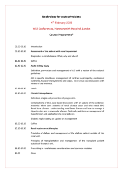

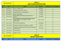
© Copyright 2026