
Acta Dermato-Venereologica - Mycobacterium chelonae Infection
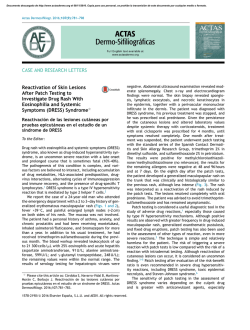
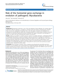
Acta Derm Venereol 2011; 91: 61–106 LETTERS TO THE EDITOR Mycobacterium chelonae Infection Associated with Tattoos Isabel Rodríguez-Blanco1, Luis Casas Fernández1, José Manuel Suárez Peñaranda2, Maria Luisa Pérez del Molino3, Jaime Esteban4 and Manuel Almagro5 Dermatology Department, Hospital da Barbanza, Ribeira, Oleiros s/n, ES-15993 La Coruña, 2Department of Pathology, Clinical University Hospital and School of Medicine, 3Microbiology Department, Clinical University Hospital, Santiago de Compostela, 4Microbiology Department, IIS-Fundación Jiménez Díaz, Madrid, and 5Dermatology Department, Hospital Universitario, A Coruña, Spain. E-mail: [email protected] Accepted September 27, 2010. 1 Mycobacterium chelonae is a non-tuberculous Mycobacterium spp. classified in the rapidly growing mycobacteria group. M. chelonae has been isolated from various environmental sources, including tap water, water tanks, industrial sources, and even medical instruments. Disruption of the skin barrier caused by trauma, surgical intervention or cosmetic procedures facilitates the entry of different bacteria that can produce localized or, usually in immunosuppressed patients, systemic infections. We describe here an outbreak of M. chelonae infection in five immunocompetent patients who were being tattooed by the same artist. Case reports Five otherwise healthy patients attended our clinic between September 2008 and April 2009 because of the presence of skin lesions in tattooed areas, all of which had been recently administered in the same tattoo parlour. The series includes 3 men and 2 women with a mean age of 21 years (age range 18–23 years). More than one type of ink was used in every patient, but in all patients the lesions were restricted to the grey areas of the tattoos. The delay in onset of the clinical lesions varied from 3 to 30 days. Lesions consisted of red papules with remarkable superficial hyperkeratosis in most cases. Patient 3 also presented with frank pustules (Figs 1 and 2). Histopathological examination showed a dense lymphohistiocytic inflammatory infiltrate mainly involving the superficial dermis in all cases. In cases 2 and 3, well-developed granulomas were easily found, with many Langhans-type giant cells and, occasionally, central necrosis. In cases 4 and 5, granulomas were also seen, but these were made up only of epithelioid histiocytes with few, if any, giant cells. Suppuration, with the presence of Fig. 1. Red hyperkeratotic papules restricted to the grey areas of the tattoos with presence of scattered pustules in patient 3. © 2011 The Authors. doi: 10.2340/00015555-1034 Journal Compilation © 2011 Acta Dermato-Venereologica. ISSN 0001-5555 Fig. 2. Red hyperkeratotic papules restricted to the grey areas of the tattoos in patient 4. polymorphonuclear cells, was noted in some of the granulomas, but none showed central necrosis. In case 3, histopathological examination revealed a dense, band-like, lympho-histiocytic infiltration in the superficial dermis. Only after serial sections and careful scrutiny, poorly-formed, histiocytic, granulomas were seen. These showed neither central necrosis nor suppuration. Acid-fast staining for bacilli was negative in all cases. Paraffinized samples were processed according to the technique described by Loeschke et al. (1). After deparaffinization, DNA was extracted and processed using GenoType CM Kit (Hain, Germany), according to the supplier’s protocol, except for a prolonged PCR second cycle (25 instead of 20 of the following cycles: 25 s at 95ºC, 40 s at 53ºC and 40 s at 70ºC). PCR products were then hybridized with the GenoType CM strips according to the manufacturer’s instructions. Hybridization was positive for positions 1, 2, 3, 5 and 10, which are the positions for identification of M. chelonae. Skin biopsy specimens were cultured in BACTEC MGIT 960 System (Becton Dickinson Microbiology Systems, Sperks, MD, USA) and Coletsos (Pronadisa/lab Conda) medium. Positive mycobacterial cultures were obtained in 3 cases (patients 1, 4 and 5). The molecular methods described above were used for the identification of M. chelonae in patients 2 and 3, being positive in both cases. The susceptibility testing was performed by the Etest® (AB Biodisk, Solna, Sweden) on Muller–Hinton agar with blood. The minimal inhibitory concentrations (MIC) of the isolates (in mg/l) were clarithromycin 2, imipenem > 32, amikacin 4, cefoxitin > 32, doxycycline > 256, cotrimoxazole > 32, and ciprofloxacin > 32. Three patients were initially treated with clobetasol propionate ointment due to suspicion of an allergic reaction to the tattoo ink. Clarithromycin 500 mg twice a day was administered in patients 1 and 4 until resolution (after 3 and 5 months, respectively). In two patients antibiotic therapy either had to be suspended after 1 week (patient 5) or reduced to a half-dose after the initial 3 weeks of therapy, for an additional 11 weeks Acta Derm Venereol 91 62 Letters to the Editor of treatment (patient 3), due to gastric intolerance. In addition, topical gentamicin twice a day was also prescribed in all patients. Patient 2 refused any treatment and did not attend the follow-up visit, but reported complete resolution of the lesions after being contacted by telephone. Two other clients of the same parlour who presented with similar lesions in the grey areas of their tattoos were evaluated in our clinic, but were lost to follow-up after the first visit. DISCUSSION Infectious complications are the most important potential sequelae in association with tattoos when the aseptic conditions of the procedure are not maintained or when the dye used for injection is not sterile. Staphylococcus and Streptococcus infections have an historical interest as causes of erysipelas, cellulitis and gangrene (2). Local skin lesions, such as verruca vulgaris (3), molluscum contagiosum (4) or superficial mycosis, have also been described. Systemic infections are the biggest concern and they include hepatitis (5), HIV (6), syphilis (5), leprosy (7) or tuberculosis (8). Atypical mycobacteria infections secondary to tattooing have been reported previously (2, 9–12). In our series, the source of infection may have been either the water used to dilute the ink for obtaining the grey colour or the containers used for mixing the ink. The owner of the establishment stated that he always used distilled water in tattoo procedures, therefore a possible origin in the tap water used to wash the mixing containers can be speculated. The detection of atypical mycobacteria in clinical samples raises concern about evaluation of their significance. These organisms are environmental; thus they could appear in these samples as the result of contamination or colonization (13). Detection of these organisms using molecular tools can increase this problem because of the high sensitivity of these techniques. However, detection of M. chelonae in extrapulmonary samples is frequently significative (14). Moreover, the detection of these organisms in an outbreak setting (such as the one described here) is difficult to interpret as potential contamination, especially because the pathology and outcome of the patients is similar to confirmed cases, and because our molecular study is a retrospective one using samples that were highly suspicious of being M. chelonae infections. There is some controversy regarding susceptibility testing in rapidly growing mycobacteria because of the difficulties in standardization and reproducibility of the results, but the American Thoracic Society recommends the broth microdilution MIC determination (15). Even when this method was not used in our patients, clari thromycin was prescribed according to the Etest® with a good clinical response in all treated patients. Although auto-resolution of an atypical mycobacterium infection in an immunocompetent patient is possible, the clearance of lesions in patient 2 could not be Acta Derm Venereol 91 confirmed as she was only contacted by telephone. A potential concern with such untreated, but apparently cured patients with subcutaneous atypical mycobacterium infection, is the possibility of reactivation if their immune status changes. We suggest that long-term follow-up of these patients would be good practice. We report here five well-documented cases and other two non-confirmed cases of M. chelonae infection related to the grey areas of tattoos administered in the same tattoo parlour. Atypical mycobacterial infection should be considered when skin lesions develop following a tattoo or any other procedure that affects the subcutaneous tissue. REFERENCES 1.Loeschke S, Goldmann T, Vollmer E. Improved detection of mycobacterial DNA by PCR in formalin-fixed, paraffinembedded tissues using thin sections. Pathol Res Pract 2005; 201: 37–40. 2.Wolf R, Wolf D. A tattooed butterfly as a vector of atypical mycobacteria. J Am Acad Dermatol 2003; 48 (5 Suppl): S73–74. 3.Miller DM, Brodell RT. Verruca restricted to the areas of black dye within a tattoo. Arch Dermatol 1994; 130: 1453–1454. 4.Pérez Gala S, Alonso Pérez A, Ríos Buceta L, Aragüés Montañés M, García Díez A. Molluscum contagiosum on a multicoloured tattoo. J Eur Acad Dermatol Venereol 2006; 20: 221–222. 5.Nishioka Sde A, Gyorkos TW. Tattoos as risk factors for transfusion-transmitted diseases. Int J Infect Dis 2001; 5: 27–34. 6.Doll D. Tattooing in prison and HIV infection. Lancet 1988; 1: 66–67. 7.Ghorpade A. Inoculation (tattoo) leprosy: a report of 31 cases. J Eur Acad Dermatol Venereol 2002; 16: 494–499. 8.Ghorpade A. Lupus vulgaris over a tattoo mark-inoculation tuberculosis. J Eur Acad Dermatol Venereol 2003; 17: 569–571. 9.De Quatrebarbes J, Pestel-Caron M, Duval-Modeste A, Abboud P, Etienne M, Caron F, et al. Epidemie a Mycobacterium chelonae chez un tatoueur. Ann Dermatol Venereol 2005; 132 (Suppl 9): S224–225. 10. Kluger N, Muller C, Gral N. Atypical mycobacteria infection following tattooing: review of an outbreak of 8 patients in a French tattoo parlor. Arch Dermatol 2008; 144: 941–942. 11. Preda VA, Maley M, Sullivan JR. Mycobacterium chelonae infection in a tattoo site. Med J Aust 2009; 190: 278–279. 12. Drage LA, Ecker PM, Orenstein R, Phillips PK, Edson RS. An outbreak of Mycobacterium chelonae infections in tattoos. J Am Acad Dermatol 2010; 62: 501–506. 13. De Groote MA, Huitt G. Infections due to rapidly growing mycobacteria. Clin Infect Dis 2006; 42: 1756–1763. 14. Esteban J, Martín-de-Hijas NZ, Fernandez AI, FernandezRoblas R, Gadea I; Madrid Study Group of Mycobacteria. Epidemiology of infections due to nonpigmented rapidly growing mycobacteria diagnosed in an urban area. Eur J Clin Microbiol Infect Dis 2008; 27: 951–957. 15. Griffith DE, Aksamit T, Brown-Elliott BA, Catanzaro A, Daley C, Gordin F, et al. An official ATS/IDSA statement: diagnosis, treatment, and prevention of nontuberculous myobacterial diseases. Am J Respir Crit Care Med 2007; 175: 367–416.


© Copyright 2026