
Programa - Santiago de Cuba
General Program
Monday November the 7th, 2016
4:00 p.m.-7:00p.m Registration at Hotel Villa San Juan
Tuesday, November 8th, 2016
9:15-9:30 a.m.
Inaugural Ceremony at Hotel Villa San Juan
9:30-10:20 a.m.
Inaugural Conference
10:20 a.m.-11:30 a.m.
Work in sessions
11:30 a.m. Snack
12:00 m -3:00 p.m.
Work in sessions
3:10 p.m.-5:00p.m.
City Tour
9:00 p.m.
Welcome Cocktail
Wednesday, November 9th, 2016
9:00-10:00 a.m.
Plenary Conference
10:10 a.m.-11:30 a.m.
Work in sessions
11:30 a.m. Snack
12:00 m -3:00 p.m.
Work in sessions
3:10 p.m.-5:00p.m.
City Tour
9:00 p.m.
Gala Concert
5
Thursday, November 10th, 2016
9:00-10:00 a.m.
Closing Conference
10:10 a.m.-11:30 a.m.
Posters
11:30 a.m.
Closing remarks
12:30 p.m
Farewell Snack
9:00 p.m.
Farewell Party, Tropicana Santiago
6
Scientific Program
Monday November the 7th, 2016
Pre-congress Teaching Course on Neuroradiology
Place: Hotel Villa San Juan
4:30 -6:00 p.m.
Neuroimaging of epilepsy. Doris D.M Lin. Johns Hopkins University
School of Medicine, Department of Radiology, Division of
Neuroradiology. 4:30 -6:00 p.m.
Tuesday, November 8th, 2016
Sala
Comissions
No 1
Epilepsy I
No 2
Non Epileptic Paroxysms1
Neuromuscular diseases2
No 3
Cerebrovascular Diseases
No 4
Neurosurgery
Chair
Margarita M. Báez Martín
Elisabeth Colina
José Miguel Láinez 1
Osvaldo Aguilera2
Oscar Medina2
Fahmi Yusuf Khan
Pablo García Bermejo
Rafael Domínguez Peña
Ricardo Hodelin Tablada
7
Wednesday, November 9th, 2016
Sala
Comisiones
Coordinadores
No 1
Epilepsy II(Mechanism-
No 2
Brain and Cognition
No 3
CNS Injury-infection
Neuroradiology2
No 4
Degenerative Diseases
Lilia M. Morales
Danielle Nolan
Erislandy Omar
Suzie Nall
Magnus Gisslén1
Jorge A. Bergado1
Solangel Bolaños2
Doris M Lin2
Management)
1
Serenella Servidei
Ernesto Simón
Thursday, November 10th, 2016
Sala
Comisión
Posters
Posters
Coordinadores
Osiel Gámez Rodríguez
Ricardo Hodelín Tablada
Mavis Casamajor
8
IDIOMAS OFICIALES DEL EVENTO: Castellano e Inglés
Tuesday, November 8th, 2016
9:30-10:20 a.m. Inaugural Conference:
Is Tropical Neurology Specific?
Professor Michel DUMAS, Institut de Neurologie Tropicale – Faculté de
Médecine – Université de Limoges – France
10:25 AM-3:00 P.M Commissions
Commission 1 Epilepsy (I)
Lugar: Sala 1
Chair persons: Margarita M. Báez Martín. CIREN, Havana
Elisabeth Colina. Hospital “S.Lora” Santiago de Cuba
10:25 - 11:00 a.m. Evaluación de sistemas sensoriales en pacientes con
epilepsia del lóbulo temporal mesial fármaco-resistente sometidos a
tratamiento quirúrgico. Margarita M. Báez Martín. CIREN. Habana, Cuba
11:10 - 11:30 a.m. Evaluación Prequirúrgica en Niños con Epilepsias
Farmacorresistentes. Resultados del CIREN. Lilia Morales Chacón, CIREN.
Habana, Cuba.
11:30-12:00 m
Snack break
12:00-12.20 p.m. Neuroinflamación y estrés oxidativo en pacientes con crisis
parciales complejas farmacorresistentes. L Lorigados, CIREN. Habana
12:30-12.50 p.m. Semiological Approach to Epilepsy Surgery
Charles Akos Szabo. University of Texas Health Science Center at San Antonio
1:00-1.20 p.m A Clinician's Guide to the Genetics of Epilepsy.
Danielle Nolan. University of Michigan.
9
1:30-2:10 p.m. Effects of Sleep and Circadian Rhythms on Epilepsy
Milena Pavlova. Medical Director, Faulkner Neurophysiology and Sleep Testing
Center, Department of Neurology, Brigham and Women’s Hospital
Harvard Medical School.
2:15-3:00 p.m Long-term outcomes of epilepsy surgery in adults. Kristina
Malmgren. Sahlgrenska University Hospital. Sweden (Plenarium)
Commission 2
Non Epileptic Paroxysms. Chair person: Miguel JA Láinez; MD. PhD,
FAAN,FANA,FAHS. Valencia, Spain
Neuromuscular diseases. Chairs person: Osvaldo Aguilera, Cuba, Oscar
Medina, México.
Lugar: Sala 2
10:20 - 11:20 a.m. Neuromodulation Techniques in Headache.
Prof. Miguel JA Láinez; Valencia, Spain.
11:30-12:00 m
Snack break
12:10-12.50 p.m. Evaluación de la Ficocianobilina y sus combinaciones en un
modelo animal de Esclerosis Múltiple.Nancy Pavón Fuentes. CIREN
1:00-1.40 p.m. SubPoblaciones Linfocitarias y sus Efectos Adversos en
Pacientes con Esclerosis Multiple Remitente-Recurrente Tratados con
FINGOLIMOD: Experiencia en Nuestro Hospital. Oscar Medina Carrillo.
Commission 3
Cerebrovascular Diseases. Chair persons: Pablo García Bermejo,HMC, Doha
Fahmi Khan, HMC, Doha
Lugar: Sala 3
10:20 - 10:40 a.m. Fístula Carótido-cavernosa. Presentación de un caso
insólito Autora: Dra. Damaris Fuentes Pelier. Hospital General Dr, Juan Bruno
Zayas Alfonso
10
10:40 - 11:00 a.m. Evidence Based Medicine in Childhood Arterial Ischemic
Stroke: New Insights and Challenges. Dr. Stéphane Darteyre. Department of
Pediatrics, French Polynesia Hospital. Tahiti, French Polynesia.
11:10-11:20 a.m. Rescue therapy for mechanical thrombectomy refractory
occlusions with detachable stent-retrievers and GP IIb/IIIa inhibitors.
Dr.Pablo Garcia-Bermejo, Hamad Medical Corporation. Department of
Medicine. Doha, Qatar
11:30-12:00 m Snack break
12:00-12.50 p.m Stroke in Minorities. Dr. Fahmi Yousef Khan. Department of
Medicine, Hamad Medical Corporation, Doha, Qatar
Commission 4 Neurosurgery
Chair persons: Rafael Domínguez Peña, Ricardo Hodelín Tablada; Cuba
Lugar: Sala 4
10:20 - 11:20 a.m. Las alteraciones de conciencia desde Victor Horsley hasta
Joseph Giacino. DrC. Ricardo Hodelín Tablada. Hospital Provincial Clínico
Quirúrgico “Saturnino Lora”. Santiago de Cuba.
11:30-12:00 m Snack break
12:00-12.40 p.m The Role of Cuban Neurosurgeons in the Development of
Neurosurgery in Ethiopia. Dr. Zenebe Gedlie Damtie. School of Medicine, Addis
Ababa University. Ethiopia.
12:40-1.30 p.m. Visión de Harvey Cushing sobre los traumatismos
craneoencefálicos.Dr. C. Ricardo Hodelín Tablada
Hospital Provincial Clínico Quirúrgico “Saturnino Lora”. Santiago de Cuba.
1:40-2:10 p.m. Tumores cerebrales asociados a epilepsia de larga evolución.
Experiencia en el Centro Internacional de Restauración Neurológica.
Bárbara O. Estupiñán Díaz. Proyecto Cirugía Epilepsia. CIREN
11
Wednesday, November 9th, 2016
9:00-10:00 AM: Special Conference (Plenarium) Sala 1
Neurostimulation of the nervus hypoglossus as a salvage treatment option in
obstructive sleep apnea for CPAP intolerance.
Armin Steffen, Otorhinolaryngology, University of Luebeck, Germany
Lugar: Salón plenario (Sala 1)
Commission 1 Epilepsy (II)
Lugar: Sala 1
Chair persons: Lilia M. Morales, CIREN, Havana, Danielle Nolan, University of
Michigan
10:10 - 10:30 a.m. Bridging epilepsy treatment gap in developing countries
based on Damtie’s `D model. Dr. Zenebe Gedlie Damtie. School of Medicine,
Addis Ababa University. Ethiopia.
10:40 - 11:00 a.m Cognitive Aspects of Antiepileptic Drugs (and yes, that will
include cannabinoids).David E. Mandelbaum, Alpert Medical School of Brown
University, Division of Child Neurology, Rhode Island and Hasbro Children's
Hospitals.
11:10 - 11:30 a.m. Natural history of epilepsy in a cohort of 127 children in
Santiago de Cuba: 2 years follow-up. Francisco Ruiz Miyares. Hospital Infantil
Sur. Santiago de Cuba.
11:30-12:00 m
Snack break
12:10-12.50 p.m. Síndrome de West: Características clínicas, terapéutica,
evolución clínica y factores pronósticos. Dr. Ernesto Portuondo Barbarrosa.
Hospital Pediátrico docente Centro Habana. La Habana, Cuba.
1:00-1:20 p.m Trastornos de la salud reproductiva en la mujer con epilepsia
en edad fértil. Dr. Juan Miguel Riol Lozano Servicio de Neurología. Hospital
Hermanos Ameijeiras.
2:00-2:20 p.m. Semiology and evolution of febrile crisis at the Hospital
Pediátrico Universitario Centro Habana. Dr. Ernesto Portuondo Barbarrosa.
Hospital Pediátrico docente Centro Habana. La Habana, Cuba.
12
Commission 2
Brain and Cognition. Chair: Erislandy Omar, ISPJAM, Santiago de Cuba; Susan
W. Nall, Ph.D. Professor Emeritus, Southern Illinois University Edwardsville.
USA
Lugar: Sala 2
10:10 - 10:30 a.m.
"Early Literacy: Research, Implications for Practice in Early Education
Programs, and Long Term Significance" Susan W. Nall, Ph.D. Professor
Emeritus, Southern Illinois University Edwardsville. USA
10:40 - 11:00 a.m. Epilepsia y sociedad
Dr. Osvaldo R. Aguilera Pacheco, Damaris González Vidal. Santiago de Cuba
11:10 - 11:30 a.m. Consecuencias Neuropsicológicas Relacionadas con la
Cirugía de la Epilepsia. Ma. Eugenia Garcia, CIREN, Habana.
11:30-12:00 m Snack break
12:10-1.00 p.m. Estimulación cognitiva temprana de funciones ejecutivas en
daño cerebral focal. Evidencias empíricas en pacientes con afasia secundaria
a enfermedad cerebrovascular isquémica.
Erislandy Omar Martinez.ISPJAM.Santiago de Cuba
Commission 3 CNS Injury. Chair persons: Magnus Gisslén
University of Gothenburg, Dept. of Infectious Diseases, Sahlgrenska University
Hospital, Gothenburg, Sweden. Jorge A. Bergado, CIREN, Havana, Cuba
Neuroradiology. Chair persons: Solangel Bolaños, Hospital Saturnino Lora,
Santiago de Cuba, Doris M Lin, Johns Hopkins University School of Medicine,
Department of Radiology, Division of Neuroradiology.
Lugar: Sala 3
10:10 - 10:50 a.m. Cerebrospinal Fluid and Blood Biomarkers of Neuronal
Injury in HIV. Magnus Gisslén. University of Gothenburg, Dept. of Infectious
Diseases, Sahlgrenska University Hospital, Gothenburg, Sweden
13
11:00 - 11:20 a.m Character of CSF inflammation through the course of HIV
infection. Richard W. Price, MD. Professor Emeritus (Active) Neurology, UCSF
11:30-12:00 m Snack break
12:10-12.40 p.m. Electrophysiology, field potentials and the study of
synaptic plasticity. J. A. Bergado. Centro Internacional de Restauración
Neurológica (CIREN) Ave. 25 • 15805 Playa 11300 La Habana, Cuba.
12:45-1.10 p.m. Neuroimaging of Genetic Generalized Epilepsies (GGE): New
Insights into Underlying Mechanisms. Charles Akos Szabo. University of Texas
Health Science Center at San Antonio.
1:15-1.50 p.m. MR imaging and spectroscopy at 7 Tesla. Peter B. Barker,
Johns Hopkins University School of Medicine, Department of Radiology,
Division of Neuroradiology.
2:00-2:50 p.m. Demyelinating central nervous system diseases: diagnosis and
management – state of the art 2016. Friedemann Paul. Charité, Berlin,
Germany (Plenarium)
Commission 4
Degenerative Diseases-Neuropediatrics: Chair persons: Dr.Ernesto Simón,
Hospital “ S.Lora”, Santiago de Cuba, Serenella Servidei. Institute of Neurology,
Catholic University, Rome.
Lugar: Sala 4
10:10 - 10:30 a.m. Morbilidad por trastornos del movimiento en la consulta
de Neurología . Hospital General Docente “Dr. Juan Bruno Zayas Alfonso”,
Santiago de Cuba, Cuba. Dra. Mónica Rodríguez Montalván. Hospital General
Docente “Dr. Juan Bruno Zayas Alfonso”
11:00 - 11:20 a.m. Caracterización de los síntomas no motores en la
Enfermedad de Parkinson.Dr.Ernesto Simón Pérez, Hospital “ S.Lora”,
Santiago de Cuba
11:30-12:00 m
Snack break
14
12:00-12.30 p.m. Prediction of Parkinson's disease through RBD and
neuroimaging. K.L. Leenders. UMCG, Groningen. The Netherlands.
12:40-1.30 p.m. Central Nervous System involvement in Mitochondrial
Encephalomyopathie. Serenella Servidei, Institute of Neurology, Catholic
University, Rome
Thursday, November 10th, 2016
9:00-10:00 AM: Final Conference (Plenarium)
“Advances in the Treatment of Epilepsy: Does Mechanism of Action matter?
Barry Gidal. University of Wisconsin-Madison, School of Pharmacy and
Department of Neurology.
10:00-11:00 a.m.: Posters
11:10-12:00 m. Closing Ceremony.
15
Pre-congress Teaching Course
Neuroimaging of Epilepsy
Doris D.M. Lin, MD, PhD
Johns Hopkins University School of Medicine, Department of Radiology,
Division of Neuroradiology
Abstract
Neuroimaging is an integral part of clinical evaluation of epilepsy, and
fundamental in delineating structural abnormalities for surgical
intervention. The common causes of epilepsy presenting in childhood
and young adults that often require surgical intervention include
congenital or developmental malformations (such as focal cortical
dysplasia, hemimegalencephaly, malformation of cortical development
and lissencephaly), phakomatoses (including tuberous sclerosis and
Sturge-Weber
syndrome),
hippocampal
sclerosis,
vascular
malformations and tumors. MRI is the most important imaging modality
in depicting the site and type of structural abnormalities in these entities,
and examples will be shown. However, not all lesions are readily
identified on conventional MRI, and complementary information may be
obtained by advanced imaging techniques such as MR spectroscopy and
DTI, and nuclear medicine techniques such as SPECT and PET. Mesial
temporal sclerosis is the most frequent pathological finding in temporal
epilepsy but may be challenging to diagnose; current advances in imaging
including high-field (7T) structural imaging and quantitative analysis will
be presented.
16
Conferences
Is Tropical Neurology Specific?
Michel DUMAS
UMR Inserm 1094 « NeuroEpidémiologie Tropicale »
Institut de Neurologie Tropicale – Faculté de Médecine – Université de Limoges –
France
Abstract
Only countries situated between the Tropic of Cancer and the Tropic of
Capricorn belong to the real tropical parts of the world. But often, the
“tropical” concept also concerns regions situated outside of the tropics,
because of environmental, cultural and socioeconomic similarities. The
neurological affections and their consequences are closely connected to
this tropical environment.
Actually, a deep and relatively quick mutation in the environmental
conditions and in the neurological practices in tropical countries occurs,
that raises the question whether the tropical neurology is still specific.
Indeed, this concept is now obsolete, because of the gradual attenuation
or even disappearance of certain environmental conditions such as poor
socio-economic status, infectious diseases. Ubiquitous diseases, such as
high blood pressure, diabetes, cancer, dementia, are now growing quickly
in tropical regions. Moreover, several infectious diseases, such as
dengue, formerly encountered only in the tropics, have now become
ubiquitous.
Nevertheless, despite these considerations, tropical neurology still
remains specific and original because: - cultural peculiarities that have
neurological consequences will persist for a long time, - there is a specific
common denominator in all tropical regions, and this will not change: the
intense solar radiations and UVB rays, which could have many
17
consequences. Among them, the characteristic of inhibiting the
functioning of the dermis Langerhans cells, with all the immunological
consequences ensuing, particularly the low prevalence of multiple
sclerosis in the tropics.
So YES, we must consider that tropical neurology is still specific, and as
such, deserves to be recognized as a speciality of neurology.
Long-term outcomes of epilepsy surgery in adults
Kristina Malmgren. Sahlgrenska University Hospital.Sweden
Abstract
Epilepsy surgery is an efficacious treatment for selected persons with
drug-resistant focal epilepsy, rendering many seizure-free and others
significantly improved. There is Class I evidence for short-term efficacy of
epilepsy surgery from two randomized controlled studies of temporal lobe
resection. In order for patients to make an informed decision about the
treatment option of epilepsy surgery, they also need data on the
probability of long-term remission or improvement. Long-term longitudinal
observational studies are necessary in order to obtain valid outcome data.
From a number of such studies the proportion of patients who have been
continuously free from seizures with impairment of consciousness since
resective surgery seems to be 40-50% after 10 years while a higher
proportion have been seizure-free at least a year at each time-point
assessed. The best longitudinal data are in patients who have undergone
temporal lobe resection and in whom the histopathology was mesial
sclerosis, and in these patients the majority of relapses occur within five
years. Whether this course is applicable to other resection types and
pathologies is not clear. There is much less information on the longitudinal
course in patients who have undergone other resection types and have
other causes. For many resection types the numbers of patients in single18
centre long-term follow-ups is limited and for almost all studies there is a
lack of controls. Multicentre observational studies following both operated
and non-operated patients are needed in order to obtain more robust
data.
Effects of Sleep and Circadian Rhythms on Epilepsy
Milena Pavlova, M.D.
Medical Director, Faulkner Neurophysiology and Sleep Testing Center
Department of Neurology, Brigham and Women’s Hospital
Harvard Medical School
Abstract
Epilepsy and sleep have many interactions. There are multiple epilepsy
syndromes that present with either exclusively or predominantly nocturnal
seizures. Particularly interesting is the association of seizure propensity
and sleep stage, and the fact that seizures are very rare in REM sleep.
Furthermore, sleep loss, and sleep disorders may impact epilepsy.
Particularly robust is the effect of obstructive sleep apnea, a fairly
common sleep disorder. Last but not least, circadian rhythms may have
an interesting effect on seizure propensity. Studies in children and in
adults indicate that the time when seizures occur may not be random and
depends on the epileptogenic region, with frontal lobe seizures being
more frequently occurring from sleep and during the early morning hours,
and temporal lobe seizures occurring more frequently from wakefulness
and in the mid to late afternoon.
A Clinician's Guide to the Genetics of Epilepsy.
Danielle Nolan M.D.
Departments of Pediatrics and Pediatric Neurology, University of
Michigan,
Ann Arbor MI USA
19
Abstract
Genetic advances are reshaping our knowledge of the etiology of epilepsy
and providing new approaches to treatment. Previously, >75% of epilepsy
syndromes were considered to be idiopathic. In contrast, we now
recognize that genetic factors contribute to the majority of epileptic
syndromes. Genetic analysis is facilitated by syndromic classification of
epilepsy (e.g. epileptic encephalopathy, myoclonic seizures, generalized
epilepsy with febrile seizures or GEFS+). In some occasions,
identification of the
genetic cause indicates specific treatment
approaches. For instance, migrating partial epilepsy of infancy (i.e. MPEI
or EIEE14) is characterized by progressive pharmacoresistant epilepsy.
When a pathogenic KCNT1 variantis identified, administration of quinidine
can be beneficial. As another example, the occurrence of POLG gene
variation in several epilepsy syndromes contraindicates the use of
valproate which can precipitate liver failure. Genetic advances are also
providing fresh insights into focal epilepsy.
Previously, epilepsy surgery was a mainstay treatment for
pharmacoresistant lesion-associated epilepsies. Recent genetic studies
have implicated the GATOR1 complex (involved in the mTOR pathway)
in focal cortical dysplasia. This suggests a new therapeutic target for
lesions in which surgery carries a high risk. The discovery of more than
100 genes has defined dozens of diverse biochemical pathways that
contribute to various epilepsy syndromes. Although there is some
pathway sharing between various types of epilepsy (e.g.. the GABA
receptor signaling pathway), it is becoming clear that different
biochemical pathways underlie the same clinical syndrome. For example,
generalized epilepsy with febrile seizures plus (GEF+) is a phenotypic
syndrome with wide genetic heterogeneity. Implicated genes range from
SCN1A (a voltage-gated sodium channel) to GABRG2 (a GABA
receptorinvolved in GABAergic neurotransmission) to STX1B (a syntaxin
20
implicated in the excitatory pathway of synaptic transmission). As genetic
testing of epilepsy subjects expands and epilepsy gene mutation
registries become more informative, genotype-phenotype correlations
become more specific. As a result, the ability to counsel individuals and
families improves. An overview into recent advances into the genetic
causes of epilepsy and the implicated molecular pathways is presented,
together with a guide for clinicians in the evaluation of epilepsy patients
for which genetic factors are likely causative
Electrophysiology, field potentials and the study of synaptic
plasticity
J. A. Bergado, W. Almaguer
Centro Internacional de Restauración Neurológica (CIREN) Ave. 25 •
15805 Playa 11300 La Habana, Cuba
Abstract
Electrophysiology developed to constitute a tool for clinical and
experimental Neurociences. The study of field potentials allows the
investigation of the functional properties of neuronal populations and their
relationship to neural processes or mental states. Monosynaptic evoked
potentials in the hippocampus is a widely used method, due mainly to its
simplicity and low technical requirements, allowing however statements
and interpretations at cellular level, especially in relation to modifications
in synaptic efficacy. Synaptic plasticity is one of the mechanisms that
allow the adaptive properties of the Nervous System. Our group at CIREN
have studied the links between neural plasticity and affective factors, and
developed a model based on the synaptic tagging theory to explain those
interactions. New investigations suggest a role for neurotrophines and
plasticity related genes in the restoration of memory functions by
stimulation of the amygdala, in rats affected by lesions of the fimbria-fornix
21
system. We are currently searching for strategies to apply these
mechanisms in clinical practice.
Las alteraciones de conciencia desde Victor Horsley hasta Joseph
Giacino
DrC. Ricardo Hodelín Tablada
Hospital Provincial Clínico Quirúrgico “Saturnino Lora”.
Resumen: Los estados de alteraciones de conciencia se conocen desde
la antigüedad pero las referencias en la literatura científica comienzan a
publicarse a fines del siglo XIX. Establecer las diferencias entre estos
múltiples estados constituye un verdadero problema científico. De su
adecuado diagnóstico dependerá el tratamiento y seguimiento de los
enfermos. En el presente trabajo el autor realiza un bosquejo histórico
desde 1886 en que Víctor Horsley analiza el nivel de conciencia hasta el
2002 que Joseph Giaciono y sus colaboradores describen el estado de
mínima conciencia. Se establecen los criterios del estado vegetativo
persistente descrito por Brian Jennett y Freud Plum en 1972, así como
los criterios del estado de mínima conciencia. Se conceptualiza la muerte
encefálica como el cese de las funciones de todo el encéfalo y se
discuten los criterios establecidos en Cuba para su diagnóstico. Otras
alteraciones de conciencia que se revisan son el coma, el mutismo
acinético y el síndrome de enclaustramiento. El diagnóstico diferencial
entre las diferentes alteraciones de conciencia se establece en base a
los ciclos sueño vigilia, la apertura ocular, la función motora, la función
auditiva, la función visual y la comunicación, asimismo se plantea el
tiempo necesario para el diagnóstico y el pronóstico neurológico. Se
considera que los modernos descubrimientos de funciones cognitivas
residuales y nuevos correlatos neuronales han contribuido a esclarecer
22
importantes incógnitas. Se concluye que a pesar de los avances en el
conocimiento de las alteraciones de conciencia, estas entidades
constituyen en la actualidad un verdadero reto para los neurocientíficos.
Visión de Harvey Cushing sobre los traumatismos
craneoencefálicos
Dr. C. Ricardo Hodelín Tablada
Hospital Provincial Clínico Quirúrgico “Saturnino Lora”
Resumen
Introducción: Harvey Williams Cushing, invitado por el Profesor William
Williams Keen, escribió un capítulo en el libro “Cirugía. Tratado teóricopráctico de patología y clínica quirúrgicas”, que traducido del inglés, se
publicó en idioma español, en 1912, en Barcelona, España. Objetivo:
Analizar los postulados de Cushing sobre los traumatismos
craneoencefálicos y destacar su vigencia más de un siglo después de su
publicación. Resultados: Se comenta sobre la edición y sus
colaboradores. En relación con el contexto histórico se reseña que al
publicarse el libro ya Cushing era reconocido como un prestigioso
neurocirujano que ese propio año tomó posesión como Jefe del Servicio
de Clínica Quirúrgica en el Peter Bent Brigham Hospital. Se analiza la
propuesta de clasificación de las fracturas del cráneo según el
mecanismo de producción, según exista o no herida comunicante con el
foco de fractura, según la forma que tienen los fragmentos y según su
situación. Se comenta el excelente esquema donde el autor muestra el
mecanismo de las fracturas por depresión. Se relacionan los
conocimientos de neurofisiología que demuestra Cushing y la teoría
propuesta para explicar las lesiones a distancia debido a los efectos de
contragolpe. Se destacan la vigencia de sus argumentos, sus
23
impresionantes análisis científicos e ilustraciones médicas entre otros
elementos. Se subrayan otros méritos como el adecuado lenguaje que
sin dejar de ser científico, es ameno y logra trasmitir con claridad todas
las ideas. Conclusiones: Los postulados sobre los traumatismos
craneoencefálicos propuestos por Cushing se mantienen vigentes más
de cien años después de publicados.
Advances in the Treatment of Epilepsy: Does Mechanism of Action
matter?
Barry Gidal. University of Wisconsin-Madison, School of Pharmacy and
Department of Neurology
Abstract
Over the past 20 years, a number of new antiepileptic drugs (AEDs) have
been introduced into the global market. While most of these newer drugs
are molecularly distinct, some may more rightfully be considered analogs
or subtle derivatives of existing agents. In many cases, these
modifications to existing drugs does not result in significantly different
efficacy, but may afford improved tolerance and medication adherence.
In some cases however, improved pharmacokinetic profiles may have the
additional benefit of improved anti-seizure properties as well. During this
same time period, we have also seen major advances in our
understanding of the neurophysiological basis for epilepsy, as well as the
discovery of new molecular targets for AEDs.The objective of this lecture
will be to review the basic neuropharmacological basis for the
pharmacotherapy of focal seizures, as well as to discuss the recent
additions to our therapeutic armentarium including lacosamide,
eslicarbazepine, breveracetam and perampanel. Specifically, this lecture
will address the following questions. First, is it still reasonable to still
classify AEDs by “drug class” second is new the same as novel, and
finally, is there a true basis for “rational polytherapy”.
24
Central Nervous System involvement in Mitochondrial
Encephalomyopathies
S. Servidei, G. Primiano, D. Sauchelli, C. Cuccagna, D. Bernardo, C.
Sancricca, C. Vollono
Institute of Neurology, Catholic University, Rome
Abstract
The term <Mitochondrial Encephalomyopathies> (MEs) was coined by
Shapira in 1977 to describe the simultaneous involvement of central
nervous system (CNS) and skeletal muscle. The CNS is metabolically
very demanding and therefore vulnerable to defects of the mitochondrial
respiratory chain. Cerebellar involvement, leukoencephalopathy, bilateral
striatal necrosis are all manifestations of specific mitochondrial disorders.
Subclinical CNS involvement is also present in the apparently pure
myopathic Progressive External Ophtalmoplegia (PEO). Epilepsy and
migraine are common events, particularly in MERRF and MELAS
syndromes, and epilepsy strongly influences course and prognosis of
mitochondrial diseases, often triggering potentially life-threatening
conditions such as metabolic crisis or SLEs.
In our cohort of 93 mitochondrial patients (age 16- 78), migraine was
reported in 35.5% with a much higher prevalence compared to general
population independently from gender, genotype or phenotype.
Migraine without aura was the most common headache (84.8%).
Mitochondrial patients with migraine showed (vs non migraineurs)
younger age, significantly increased prevalence of epilepsy (p=0.0103),
myoclonus (p=0.0309), stroke-like episodes (p=0.0290), EEG focal slow
abnormalities (p=0.0359) and EEG epileptic focal abnormalities
(p=0.0425). In the same series, 40.5% of the 93 patients showed
epileptiform abnormalities consisting of focal or multi-focal slow wave
activities and spikes and 21.5% presented clinically manifested epilepsy
(partial seizures, with or without secondary generalization), mostly, but
25
not only, MELAS and MERRF. Thus, migraine and epilepsy are not
merely phenotypic aspects of specific MEs but rather the expression of
vulnerability of CNS probably directly related with defects of respiratory
chain . Our data support the hypothesis that mitochondrial dysfunction
plays a key role in the pathophysiology of migraine and epilepsy.
Stroke-Like episodes (SLEs) are the hallmark of MELAS, ma can be
observed even in other MEs, as POLG1-related disorders. MELAS is
characterized by sudden neurological deficits similar to vascular strokes,
but the pathophysiology of SLEs is still debated. Out of 41 SLEs in our
patients with A3243G MELAS mutation, 64% followed migraine and 74%
were associated with seizures. Attacks involved occipital (62%), parietal
(59%) and temporal (51%) lobes. Acute lesions were bilateral in 66% of
SLEs and symmetric at onset in 51% with a very high incidence,
compared with vascular strokes, of bilateral clinical syndromes as cortical
visual loss or cortical deafness. Brain MRI demonstrated cortical and
subcortical lesions with non-vascular distribution, migrational behavior
and a mixed pattern of restricted and increased diffusion on ADC maps.
Follow-up MRIs demonstrated partial reversibility of the lesions, with
subsequent development of pseudolaminar necrosis and gliosis.
However cerebral and/or cerebellar atrophy by far exceeded the
extension of SLEs demonstrating two mechanisms playing in MELAS
patophysiology: abrupt loss of function due to cell injury followed by partial
recovery and an independent slowly progressive degenerative process.
Beside classical CNS manifestations, dementia and extrapyramidal signs
may be also present in mitochondrial disorders, sometime dominating the
phenotype. In our patients cognitive impairment was frequent revealing a
consistent pattern independent from phenotype. Mental control, short
term memory and visual selective attention were in fact selectively
impaired in 75% of the patients, but global intellectual decline or
dementia, according to DSM-5 criteria, was also present in a minority of
26
cases, including Alzheimer’s like syndromes. We did not found any strict
correlation between neuropsychological profile, and age, clinical
phenotypes and genetics, again supporting the idea that even cognitive
deficits are linked to mitochondrial dysfunction and not to the effects of
specific genetic changes.
As far brain imaging, in our cohort, abnormal MRI findings, excluding
SLEs, consisted in cerebral cortical atrophy (35 patients), cerebellar
atrophy (24 patients), brain stem atrophy (11 patients), white matter
abnormalities (45 patients) and signal changes in deep grey matter (25
patients). Brain atrophy was global, severe and progressive in MELAS,
was milder and quite stable in MERRF and mostly absent in PEO. The
study by Proton Magnetic Resonance Spectroscopy (H1-MRS) revealed
normal spectra in all patients with single or multiple deletions of
mitochondrial DNA at any stage of the disease, independently from
severity of the phenotype and CNS involvement. Instead, in patients
carrying A8344G or the A3243G non-MELAS patients we found increased
peak of choline-containing compounds (Cho), variable reduction of Nacetyl-L-aspartate (NAA) and no Lactate (LA) peak in brain, while in a
minority of these patients LA was present in CSF. In MELAS, LA was
instead found in both affected and non-affected brain areas and in CSF.
Neuroimaging, MRI spectroscopy and functional studies, as in vivo
biomarkers, may help to better characterized MEs. MRI can identify
common pattern in patients with MD, but only the integration with MRS,
can provide useful information to guide genetic studies and follow-up in
this heterogeneous group of disorders. Moreover, mitochondrial
abnormalities may also occur in common neurodegenerative diseases,
implying a mechanistic link between mitochondrial dysfunction and
neurodegeneration. Thus, there is a strong need of biomarkers. The
appropriate use of reliable biomarkers may in fact be helpful a) to identify
primary mitochondrial disorders among common diseases b) to
27
characterize mitochondrial dysfunction in neurodegenerative diseases c)
for diagnosis and disease monitoring d) in defining natural history e) to
characterize and better understanding mechanisms of mitochondrial
dysfunction that lead to heterogeneous phenotypes f) to orient therapeutic
strategies.
Neuromodulation Techniques in Headache
Prof Miguel JA Láinez; MD. PhD, FAAN, FANA, FAHS.
Valencia, Spain
Abstract
The use of neuromodulation for the treatment of headache is not new; in
fact, it uses dates back to Roman times. In the modern time, the use of
electrical stimulation in the treatment of refractory headache disorders
start with the use of occipital nerve stimulation in the occipital neuralgia
and the hypothalamic stimulation (DBS) in cluster patients. Several series
have been published evaluating the efficacy of DBS in the management
of refractory cluster patients. Today DBS is considered a technique with
good short and long-term efficacy results, but as very invasive procedure.
Occipital Nerve stimulation has been used in the treatment of chronic
migraine with good results in open studies and no so impressive data in
clinical trials. In trigeminal autonomic headaches, the results have been
better and it could be one of the first line therapies in refractory cluster
patients. One of the problems is that the rate of complications related with
the device is not low.
Another target for electrical stimulation has been the sphenopalatine
ganglion with a new implantable microstimulator; the results in the first
trial in cluster headache have been good with acute and potentially
preventive effect and low rate of device related complications. A trial in
migraine is ongoing.
28
Other non-invasive neuromodulation technique like the transcutaneous
vagus nerve stimulation has been tried in several headache syndromes
with positive results and very good tolerability.
Electrical stimulation play a role in the treatment of refractory (invasive
methods) and could be useful in usual (non-invasive) headache patients.
Neurostimulation of the nervus hypoglossus as a salvage treament
option in obstructive sleep apnea for CPAP intolerance.
Steffen A, Otorhinolaryngology, University of Luebeck, Germany
Abstract
Obstructive sleep apnea (OSA) is characterized by repetitive breathing
stops in sleep and leads to daytime sleepiness with higher risk of traffic
accidents, more cardiovascular complications such as stroke, and to
disturbed glucose metabolism. Standard treatment is positive airway
pressure therapy but a relevant portion cannot tolerate that for life time
usage. Besides dental appliances and classic sleep surgery like
tonsillectomy with UPPP, neurostimulation of the hypoglossal nerve
reaches high success rates in selected cases, even in patients with higher
overweight and more severe OSA burden. There are several implants
such as by ImThera and Inspire Medical which differ in evidence and
technical issues. Most results exist for the later with strict selection
criterias. Especially the exclusion of a complete concentric collapse at the
soft palate during drug-induced sleep endoscopy leads to high rate of
treament responders. The cuff placement of the stimulation leads at the
protruding branches of the hypoglossal nerve serves a promising tongue
motion pattern which correlates with better reduction of apneas during
sleep. The therapy adjustment of the electrode configuration and
respiration sensing settings helps to ensure treatment success and high
therapy adherence.
29
This talk covers the neurostimulation treatment, the identification of
candidates, implantation, therapy adjustment and setbacks. As a high
volume implant center, our experiences and academic projects are
shared with the audience.
30
Free Communications
Evaluación de sistemas sensoriales en pacientes con epilepsia del
lóbulo temporal mesial fármaco-resistente sometido a tratamiento
quirúrgico.
Dr.C. Margarita M. Báez Martín1, Dr.C. Lilia M. Morales1, Dr. Iván
García1, Dra. Bárbara Estupiñán1, Dr.C. Lourdes Lorigados1, Dra.
Yamila Pérez1, MsC. María E. García1, Lic. Ivette Cabrera1, Dr. Otto
Trápaga1, Dr.C. Juan E. Bender1, Dra. Judith González1, Dr. Reinaldo
Galvizu1, Lic .Karla Batista1, Lic. Rafael Rodríguez1, Dra Gladys Soto2,
Lic. Yerenia Delgado2, Téc. Yamilét Ávila2, Dr.C Lídice Galán3.
1 Centro Internacional de Restauración Neurológica, CIREN. Habana,
CUBA.
2 Centro de Investigaciones Médico-Quirúrgicas, CIMEQ. Habana,
CUBA.
3 Centro de Neurociencias de Cuba, CNC. Habana, CUBA.
Resumen
Por su relación anatómica con el lóbulo temporal, las vías auditiva y visual
son susceptibles de sufrir cambios anatómicos y/o funcionales luego de
la resección quirúrgica del lóbulo temporal utilizada como opción
terapéutica en pacientes con epilepsia del lóbulo temporal mesial
fármaco-resistente (ELTm-FR). Estudiamos una muestra de 28 pacientes
con ELTm-FR intervenidos quirúrgicamente en el Centro Internacional de
Restauración Neurológica entre los años 2002 y 2012, con el objetivo de
evaluar los posibles cambios de los sistemas sensoriales auditivo y visual
secundarios a la lobectomía temporal anterior guiada por
electrocorticografía. Se constató la existencia de alteraciones funcionales
en los sistemas sensoriales auditivo y visual antes de la intervención
31
quirúrgica en comparación con un grupo de sujetos sanos. Luego de la
resección se detectaron cambios en el funcionamiento de la vía auditiva
mediante el empleo de los potenciales evocados auditivos,
probablemente mediados por un efecto indirecto y a largo plazo de la
remoción de estructuras específicas como la amígdala y el polo temporal
medio. En la vía visual la resección de la zona epileptogénica provocó
defectos del campo visual que fueron evidenciados por las técnicas de
potenciales evocados visuales con estimulación por cuadrantes en
combinación con la perimetría, y que se correspondieron con la magnitud
del tejido neocortical removido. La lesión de la vía visual pudo ser
corroborada mediante la tractografía de la radiación óptica y el estado de
su conectividad. Estos resultados deben ser considerados para la
evaluación de pacientes con ELTm-FR que sean sometidos a tratamiento
quirúrgico.
Evaluación Prequirúrgica en Niños con Epilepsias
Farmacorresistentes. Resultados del CIREN.
Lilia Morales Chacón, Maria E Garcia, Reynaldo Galvizu, Carlos
Maragoto, Hector Vera, Maria A Ortega, Rafael Rodriguez, Carlos
Sánchez Catasus, Barbara O Estupiñan, Lourdes Lorigados, Margara M
Báez, Digna Pérez, Yunilda Rodríguez, Miriam Guevara, Abel
Sánchez.
Proyecto Cirugía Epilepsia. CIREN
Resumen
OBJETIVO: Presentar los resultados obtenidos en la evaluación
prequirúrgica de pacientes en edad pediátrica con epilepsias
farmacoresistente.
SUJETOS Y METODOS: Se evaluaron 50 niños entre 2-18 años, con
una duración media de la epilepsia 5.68 ± 4.65. En los registros de VideoEEG (V-EEG) se analizaron variables conductuales y electrográficas, y
32
se contrastaron los resultados con los obtenidos en pacientes adultos.
RESULTADOS: La eficiencia del monitoreo V-EEG fue de 0.83± 0.16
crisis/día, con diferencia estadísticamente significativa con los adultos,
p=0.02. El número de crisis/día, tanto en vigilia como en sueño fue mayor
en los niños que en los adultos, p=0.01, p=0.04 respectivamente. El
análisis de los patrones de electroclínicos permitió definir en el 54 % de
los pacientes el diagnóstico de epilepsia extratemporal, en el 45%
temporal,
10%
generalizadas.
Estos
resultados
resultaron
estadísticamente significativos al contrastarlos con los adultos. Se
muestra la relación de la zona de inicio ictal con la lesión epileptogénica
demostrada por Imágenes de Resonancia Magnética (IRM), así como la
utilización de imágenes multimodales cuando las IRM resultaron
negativas con especial énfasis en el coregistro EEG-SPECT interictal e
ictal y los métodos de solución de fuentes generadoras del EEG ictal.
Conclusiones: Los resultados obtenidos avalan la factibilidad de la
evaluación pre quirúrgico de pacientes con epilepsias farmacorresistente
para el desarrollo de la cirugía de epilepsia como alternativa terapéutica
en la edad pediátrica.
Neuroinflamación y estrés oxidativo en pacientes con crisis
parcialescomplejas farmacorresistentes
L Lorigados, ML Díaz, JM Gallardo, L Morales, B Estupiñán, ME
González, I García, M Báez, ME García, JE Bender, N Pavón, S
Orozco, L Rocha.
Resumen
Introducción
En la epilepsia farmacorresistente se ha postulado la acción de
mecanismos de muerte por apoptosis tras fenómenos de estrés
oxidativo e inflamación.
33
Objetivo: evaluar las concentraciones de NFkB y p-JNK en tejido de
pacientes con crisis parciales complejas (CPC) y determinar la relación
de las mismas con parámetros de estrés oxidativo e inflamación.
Material y método.
Se evaluaron parámetros de estrés oxidativo (malondialdehido, MDA;
superoxido dismutasa, SOD y catalasa CAT) en tejido neocortical de
pacientes con CPC y en muestras de tejido de pacientes controles
(fallecidos por causas no neurológicas).
Se determinó NFkB y p-JNK por técnica de western
blott mientras que el MDA, la CAT y la SOD fueron evaluadas por
espectrofotometría. Resultados: Se observó un incremento de las
proteínas: p-JNK y NFkB (p<0.001) y del MDA (p<0.002), y una
disminución de la CAT (p<0.002).
Existe una correlación positiva del p-JNK con la CAT (r=0.892,
p<0.04), la SOD (r=0.8801, p<0.04) y el MDA (r=0.8817, p<0.004).
Conclusiones: El NFkB y el p-JNK responden ante estímulos de estrés
oxidativo e inflamación y están implicados en la cascada molecular de
los procesos de muerte neuronal que ocurren en los pacientes con CPC
farmacorresistentes descritos anteriormente.
Consecuencias Neuropsicologicas Relacionadas con la Cirugía de
la Epilepsia.
Ma. Eugenia García, L. Morales, Sonia Salazar, L. Lorigados, M. Báez,
B. Estupiñan, J. Bender
Resumen
Introducción: Dentro de los objetivos de la evaluación neuropsicológica
en la población epiléptica resistente a tratamiento farmacológico se
encuentra el valorar, a partir de la evolución neuropsicológica postoperatoria, la influencia de la cirugía realizada con el fin de lograr un
34
control de las crisis. Objetivo: Evaluar en 33 pacientes con epilepsia del
lóbulo temporal, los efectos de la cirugía, al año de realizada, sobre las
funciones cognitivas, a partir de la comparación del rendimiento pre y
post quirúrgico y relacionar la evolución de variables neuropsicológicas
con variables clínicas. Resultados: Se encuentran mejorías en la
cognición general asociado a la disminución de las crisis posterior a la
intervención, manifiesto sobre todo en los que esta se lleva a cabo en el
hemisferio izquierdo, mejoría en la memoria en la modalidad de memoria
relacionada con hemisferio contralateral a la cirugía y mejoría en tareas
de fluencia, como función neuropsicológica relacionada con regiones
extratemporales. Conclusiones: El control o disminución de crisis,
logrado como resultado de la cirugía resulta el elemento de mayor
influencia positiva en la evolución neuropsicológica.
Semiological Approach to Epilepsy Surgery
Charles Akos Szabo. University of Texas Health Science Center at San
Antonio.
Abstract
Selection of epilepsy surgery candidates is driven mainly by electroclinical
diagnosis. While improved structural and functional neuroimaging have
enhanced the detection of epileptogenic lesions and the presurgical
mapping of functionally eloquent cortices, understanding semiology may
help to lateralize or even localize the epileptogenic zone, even in cases
with structural abnormalities or inconclusive scalp EEG recordings. This
presentation will present important, electroclinically or surgically validated
semiological features to aid diagnosis and surgical planning. The most
important semiological features to be covered are epileptic auras, tonic or
clonic motor manifestations of focal dyscognitive and secondary
generalized tonic-clonic seizures, and ictal automatisms.
Case
presentations will be presented for the audience for comment.
35
Semiología y evolución de las crisis febriles en el Hospital
Pediátrico Universitario de Centro Habana.
Dr. Ernesto Portuondo Barbarrosa
Especialista de 1er grado de MGI, Pediatría.
Diplomado en Neuropediatría y Atención práctica en cuidados
intensivos y emergencias médicas. Fellow ILAE: Tratamiento y manejo
de las Epilepsia en el niño. Epilepsias refractarias. Profesor Auxiliar de
Pediatría.
Servicio de Neuropediatría. Hospital Pediátrico Universitario de Centro
Habana.
Resumen
Objetivo: Describir la semiología, factores y criterios de riesgo de
recurrencia y desarrollo de epilepsia y la evolución clínica de un grupo
de pacientes con crisis febriles.
Método: Se realizó estudio observacional, prospectivo y descriptivo de
las características clínicas de las crisis febriles en el periodo de 01- 2005
al 12-2013. Se obtuvo de las historias clínicas y/o en consulta de
Neuropediatría: antecedentes personales y familiares relevantes,
semiología de la crisis, factores y criterios de riesgo; que permitió
clasificarlas en simples y complejas; observando la evolución de los niños
con crisis febriles recurrentes y complejas y uso de tratamiento
preventivo.
Resultados: De 313 pacientes (185 masculinos/ 128 femeninos), 68
(21.7%) tuvo su primo crisis febril antes del primer año y 245 (78.3%)
después. El 85,9% fue simple (17.5% recurrentes) y 14.1%, compleja
(p=0.000). El antecedente familiar de crisis febril (31.6%) y la primo crisis
antes del primer año (21.7%), fueron los factores de riesgo más
frecuentes de recurrencia. Los criterios de riesgo a desarrollar epilepsia
36
estuvieron por debajo del 16% y el antecedentes familiar de epilepsia
(15.6%) el más preponderante. 19 pacientes evolucionaron a Síndromes
epilépticos afines y 5 al Síndrome Dravet. El tratamiento preventivo se
utilizó en 45 pacientes (10 intermitente y 35 continuo) con diazepan oral
y ácido valproico o fenobarbital. En los pacientes con crisis febriles
simples sin uso de tratamiento continuo, el 17.5 % tuvo más 3 crisis y
ninguno después de los 6 años (p=0.000).
Conclusiones: Las crisis febriles tienen buen pronóstico a corto, mediano
y largo plazo, son dependientes de la edad y predominan las simples. El
tratamiento continuo puede ser útil en la prevención de la recurrencia y
no en el desarrollo futuro de epilepsia.
Palabras claves: crisis febril simple, compleja, factores y criterios de
riesgo.
Semiology and evolution of febrile crisis at the Hospital Pediátrico
Universitario Centro Habana.
Summary
Objective.
To describe semiology, recurrence risk factors and evolution to epilepsy
in a group of patients suffering from febrile crisis.
Method.
An observational, prospective and descriptive study of patients suffering
from febrile crisis between January 2005 and December 2013. Data was
collected from medical records of neurology clinic and admission clinical
files. Personal and family history, type of epileptic crisis and risk factors
were considered. Epileptic crisis were classified as simple or complex.
Results.
From 313 patients (185 female / 128 male), 68 (21.7%) had the first
episode before the age of one year and 245 (78.3%) after this age. The
37
85.9% were considered as simple crisis (17.5% recurrent) and 14.1%
complex (p⁼0.000). The family history of febrile crisis (31.6%) and the first
episode before the age of one year (21.7%) were the more common risk
factors associated to recurrence. The risk factors to develop epilepsy
were under 16% and the family history of epilepsy (15.6%) the most
common. 19 patients evolve to epileptic syndromes and 5 to Dravet
syndrome. Prophylactic treatment was used on 45 patients (10
intermittent and 5 permanently) with oral diazepam, phenobarbitone or
valproic acid. 17.5% of patients with simple febrile crisis without treatment
had 3 more episodes and none after 6 years of age.
Conclusions.
Febrile crisis have good prognosis at short or long time. This crisis are
age dependent and are usually simple. Continuous treatment can be
useful only to prevent recurrences but not epilepsy.
Síndrome de West: Características clínicas, terapéutica, evolución
clínica y factores pronósticos.
Dr Ernesto Portuondo Barbarrosa
Especialista de primer grado de MGI y Pediatría.
Diplomado de Neuropediatría. Fellows de Manejo y tratamiento de
Epilepsias en la infancia. ILAE 2014.Profesor auxiliar de Pediatría.
Servicio de Neuropediatría. Hospital Pedíatrico docente Centro Habana.
La Habana, Cuba.
Resumen
Objetivo: Identificar las características clínicas del Síndrome de West de
nuestros pacientes, atendiendo a la etiología, semiología,
electroencefalograma, terapéutica utilizada y efectos adversos.
Establecer los factores pronósticos que influyeron en su evolución.
38
Metodología: Se realizó un estudio descriptivo y observacional. La
recogida de datos se elaboró mediante la revisión de historia clínica y
documento según los criterios de inclusión, 45 pacientes; en el periodo
comprendido desde enero del 2010 hasta diciembre del 2015. El análisis
de los datos fue descriptivo y se estableció la significación estadística de
los posibles factores pronósticos.
Resultados: De los 45 pacientes, la etiología genética, estructural/
metabólica fue predominante. Con mayor frecuencia de la Encefalopatía
hipoxica- isquémica (33.3%) y los síndromes neurocutáneos (15.5%); p˂
0.05. El 66.6% respondió con desaparición de los espasmos en las
primeras 4 semanas con el uso combinado de hormona
adrenocorticotropa (ACTH) y Vigabatrina; p˂ 0.05. El 100% tuvo
hipertensión transitoria como efecto adverso. El 88.8% evoluciono con
retraso psicomotor moderado o grave, el 33.3% al síndrome LennoxGastaut y el 44.4% a epilepsias focales. Los factores pronósticos de
evolución desfavorable más significativos fueron, los antecedentes pre y
perinatales, la etiología sintomática, el retraso psicomotor, las crisis
epilépticas y electroencefalograma patológico previo y la edad de inicio
menor 4 meses en más del 40% y la combinación de factores con p˂
0.05.
Conclusiones: El uso combinado de ACTH y Vigabatrina puede reducir
el tiempo de desaparición de los espasmos y la Hipsarritmia. La evolución
desfavorable guarda relación con la combinación de factores pronósticos.
West Syndrome. Clinical features, therapeutics, outcome and
prognosis.
Ernesto Portuondo Barbarrosa MD.1st degree specialist on pediatrics.
Fellow on management and treatment of children epilepsy. ILAE 2014.
Associate profesor of pediatrics. Neurology ward. Hospital Pediátrico
docente Centro Habana. La Habana, Cuba.
39
Abstract
Objective: To identify clinical features of West syndrome, etiology, and
electroencephalography patterns (EEG), therapeutics, side effects and
prognostic factors.
Patients and methods: An observational study was performed based on
review of clinical files. Inclusion criteria were defined. We included 45
patients admitted from January 2010 to December 2015. Statistical
analysis was applied.
Results: Genetic, metabolic/structural etiology was predominant. There
was a high frequency of Hypoxic – ischemic encephalopathy (33.3%)
and neurocutaneous syndromes (15.5%). On 66.6% of patients
muscular spasms were absent by four weeks of treatment with
combined use of Vigabatrine and ACTH; p˂ 0.05. All patients developed
transient hypertension as a side effect. 88.8% of patients developed
moderate to severe developmental retardation, 33.3% Lennox-Gastaut
syndrome and 44.4 % focal epilepsy.
Prognostic factors related to poor outcome were prenatal and perinatal
antecedents, symptomatic etiology, neurodevelopmental retardation,
epileptic crisis, previous positive EEG and age under 4 months (40%)
and combinations of factors, p˂ 0.05.
Conclusions: Combined use of Vigabatrine and ACTH may reduce the
length of spasms and the EEG Hypsarythmic pattern. Poor outcomes
are related to a combination of prognostic factors.
Natural history of epilepsy in a cohort of 127 children in Santiago
de Cuba: 2 years follow-up.
Francisco Ruiz Miyares, Rubén Miranda Matos, Mavis Casamajor,
Damaris González Vidal. Hospital Infantil Sur. Santiago de Cuba.
Abstract
40
A hospital based study was conducted in Hospital Infantil Sur, Department
of Neurology, Santiago de Cuba in a newly diagnosed cohort of 127
epileptic patients that were followed up in a period of 2 years. Each
patient received an individualized treatment, following the ILAE criteria of
seizure and syndrome classification with regular follow-up in outpatient
department every 3 months. A detailed characterization of clinical and
epidemiologic variables with predictor value for remission, relapse or
refractoriness was the main purpose of this prospective study.
A terminal remission of 33.85% (43 patients) was achieved, meanwhile
48 suffered a relapse within the first 6 months but remitted in the last 18
months.
A total of 36 cases were considered drug-resistant (28.34%) that included
16 Symptomatic partial complex, 15 severe syndromes and 5 generalized
idiopathic.
Severe generalized syndromes (23 cases, 18.11%) were the main
source, as expected, of refractoriness, however, cases of generalized,
idiopathic epilepsies like absences, were difficult to control. Partial
idiopathic syndromes, predominated with 37.79% (48 cases) with good
control in their majority.
Diverse comorbidities were present in majorities of cases and sepsis of
CNS and respiratory sepsis interfered with the good control of seizures
and were an important associated risk factor for refractoriness, as well as
delayed motor development and mental retardation. Prenatal
hypertension, prematurity both in age and weight, were found to have
influenced the outcome negatively.
Many as 40.94% of these cases have increased number of crisis at
onset (> 1 per week) and seems to influence the outcome, however, 2
year period is of limited value.
It is strongly recommended to get started in our country a unified,
nationwide, population based study of epileptic patients with a long term
41
follow up for a meaningful answer of prevalence of childhood epilepsy and
its clinical profile.
Trastornos de la salud reproductiva en la mujer con epilepsia en
edad fértil
Dr. Juan Miguel Riol Lozano
Unidad de Monitoreo de Epilepsia y Video electroencefalografía
Servicio de Neurología
Hospital Hermanos Ameijeiras.
Resumen
Introducción. La epilepsia es una enfermedad crónica no transmisible del
sistema nervioso central, de distribución universal, que afecta del 0.51% de la población general sin distinción de género, raza, condición
socioeconómica o situación geográfica. Sin embargo las mujeres con
epilepsia tienen necesidades de salud específicas que requieren un
manejo terapéutico peculiar. Desarrollo. Los trastornos de la salud
reproductiva, la disfunción sexual, la infertilidad, el síndrome del ovario
poliquistico, la osteoporosis, la depresión y el suicidio son más frecuentes
en las mujeres epilépticas, comparadas con grupos controles integrados
por mujeres no epilépticas. Las crisis epilépticas constituyen la principal
complicación neurológica durante el embarazo, se calcula que el 0.5%
de todos los embarazos ocurren en mujeres epilépticas. Las crisis
epilépticas pueden aumentar en un tercio de las mujeres durante el
embarazo, hecho que se relaciona con el abandono del tratamiento
farmacológico por miedo a los efectos teratógenos de los FAEs y los
cambios farmacocinéticos y farmacodinámicos que acompañan al
embarazo. Informes recientes reportan una mayor incidencia de
malformaciones congénitas en hijos de madres epilépticas, sobre todo
en aquellas que presentan crisis durante el primer trimestre del
embarazo, usan esquemas terapéuticos con altas dosis de FAEs,
42
politerapia o no reciben ácido fólico profiláctico, sin embargo más del 90%
de las embarazadas epilépticas tienen niños normales. El uso de
contraceptivos hormonales, el embarazo, el parto y la lactancia materna
no están contraindicados en la mujer epiléptica. Conclusiones. El manejo
terapéutico de la mujer epiléptica en edad fértil representa un importante
dilema clínico, siendo necesario identificar tempranamente sus
necesidades de salud y realizar un abordaje multidisciplinario e
intersectorial de la mujer epiléptica en edad fértil.
Cognitive Aspects of Antiepileptic Drugs (and yes, that will include
cannabinoids)
David E. Mandelbaum, MD, PhD. Alpert Medical School of Brown
University, Division of Child Neurology, Rhode Island and Hasbro
Children's Hospitals.
Abstract
Over the past several decades there has been a veritable explosion of
choices available for the pharmacological management of seizures. As
the treatment options have increased there has been a commensurate
increased interest in the side effect profile of these drugs. For the most
part, Phase III trials reveal that there is no true “standout” with regard to
efficacy amongst the various anti-epileptic drugs (AEDs), thus, many of
the decisions regarding which AED to use for a particular patient are
driven by the side-effect profile. There are limitations in research that
interfere with conclusions about the likelihood of cognitive side effects.
Unfortunately, formal psychometric testing has not routinely been
included in the studies done as part of the FDA approval process, limiting
the information available regarding this aspect of medication effects.
Some of the Phase II and III trails have included quality of life
assessments, but this is not a substitute for objective, standardized and
quantifiable psychometric measures. The recent interest in cannabinoids
43
in the treatment of epilepsy has added yet more complexity to the choice
of anti seizure medication from the standpoint of possible adverse effects.
This presentation will review the data available on the adverse cognitive
and behavioral side effects of the various antiepileptic medications,
including cannabinoids.
Objectives:
Discuss the cognitive effects of AEDs
Explain the mechanisms of action of AEDs
Identify known and potential adverse effects of AEDs
Bridging epilepsy treatment gap in developing countries
based on Damtie’s `D model.
Zenebe Gedlie Damtie, MD, Executive Director, Christmas International
Brain & Spine. Hospital, Addis Ababa, ETHIOPIA (WFNS reference
Center for East Central & Southern Africa).Executive Director, Epilepsy
Support Association of Ethiopia, Past Vice-President, International
Bureau for Epilepsy (IBE)
Abstract
Epilepsy is the most common serious neurological disorder and is one of
the world’s most prevalent non-communicable disease. Over ⅘ of the 50
million people with epilepsy worldwide are thought to be living in the
developing countries. It is believed that around 90% of people with
epilepsy in these countries are not receiving appropriate treatment
(treatment gap).
Several factors that hinder people with epilepsy to obtain adequate care
are mentioned and very simplified model to bridge the gap is described.
44
The Role of Cuban Neurosurgeons in the Development of
Neurosurgery
In Ethiopia.
ZENEBE GEDLIE DAMTIE, MD FOUNDER OF THE TRAINING PROGRAM OF
NEUROSURGERY, DEPARTMENT OF SURGERY, SCHOOL OF MEDICINE, ADDIS
ABABA UNIVERSITY, Past President and Honorary President of Society of
Ethiopian Neurological Surgeons (SENS)
Abstract
Ethiopia, a nation of more than 90 million people faces both great
challenges and great opportunities in the pursuit of improved national
access to health care. In addition to the daunting of socioeconomic
challenges, the development of medical knowledge in Ethiopia also has
been hindered by physician emigration. Approximately 15 percent of
Ethiopian physicians now are practicing in the United States, Canada or
Australia, with another significant portion serving other portions of Africa
and the Middle East. Until the end of 1990, there were at least two Cuban
neurosurgeons among the huge number of Cuban physicians and
paramedics that constitute “Brigada Medica Cubana en Etiopia
Socialista”. It was only by the end of the downfall of Socialist regime, three
Ethiopian Neurosurgeons two of them trained in Cuba started providing
basic Neurosurgical services. The evolution of the development of
Neurosurgery is described in four periods or era in which the role of the
cuban part is strongly emphasized.
Tumores cerebrales asociados a epilepsia de larga evolución.
Experiencia en el Centro Internacional de Restauración Neurológica.
Bárbara O. Estupiñán Díaz, Iván García Maeso, Lilia M. Morales Chacón,
45
Margarita M. Báez Martín, Lourdes Lorigados Pedre, Nelson Quintanal
Cordero, José A. Prince López, María E. García Navarro, Isabel
Fernández Jiménez, Karen Alí Grave de Peralta.
Centro Internacional de Restauración Neurológica (CIREN). Ave. 25, No.
15805 e/ 158 y 160. Cubanacán. Playa. La Habana, Cuba.
Resumen
Objetivo: Evaluar la relación entre el diagnóstico patológico, el proceder
quirúrgico y la evolución clínica postquirúrgica de pacientes con tumores
cerebrales asociados a epilepsia de larga evolución incluidos en el
programa de cirugía de epilepsia del Centro Internacional de
Restauración Neurológica. Métodos: De los 47 pacientes operados,
seleccionamos los casos con diagnóstico de tumor cerebral, evaluando
los hallazgos encontrados en las imágenes de resonancia magnética. Se
efectuaron resecciones guiadas por electrocorticografía. El diagnóstico
histopatológico de tumor cerebral se realizó según la clasificación de la
Organización Mundial de la Salud. Resultados: Al efectuarse el análisis
histológico se confirmó la presencia de tumor en 6 pacientes (3
gangliogliomas, 2 astrocitomas pilocítico y 1 tumor neuroepitelial
disembrioplásico). De ellos, 3 estuvieron asociados a displasia cortical
focal (2 gangliogliomas y el tumor neuroepitelial disembrioplásico). La
evolución postquirúrgica fue satisfactoria, estando libres de crisis 5 de los
6 pacientes en su última evaluación (rango de 9 y 7 años de operados).
La paciente restante logró una reducción considerable de las crisis (5
años y medio de operada). Conclusiones: La resección de la zona
epileptogénica guiada por electrocorticografía permitió alcanzar una
buena evolución clínica, independientemente del tipo histológico del
tumor su asociación o no con displasia cortical focal.
46
Epilepsia y sociedad
Dr. Osvaldo R. Aguilera Pacheco (1), Dra. Dámaris González Vidal (2)
(1)Servicio de Neurología, Hospital Provincial Saturnino Lora, Santiago
de Cuba
(2)Servicio de Neurología, Hospital Infantil Sur Dr. Antonio M. BéguezCésar, Santiago de Cuba.
Resumen
La epilepsia es probablemente la condición neurológica más frecuente
en el mundo, que involucra entre 1-2% de la población mundial. La OMS
estima que cerca de 60 millones de personas en el mundo son
epilépticos, de ellas 40 millones viven en países subdesarrollados.
Los epilépticos sufren desde los inicios de la historia de la humanidad de
un estigma que provocó en la antigüedad y el medioevo prácticas
bárbaras y anticientíficas. A pesar de los avances en el conocimiento de
la génesis de la enfermedad, persisten en nuestros días una serie de
mitos y actitudes que provocan que el paciente epiléptico no sea
aceptado plenamente en la sociedad moderna. Entre ellos se encuentran
sentimientos de sobreprotección por parte de la familia, rechazo por parte
de maestros y colegas, así como una serie de prácticas discriminatorias
en la edad adulta en lo referente a posibilidades de empleo y maternidad,
entre otras.
Epilepsy and Society
Dr. Osvaldo R. Aguilera Pacheco (1), Dra. Dámaris González Vidal (2)
(1)Servicio de Neurología, Hospital Provincial Saturnino Lora, Santiago
de Cuba
(2)Servicio de Neurología, Hospital Infantil Sur Dr. Antonio M. BéguezCésar, Santiago de Cuba.
47
Abstract
Epilepsy is probably the most prevalent neurological disease in the world.
It has been estimated by WHO that about 60 million people in the world
suffer epilepsy, 40 million of them in underdeveloped countries.
Epileptic patients carry the stigma of the disease from the beginning of
times and such thoughts were responsible of barbarian and anti-scientific
attitudes in ancient times and in the Middle Age. We have walked so much
in the knowledge of epilepsy genesis, but at present there are still so
many myths and wrong attitudes, so epileptic patients are not fully
accepted in our days. We may highlight rejection or overprotective
feelings at home or school and exclusion practices in the fields of
maternity or job, among others.
Morbilidad por trastornos del movimiento en la consulta de
Neurología
Dra. Mónica Rodríguez Montalván, Dr. Osiel Gámez Rodríguez, Dr.
Maikel Correa Sánchez. Hospital General Docente “Dr. Juan Bruno
Zayas Alfonso”, Santiago de Cuba.
Resumen
Se realizó un estudio observacional de tipo descriptivo transversal en la
consulta de Neurología del Hospital General Santiago entre los años
2007- 2009, con el objetivo de describir la morbilidad de los pacientes
con trastornos del movimiento. El estudio estuvo conformado por 175
pacientes. Los resultados obtenidos muestran que: las patologías más
frecuentes fueron la Enfermedad de Parkinson, el Temblor Esencial y las
Distonías, con un inicio a partir de la quinta- sexta década de la vida,
frecuentemente en el sexo masculino, con un predominio en los mestizos,
predominan el temblor de reposo y la disfunción en la inhibición recíproca
de músculos agonistas y antagonistas en la neurofisiología.
48
Prediction of Parkinson's disease through RBD and neuroimaging
K.L. Leenders. UMCG, Groningen. The Netherlands.
Abstract
Objective
Idiopathic rapid eye movement sleep behavior disorder (iRBD) is a wellknown risk factor for Parkinson’s disease, and provides an opportunity to
test biomarkers in the prodromal stage. Recently, expression of the
Parkinson’s disease related metabolic pattern (PDRP) was used to
predict conversion from iRBD to Parkinson’s disease. We compared
PDRP expression in iRBD to striatal dopamine transporter (DAT) binding
and olfaction.
Methods
PDRP expression z-scores were computed in 18F-FDG PET brain scans
of 30 iRBD patients and 19 controls. PDRP z-scores were compared to
z-scores in Parkinson’s disease (n=14) and dementia with Lewy bodies
(n=14). Based on prior research, a cut-off z-score of 1.8 was used to
indicate 100% specificity for Parkinson’s disease.
In 21/30 iRBD patients, DAT imaging with 123I-FP-CIT SPECT was
performed. The relationship between pattern expression, DAT binding
and olfactory function (Sniffin’ Sticks test) was studied.
Results
PDPRP expression was higher in iRBD compared to controls (P=0.003),
but lower compared to Parkinson’s disease. 17 iRBD patients (57%) had
a z-score≥1.8. PDRP z-scores correlated to putamen DAT binding (r=0.61, P=0.005). Loss of striatal DAT binding was observed in 9/21
patients (43%). Interestingly, of the 12 iRBD patients with a normally rated
123I-FP-CIT SPECT scan, 5 (42%) expressed the PDRP (z-score≥1.8).
Olfactory dysfunction was observed in 23/26 iRBD subjects. There was
no significant relationship between olfaction and PDRP z-scores or DAT
binding.
49
Interpretation
The PDRP is a suitable biomarker for neurodegeneration in prodromal
Parkinson’s disease, and its expression may be detected before the
nigrostriatal pathway is affected.
Abbreviations: FDG PET = [18F]-Fluoro-deoxyglucose Positron Emission
Tomography; FP-CIT SPECT = ¹²³I-2β-carbomethoxy-3β-(4-iodophenyl)N-(3-fluoropropyl)-nortropane Single Photon Emission Computed
Tomography.
Caracterización de los síntomas no motores en la Enfermedad de
Parkinson
Dr. Ernesto Simón Pérez, Dra. Idoris Núñez Lahera, Dr. Osvaldo R.
Aguilera Pacheco. Hospital Provincial Docente” Saturnino Lora”.
Santiago de Cuba
Resumen
La enfermedad de Parkinson es la segunda enfermedad
neurodegenerativa más frecuente después de la enfermedad de
Alzheimer, aparece generalmente en la sexta década de vida y se
caracteriza por síntomas motores y no motores, estos últimos son
frecuentes, discapacitantes y en ocasiones poco explorados.
Se realizó un estudio observacional, descriptivo y transversal de 44
pacientes con diagnóstico de Enfermedad de Parkinson que asistieron a
la consulta de Trastornos del Movimiento del Policlínico de
Especialidades de Santiago de Cuba desde agosto de 2013 hasta julio
de 2015 con objetivo de caracterizar los síntomas no motores en cuanto
a variables demográficas (edad, sexo, color de la piel), estado motor,
síntoma inicial, tiempo de evolución de la enfermedad y se aplicó además
el Cuestionario de Síntomas No Motores (PDNMSQ). Conclusiones: la
mayoría de los pacientes tenían entre 50 y 74 años, sexo masculino, color
de la piel blanca, debut con temblor de reposo unilateral. Los síntomas
50
más frecuentes fueron depresión, nicturia, ansiedad, síndrome de
piernas inquietas, estreñimiento, insomnio, trastornos de comportamiento
durante el sueño REM, hipomnesia de fijación, sueños vívidos y disfagia.
El promedio de síntomas por paciente fue de 7,45 y estos fueron menos
frecuentes al inicio de la enfermedad.
Cerebrospinal Fluid and Blood Biomarkers of Neuronal Injury in
HIV
Magnus Gisslén
University of Gothenburg, Dept. of Infectious Diseases, Sahlgrenska
University Hospital, Gothenburg, Sweden
Abstract
Neurological complications are common in patients with HIV and the
prevalence of neurocognitive impairment is high also among those on
suppressive antiretroviral treatment (ART). Sometimes neurocognitive
complications may be ascribed to CNS injury that occurred before
treatment initiation (inactive disease), especially in patients with a low
CD4 cell nadir; and other times to ongoing neuronal injury accompanied
by chronic intrathecal immunoactivation (active disease). Cerebrospinal
fluid (CSF) biomarkers of viral replication, immunoactivation, and
neuronal injury provide an objective means of measuring ongoing HIV
CNS infection and inflammation along with its effect on brain cells. This
presentation will consider the usefulness of CSF biomarkers of neuronal
injury in measuring HIV CNS disease, particularly in HIV-infected patients
on ART. Employing biomarkers for differential diagnostic purposes will
also be covered. However, the need to sample CSF limits the application
of those measurements in a number of clinical and scientific contexts. A
sensitive blood biomarker of neuronal injury continues to be sought. The
recent development of a novel ultrasensitive Single Molecule Array
(Simoa) immunoassay for the quantification of the neurofilament light
chain protein (NFL) in blood will be presented.
51
Character of CSF inflammation through the course of HIV infection
Richard W. Price, MD.Professor Emeritus (Active) Neurology, UCSF
Neurology Service, Building 1, Room 123, Box 0870
San Francisco General Hospital, 1001 Potrero Ave. San Francisco, CA
94110
Abstract
One of the salient features of HIV infection is immune activation and
establishment of a chronic inflammatory state. Systemically, this can be
detected in activation profiles of T-lymphocytes and elevated levels of
inflammatory biomarkers in blood, and is implicated in the pathogenesis
of some of the ‘non-AIDS’ morbidity of infection, including cardiovascular
disease and some neoplasms. Chronic inflammation is likewise detected
in the CNS with elevated levels of inflammatory biomarkers and has been
speculated to play a role in HIV-related brain injury. To better define the
evolving character of central nervous system (CNS) inflammation
accompanying HIV infection and compare it to systemic inflammation, we
assessed a panel of 10 soluble inflammatory biomarkers in cerebrospinal
fluid (CSF) and blood in grouped subjects representing the spectrum of
systemic HIV progression, development of CNS injury and viral
suppression. We found that HIV infection was associated with a broad
CSF inflammatory response through its full course. Its character diverged
from that of systemic inflammation, and changed with systemic disease
progression and development of neurological injury. Our findings suggest
separate evolution of at least two CNS inflammatory components in those
without overt HIV-associated dementia (HAD), one related to lymphocytic
inflammation that also associated with CSF pleocytosis and HIV RNA
levels and another related to macrophage responses that associated with
brain injury. Both of these components were then present in HAD in which
CSF inflammation was most prominent and also accompanied by bloodbrain barrier disruption, setting this clinical presentation apart as an
52
advanced and marked CNS inflammatory process. Suppression of HIV
replication by ART or endogenously in elite controllers was associated
with reduced CSF inflammation, though not fully to levels found in HIV
uninfected controls. Overall, HIV infection is accompanied by a complex
evolving pattern of CNS inflammation that likely relates to interactions of
progressive systemic immune and inflammatory reactions, CNS virus
populations and CNS injury.
MR imaging and spectroscopy at 7 Tesla
Peter B. Barker,MD, PhD
Johns Hopkins University School of Medicine, Department of Radiology,
Division of Neuroradiology
Abstract
Clinical MRI studies are generally performed at field strengths of either
1.5 or 3T. Increasing the magnetic field strength increases the size of the
MR signal, allowing images to be recorded at higher spatial resolution, or
in a shorter scan time with the same quality. For MR spectroscopy (MRS),
there is an additional advantage that the spectral resolution increases,
allowing more compounds to be detected, and more accurately
quantified. However, many technical challenges have to be overcome in
order to fully realize the expected advantages of higher magnetic fields.
This presentation will review MRI and MRS for studies of the human brain
at 7T. Some technical issues will be discussed, as well as example
applications in neurodegenerative disease (Huntington’s disease) and
schizophrenia.
Neuroimaging of Genetic Generalized Epilepsies (GGE): New
Insights into Underlying Mechanisms.
Charles Akos Szabo. University of Texas Health Science Center at San
Antonio
53
Abstract
While our knowledge of the causes and mechanisms underlying focal
epilepsies has evolved with the development of epilepsy surgery, our
understanding of the mechanisms is still lagging. Functional and
structural neuroimaging studies of human Genetic Generalized Epilepsies
(GGE) from the past two decades has provided important insights into
alteration of gray matter volume, microstructural integrity and functional
connectivity associated with GGE. However, there is still a need for
correlation of neuroimaging with pathology and electrophysiology to
corroborate proposed mechanisms, which may be best served by animal
models, such as the rodent and baboon models of GGE.
Evaluación de la Ficocianobilina y sus combinaciones en un
modelo animal de Esclerosis Múltiple.
Nancy Pavón Fuentes, Giselle Pentón Rol, Javier Marín Prida, Lourdes
Lorigados Pedre, William Almaguer Melián, Lisette Blanco Lezcano.
Resumen
La esclerosis múltiple es una enfermedad inflamatoria crónica del
sistema nervioso central caracterizada por áreas generalizadas de
desmielinización focal. El modelo animal principalmente utilizado es la
encefalomielitis autoinmune experimental. El objetivo de este estudio fue
demostrar los efectos del tratamiento con Ficocianobilina y sus
combinaciones. La PCB es una biliproteína derivada de la cianobacteria
Spirulina platensis. Se indujo EAE en ratones C57BL / 6 con MOG35-55
en adyuvante completo de Freund/emulsión de Mycobacterium
tuberculosis y la toxina pertussis. En este modelo fue evaluado el efecto
de la PCB/IFNbeta en comparación con los ingredientes activos de forma
independiente. La progresión clínica diaria fue evaluada, y se
determinaron, mediante ELISA, las concentraciones de CCL5, CXCL10,
IFN-γ, CXCL2, CCL2, IL-17A, IL-6 e IL-10 en homogeneizados de tejido.
Los ratones con EAE tratados con la combinación PCB/IFN-beta
54
mostraron una disminución significativa de la puntuación clínica. Por otro
lado, el tratamiento de combinación PCB/IFN-beta indujo una
disminución significativa de los niveles de IL-10 y IL-17 en
homogeneizados de cerebro. Conclusión: La combinación PCB/IFN-beta
aminora la progresión clínica de la enfermedad EAE, un efecto
posiblemente mediado en el cerebro por una disminución de los niveles
de IL-17A e IL-10.
Sub-poblaciones Linfocitarias y sus Efectos Adversos en
Pacientes con Esclerosis Multiple Remitente-Recurrente Tratados
con FINGOLIMOD: Experiencia en Nuestro Hospital.
Medina Carrillo O, Guerra Mora JR, García Benítez C, González
Hernández AV, Santillán Fragoso WJ, Ochoa Cacique D.
Resumen
El fingolimod, tratamiento de segunda línea para Esclerosis Múltiple
Remitente Recurrente (EMRR), modula el receptor de la esfingosina-1fosfato (S1P) evitando la salida de linfocitos de los ganglios linfáticos;
reportándose linfopenia e infecciones por microorganismos oportunistas
secundarias(1).
Antecedentes. La linfopenia afecta más las células CD4+ que las CD8+,
disminución del cociente CD4+/CD8+ (2), debiéndose a la redistribución
celular. La tasa de células efectoras de memoria no se ve afectada (2).
Objetivo.Observar las subpoblaciones linfocitarias y la presencia de
efectos adversos en pacientes EMRR tratados con fingolimod en
nuestro hospital.
Método.Se incluyeron 23 pacientes con EMRR en tratamiento con
fingolimod, 22 mujeres y 1 hombre, con una edad media de 41.75
±10.78, tiempo medio de diagnóstico en meses de 75.8 ±51.8, y tiempo
medio de seguimiento en meses de 22.36 ±11.25. Se midieron
55
subpoblaciones linfocitarias al inicio y ulteriormente cada 3 meses.
Resultados reportados en media ± desviación estándar.
Resultados.
Se registraron las siguientes cuantificaciones: CD4 727.41 ±407.1, CD8
366.58 ±119.62, relación CD4/CD8 1.88 ±0.7 al inicio del tratamiento;
CD4 190.25 ±238.5, CD8 200 ±152.4, relación CD4/CD8 0.9 ±0.7 a los 3
meses; CD4 65 ±27.6, CD8 88 ±28.17, relación CD4/CD8 0.8 ±0.4 a los
6 meses; CD4 45 ±26, CD8 172.5 ±90.5, relación CD4/CD8 0.5 ±0.4 a
los 9 meses; CD4 22.25 ±5.26, CD8 148.75 ±71.23, relación CD4/CD8
0.25 ±0.2 a los 12 meses; CD4 31.33 ±13.47, CD8 79 ±24.26, relación
CD4/CD8 0.4 ±0.08 a los 15 meses; CD4 17.5 ±2.5, CD8 142.5 ±115.5,
relación CD4/CD8 0.4 ±0.3 a los 18 meses; CD4 36.5 ±4.5, CD8 123 ±26,
relación CD4/CD8 0.3 ±0.1 a los 21 meses.
Conclusiones.Existe un descenso de las subpoblaciones de CD4+ y
CD8+ en pacientes tratados con fingolimod, más evidente en las CD4+.
A pesar de esto no se presentaron infecciones por microorganismos
oportunistas.
56
Posters
Caracterización clínica de la esclerosis múltiple según los
criterios de McDonald 2010.
Dr. Roberto Lotti Mesa.
Hospital ”Celia Sanchez Manduley”. Manzanillo
Resumen
Se realizó un estudio observacional, descriptivo de corte transversal
sobre el comportamiento de la esclerosis múltiple para la caracterización
clínica de los pacientes atendidos en la consulta de neurología del
hospital “Juan Bruno Zayas” de Santiago de Cuba en el período
comprendido desde Octubre de 2012 a Octubre de 2014, utilizando los
criterios de McDonald 2010.
La muestra estuvo constituida por un total de 30 pacientes. La esclerosis
múltiple representó el 28.3 % del total de casos atendidos. El género más
afectado fue el femenino. La forma clínica más frecuente fue brotes y
remisiones.
Recomendamos realizar un estudio analítico multivariado, con una
casuística más amplia que nos permita determinar parámetros
neuroepidemiológicos adecuados, así como continuar aplicando los
criterios de McDonald 2010 en el diagnóstico de la esclerosis múltiple
como herramienta útil para un diagnóstico precoz y específico.
Epilepsia en el anciano. Prevalencia y etiología en la población
urbana del municipio de Consolación del Sur, Provincia Pinar del
Río, Cuba.
Dr. Juan Miguel Riol Lozano 1 Dr. José Nelet Rodríguez Gracia 2Dr.
57
Ernesto Cruz Menor; Dra. Marlen Cruz Menor 3; Dra. Ana Lis de Paula 4
1- Unidad de Monitoreo de Epilepsia y Videoelectroencefalografía
Servicio de Neurología
Hospital Docente Clínico Quirúrgico Hermanos Ameijeiras. La Habana
2- Servicio de Neurología. Hospital Universitario Abel Santamaría. Pinar
del Rio.
3- Servicio de Neurofisiología. Hospital Universitario Abel Santamaría.
Pinar del Rio.
4- Servicio de Neurorradiología. Hospital Universitario Abel Santamaría.
Pinar del Rio.
Resumen
Introducción. La epilepsia es una enfermedad crónica no transmisible que
afecta del 0.5- 1% de la población general. La epilepsia es la tercera
enfermedad neurológica más frecuente en el anciano, solamente
superada por la enfermedad cerebrovascular y las demencias. Informes
recientes sugieren un incremento de la prevalencia e incidencia de las
epilepsias en el anciano. Objetivos: Conocer la prevalencia y el
comportamiento clínico- etiológico de la epilepsia en ancianos, en la
población urbana del municipio Consolación del Sur, Provincia Pinar del
Río. Métodos: El municipio Consolación del Sur tiene una población
urbana de 87.419 habitantes, el 14.30% de esta población tiene más de
60 años. Se realizó un estudio prospectivo, descriptivo y de localización
de casos con epilepsia en ancianos, en la población urbana de
Consolación del Sur desde 1 de Enero de 2015 – 1 de Enero de 2016.
Se diseñó un formulario estructurado de registro de información que
incluía los datos: sociodemográficos, clínicos, paraclínicos y
terapéuticos. Los datos finales se almacenaron en una base de datos
para su posterior procesamiento estadístico. Resultados: La prevalencia
de epilepsia en ancianos fue de 16.9 casos x 1000 habitantes, límites de
58
edad 61-102 años, media de edad 75.6 años, con predominio en el sexo
masculino. La epilepsia fue más frecuente en el grupo de 70-80 años de
edad. Predominaron las crisis parciales complejas (52.3%) con o sin
generalización secundaria. Las principales causas de epilepsia
sintomática fueron: enfermedad cerebrovascular (42.5%), enfermedades
degenerativas (14.7 %), y tumores cerebrales (4.3%).Conclusiones: La
epilepsia en el anciano representa un problema de salud creciente, de
gran importancia epidemiológica por el envejecimiento progresivo de la
población cubana. La edad y la enfermedad cerebrovascular constituyen
los principales factores de riesgo de crisis epilépticas en el anciano.
Manifestaciones neurológicas de la enfermedad renal crónica
Dra. Ariatna Galano Estévez(1), Dr. Osvaldo R. Aguilera Pacheco(1),Dra.
Elizabet Colina Avila(1), Dra. Soraida Rodríguez Bell(2)
(1) Servicio de Neurología.
(2) Servicio de Nefrología.
Hospital Provincial Saturnino Lora, Santiago de Cuba
Resumen
La enfermedad renal crónica (ERC) es un problema de salud a nivel
mundial, con incremento sostenido del número de pacientes tanto en
países desarrollados como en vías de desarrollo. Esto trae como
consecuencia la necesidad cada vez mayor de recurrir a procedimientos
de diálisis y/o trasplante renal y el aumento progresivo del costo de
atención.
Los pacientes con ERC pueden tener una variedad de trastornos
neurológicos, tanto a nivel central como periférico. En la etapa
comprendida entre diciembre del 2014 a diciembre del 2015, se
realizaron tratamiento dialítico 102 enfermos en el Servicio de Nefrología
del Hospital Provincial Saturnino Lora, de ellos 48 presentaron algún tipo
de afectación neurológica. Estas alteraciones consistieron básicamente
59
en manifestaciones de tipo polineuropático, cuadros convulsivos de
diverso grado, alteraciones del nivel de conciencia y trastornos del
lenguaje, entre otros. Entre los exámenes complementarios, el más útil
fue la Tomografía Axial Computarizada que mostró elementos de
enfermedad cerebrovascular reciente en el 22,5% de los casos
estudiados. Estos resultados confirman la necesidad de pesquisar
activamente las manifestaciones neurológicas de la ERC a fin de
detectarlas precozmente y minimizar su efecto sobre estos pacientes.
Neurological manifestations in chronic renal failure
Dra. Ariatna Galano Estévez(1), Dr. Osvaldo R. Aguilera Pacheco(1),Dra.
Elizabet Colina Avila(1), Dra. Soraida Rodríguez Bell(2)
(3) Servicio de Neurología.
(4) Servicio de Nefrología.
Hospital Provincial Saturnino Lora, Santiago de Cuba
Abstract
Chronic renal failure is a widespread disease with increasing number of
patients in developed or underdeveloped countries. The need of dialysis
or transplant procedures is growing together with the costs of medical
attention. Patients with chronic renal failure may have a constellation of
manifestations both in central or peripheral nervous system. One hundred
and two patients had dialysis in Nephrology Service of Hospital Provincial
Saturnino Lora and 48 of them had some kind of neurological complaint.
They were mainly peripheral neuropathy, seizures, fluctuations of the
consciousness level, speech disturbances, etc. Computed Tomography
showed recent stroke in 22,5% of individual tested. The results show the
need to search for these neurological manifestations in all patients
suffering chronic renal failure.
Manifestaciones neurológicas de la neoplasia de pulmón
Dra. Annye Alarcón Segura(1), Dr. Osvaldo R. Aguilera Pacheco(1),
60
Dra. Zoraida Acosta Brooks(2)
(1) Servicio de Neurología.
(2) Servicio de Medicina Interna.
Hospital Provincial Saturnino Lora, Santiago de Cuba
Resumen
Se realizó un estudio observacional, descriptivo y de serie de casos para
determinar
las
manifestaciones
neurológicas
en
pacientes
diagnosticados con cáncer de pulmón de mayo del 2015 a abril de 2016
en los hospitales Saturnino Lora y Dr. Juan Bruno Zayas Alfonso de
Santiago de Cuba. La muestra estuvo constituida por 21 pacientes, la
mayor parte con tiempo de diagnóstico inferior a 3 meses.
En los pacientes estudiados predominó el sexo masculino y el grupo de
edad de 65 y más años. A pesar de que el 42,8% de los pacientes no
tenían síntomas, todos los pacientes tuvieron examen neurológico
anormal, mostrando signos clínicos y neurofisiológicos de neuropatía
periférica. En dos de los enfermos se diagnosticó además la presencia
de una metástasis cerebral. Estos resultados sugieren la necesidad de
pesquisar precozmente las manifestaciones neurológicas, muchas veces
inadvertidas, en este tipo de enfermos.
Neurological manifestations in lung cancer
Dra. Annye Alarcón Segura(1), Dr. Osvaldo R. Aguilera Pacheco(1),
Dra. Zoraida Acosta Brooks(2)
(3) Servicio de Neurología.
(4) Servicio de Medicina Interna.
Hospital Provincial Saturnino Lora, Santiago de Cuba
Abstract
We performed an observational, descriptive study in order to find out
neurological manifestations in lung cancer from May, 2015 to April, 2016
in two general hospitals at Santiago de Cuba. The final sample was 21
patients, most of them with diagnosis time less than three months.
61
The majority of patients were male, the age prevalent 65 or more years.
All patients had abnormal neurological examination suggesting peripheral
neuropathy, in spite of 42,8% of them didn’t have any symptom. Two of
the individual had also a brain metastatic tumor. The results show the
need to search for these neurological manifestations in all patients
suffering lung cancer.
Caracterización de pacientes con complicaciones neurológicas en
el posoperatorio de Cirugía Cardiovascular
Dra. Liliana Renté Cantillo (1), Dr. Rafael Antonio Martín Torres (2).
(1) Servicio de Neurología
(2) Servicio de Cirugía Cardiovascular
Hospital Provincial Saturnino Lora, Santiago de Cuba
Resumen
Se realizó un estudio descriptivo y transversal en el período comprendido
entre enero de 2009 hasta abril de 2014, que incluyó a 72 pacientes
operados de Cirugía Cardiovascular en el Cardiocentro del Hospital
“Saturnino Lora “de Santiago de Cuba que presentaron alguna
complicación neurológica, con el objetivo de caracterizarlos según
variables epidemiológicas, clínicas, diagnósticas y evolutivas
seleccionadas e identificar los factores de riesgo mayormente asociados
a las complicaciones neurológicas.
La presencia de Clase Funcional II-IV, el tiempo de by-pass mayor de 90
minutos y el bajo gasto cardiaco fueron los factores de riesgo
preoperatorios, transoperatorios y post operatorios más frecuentes,
presentes en más del 50% de los pacientes. La Encefalopatía Post
Circulación extracorpórea y la Encefalopatía Hipóxico Isquémica severa
fueron las complicaciones más frecuentes.
Las complicaciones neurológicas en la cirugía cardiovascular parecen
ser un fenómeno multifactorial, cuya aparición y evolución podría estar
influenciada por los antecedentes del paciente.
62
Neurological complications in cardiac surgery
Dra. Liliana Renté Cantillo (1), Dr. Rafael Antonio Martín Torres (2).
(3) Servicio de Neurología
(4) Servicio de Cirugía Cardiovascular
Hospital Provincial Saturnino Lora, Santiago de Cuba
Abstract
We performed a descriptive study in patients that underwent cardiac
surgery from January, 2009 to April, 2014. The final sample was 72
individuals who had some kind of neurological complication
The risk factors most prevalent were Functional Class II-IV, low cardiac
output and by-pass time more than 90 minutes; they were present in more
than 50% of patients. The most frequent neurological complications were
Hypoxic-ischemic encephalopathy and extra-corporeal circulation
encephalopathy.
Neurological complications in cardiac surgery seem to be a multifactorial
phenomenon, and the previous status of the patient could strongly
influence their evolution.
Anticonvulsant Effects of Valerian extracts.
José G. Ortiz. Dept. of Pharmacology UPR School of Medicine, San
Juan, Puerto Rico 00936-5067
Abstract
Purpose: Anticonvulsant properties have been attributed to Valeriana
officinalis extracts. Our aim was to examine the anticonvulsant properties
of valerian extracts in adult Danio rerio (zebrafish).
Methods: Zebrafish were pretreated with anti-epileptic drugs (AEDs),
valerian extracts: aqueous (ValH2O) or ethanolic (ValEtOH), valerenic
acid (VA) or a mix (phenytoin or clonazepam with valerian extracts or
valerenic acid). Seizures were then induced with pentylenetetrazole
(PTZ) in the water tank. The latency period was defined from initial
63
exposure until the wild jumping immediately followed by the loss of
posture.
Results: Valerian extracts and VA had anticonvulsant properties similar
to common AEDs such as phenytoin, gabapentin or valproate. Ethanolic
valerian extracts were more potent than aqueous extracts. Valerenic acid
is more potent than valerian extracts. A mix of clonazepam with valerian
(aqueous or ethanolic) extracts or valerenic acid significantly increased
the latency. However, phenytoin showed an exclusive interaction with the
ethanolic valerian extracts.
Conclusion: Valerian extracts and valerenic acid have anticonvulsant
properties similar to AEDs in adult zebrafish. The beneficial interaction of
valerian extracts with phenytoin or clonazepam could be a possible
adjuvant therapy to reduce drugs adverse effect in epilepsy patients.
Bases neurales en la generación de los Potenciales evocados
auditivos de estado estable a múltiples frecuencias.
Arquímedes Montoya Pedrón. Hospital “Juan Bruno Zayas”. Santiago
de Cuba
Resumen
La presente investigación estudia las bases neurales involucradas en la
generación de los Potenciales Evocados Auditivos de estado estable a
múltiples frecuencias. Se diseña un protocolo de registro multicanal de
alta resolución espacial para los Potenciales Auditivos de estado estable
a múltiples frecuencias y se aplica el Método Bayesiano para la
estimación de sus neurogeneradores, definiendo restricciones a priori
para focalizar el cálculo inicial de las fuentes en el sistema auditivo. Los
resultados muestran que el lóbulo temporal superior de ambos
hemisferios y el tallo cerebral resultan activados en respuesta a tonos
modulados en amplitud. Además, se describe el mapa tonotópico de la
corteza auditiva primaria, con áreas de activación posteromediales a las
altas frecuencias portadoras y anterolaterales para las bajas frecuencias.
64
Así mismo, se demuestra la sincronización y reactividad de la banda
gamma del EEG a nivel del lóbulo temporal superior izquierdo en
respuesta a tonos modulados en amplitud. Los presentes resultados
obtienen una nueva metodología para la evaluación del sistema auditivo,
que puede ser aplicada en la caracterización de la reorganización cortical
y neuroplasticidad en hipoacusias neurosensoriales severas y otras
deprivaciones sensoriales.
Caracterización clínico- epidemiológica de las Ataxias Hereditarias
en Santiago de Cuba
Dra. Ilya Sagaró Zambrano, Dr. Osiel A. Gámez Rodríguez
Hospital CQD” Juan Bruno Zayas”
Resumen
Las Ataxias Hereditarias Espinocerebelosas, son enfermedades
heredodegenerativas de carácter hereditario autosómico dominante,
recesivo o ligado al cromosoma X, aunque pueden observarse casos
esporádicos.
Clínicamente se caracterizan por ataxia de la marcha, dismetría,
adiadococinesia, disartria, alteraciones de los reflejos profundos y del
tono muscular.
Se realizó un estudio en dos fases, una descriptiva con el fin de
determinar la prevalencia de las Ataxias Hereditarias Espinocerebelosas
en la población de Santiago de Cuba, así como describir las
características clínicas de estos pacientes y la segunda fase fue analítica,
con un diseño de casos y controles anidados para establecer las
diferencias entre patrones genéticos, neurofisiológicos, imagenológicas
y de la marcha de estos pacientes durante el periodo comprendido entre
los años 2008 al 2010.
Los resultados arrojaron que Santiago de Cuba tiene una prevalencia de
4,22, los municipios con mayor tasa los ocupan Mella y II Frente.
65
Se encontraron hasta el momento 14 familias, 45 viven actualmente en
la provincia. Los síntomas y signos más relevantes fueron la ataxia de la
marcha, la dismetría, la disartria y la disdiadococinesia.
El estudio genético y molecular arrojó que en las familias se observó el
fenómeno de anticipación genética en todas las generaciones. El
Potencial Evocado Visual tuvo una prolongación significativa de latencia.
En las mediciones morfométricas realizadas al tallo cerebral se encontró
disminución de tamaño de la protuberancia. El estudio de la locomoción,
la variable que más alteración tuvo fue la velocidad de la marcha,
afectándose en ambos sexos.
66


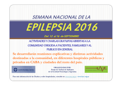
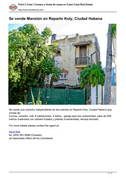
© Copyright 2026